

- •Contents
- •Preface
- •Introduction
- •The importance of cross-sectional anatomy
- •Orientation of sections and images
- •Notes on the atlas
- •References
- •Acknowledgements
- •Interpreting cross-sections: helpful hints for medical students
- •BRAIN
- •HEAD
- •NECK
- •THORAX
- •ABDOMEN
- •PELVIS
- •LOWER LIMB
- •UPPER LIMB
- •Index

UPPER LIMB
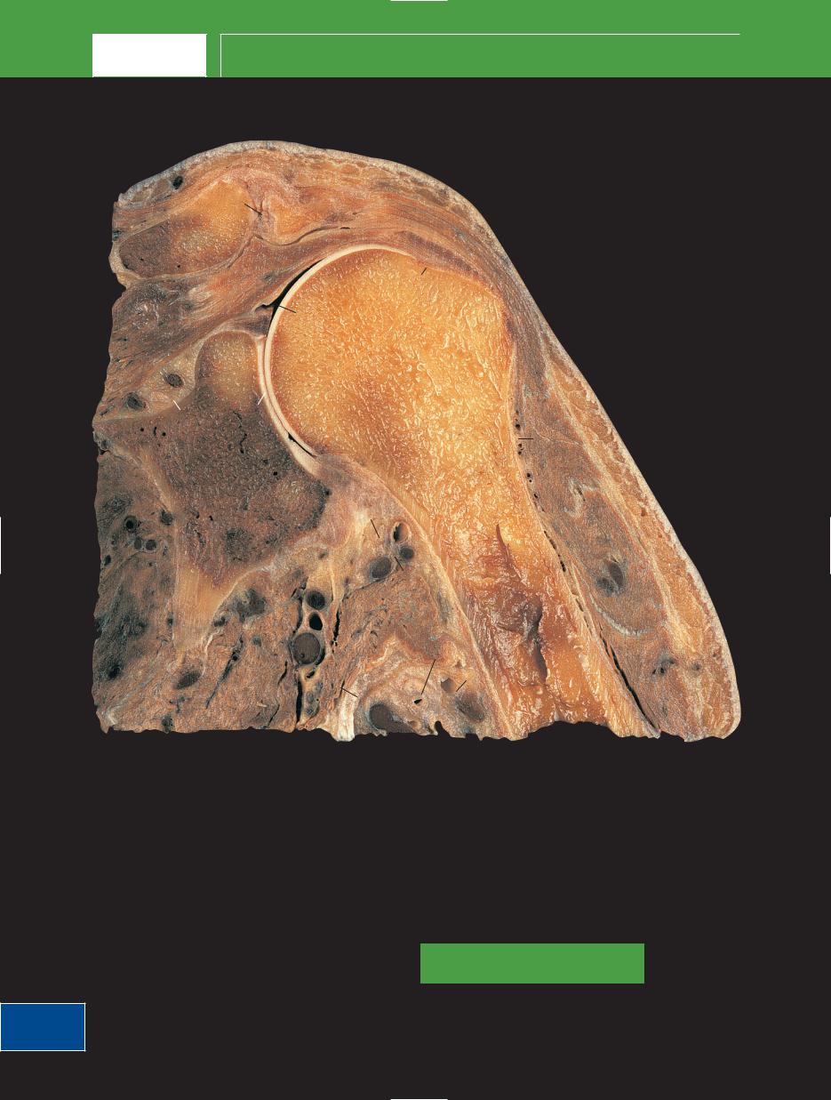
Shoulder left – Axial section 1 – Female
6
8
7 |
2 |
|
8 |
||
|
||
|
4 |
10
11
|
1 |
|
|
3 |
|
5 |
9 |
|
8 |
||
|
||
|
9 |
8
1 |
Head of humerus |
12 |
Labrum of glenoid |
2 |
Greater tubercle of |
13 |
Subscapularis tendon |
|
humerus |
14 |
Middle glenohumeral |
3 |
Glenoid fossa of |
|
ligament |
|
scapula |
15 |
Long head of biceps |
4 |
Coracoid process of |
|
tendon in bicipital |
|
scapula |
|
groove (intertubercular |
5 |
Spine of scapula |
|
groove) |
6 |
Clavicle |
16 |
Attachment of |
7 |
Subclavius |
|
coraco-acromial and |
8 |
Deltoid |
|
coraco-humeral |
9 |
Infraspinatus |
|
ligaments |
10 Subdeltoid bursa |
17 |
Lesser tubercle of |
|
11 Suprascapular artery |
|
humerus |
|
|
and vein |
18 |
Transverse humeral |
|
|
|
ligament |
|
|
|
|
238

Shoulder left – Axial section 1 – Female
UPPER LIMB
■ Section level |
■ Notes |
View
■ Orientation
Anterior
Medial 
 Lateral
Posterior
The greater tubercle of the humerus (2) is the most lateral bony landmark around the shoulder. The subacromial bursa passes below the acromion and above supraspinatus to continue into the subdeltoid bursa (10) between the upper shaft of the humerus and the deltoid muscle (8).
Infraspinatus (9), together with supraspinatus, teres minor and subscapularis, forms a protective rotator cuff around the shoulder joint, which, as can be seen in this section, has little stability afforded by either its bony configuration or its capsular strength.
The shallow glenoid is in sharp contrast to the deep acetabulum in the hip; stability has been sacrificed for mobility in order to allow a greater range of movement.
The orientation and shape of the coracoid process is an important feature; the coraco-acromial ligament can impinge on the rotator cuff.
The tendon of subscapularis attaches mainly to the lesser tubercle, but some slips attach to the floor of the intertubercular sulcus. Furthermore, the transverse humeral ligament, which retains the long head of biceps tendon, could be regarded as fibres from the subscapular’s attachment on the lesser tubercle extending on towards the greater tubercle.
818
16 |
13 |
|
|
|
|
|
|
17 |
|
2 |
|
|
|
|
|
||
4 |
|
|
15 |
8 |
|
|
|
|
|||
|
|
|
|
|
|
13 |
|
14 |
|
1 |
|
|
|
12 |
|
|
|
|
|
|
|
|
12
3
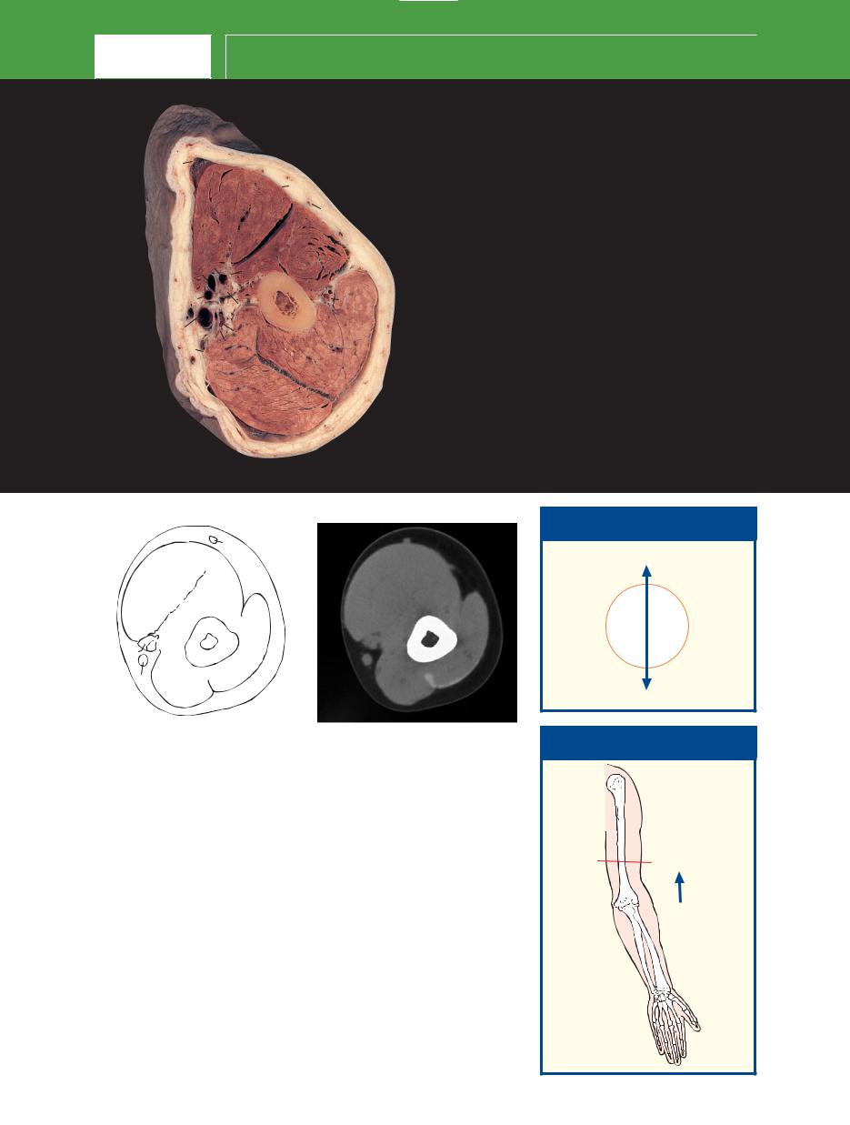
Axial magnetic resonance image (MRI)
8
9
239
UPPER LIMB
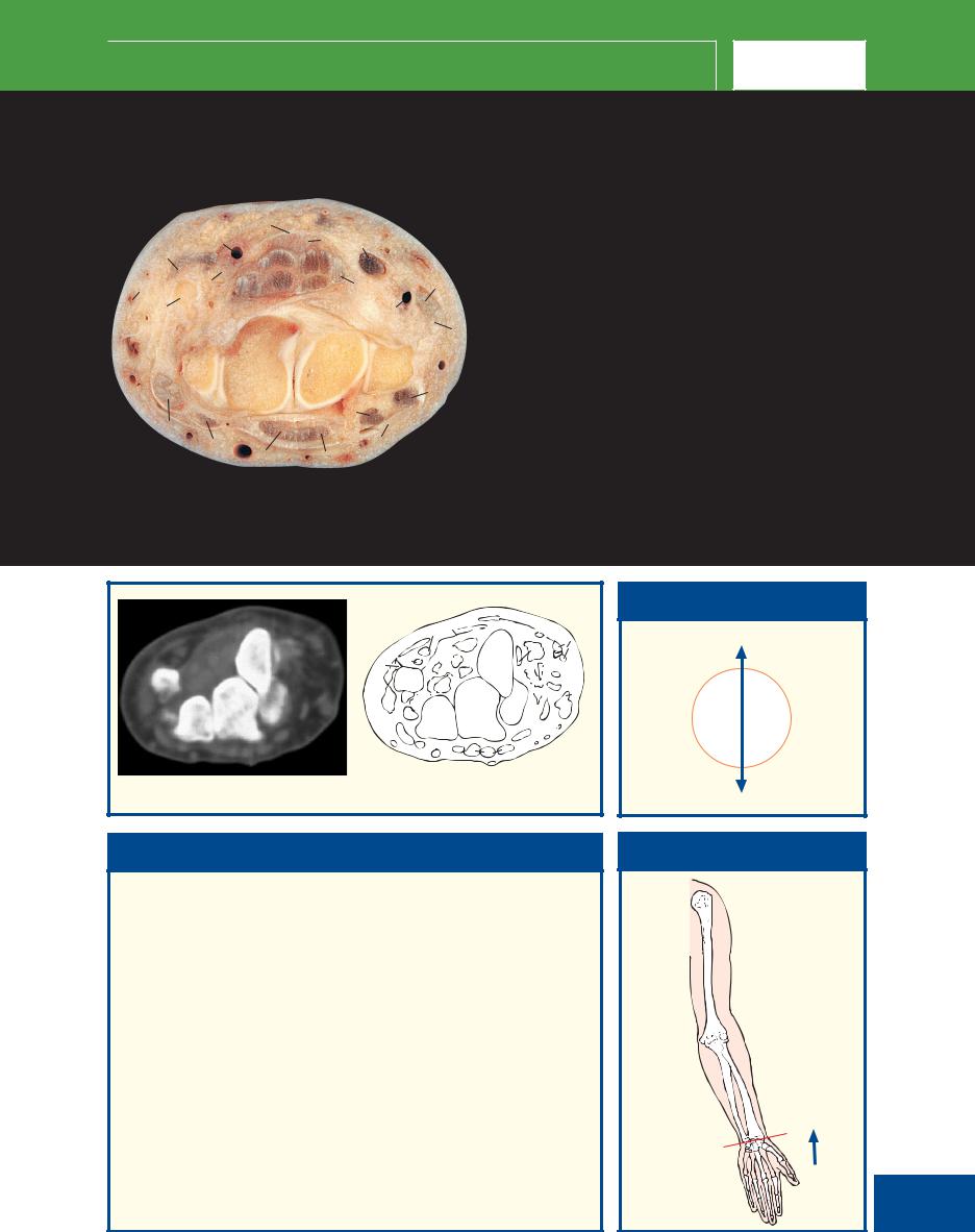
Shoulder left – Coronal section 1 – Male
2
3
1
7
4
6 |
8 |
5
20 |
21 |
|
|
|
|
|
|
10 |
|
19 |
|
|
|
10 |
|
18 |
9 |
|
11 |
22
|
|
12 |
|
|
13 |
|
|
15 |
17 |
16 |
14 |
|
||
|
|
1 |
Clavicle |
13 |
Latissimus dorsi |
2 |
Acromioclavicular joint |
14 |
Brachial artery and vein |
3 |
Acromion of scapula |
15 |
Nerves of brachial plexus |
4 |
Supraspinatus |
16 |
Tendon of teres major |
5 |
Glenoid labrum |
17 |
Teres minor |
6 |
Shoulder joint cavity |
18 |
Long head of triceps |
7 |
Anatomical neck of humerus |
19 |
Head of scapula |
8 |
Greater tubercle of humerus |
20 |
Neck of scapula |
9 |
Deltoid |
21 |
Glenoid fossa of scapula |
10 Axillary nerve accompanied by |
22 |
Subscapularis |
|
|
posterior circumflex humeral artery |
|
|
|
and vein |
23 |
Long head of biceps tendon |
11 Shaft of humerus |
24 |
Surgical neck of humerus |
|
12 Medial circumflex artery and vein |
|
|
|
240

Shoulder left – Coronal section 1 – Male
UPPER LIMB
■ Section level
View
■ Orientation
Proximal
■ Notes
The important relationship of the supraspinatus tendon (4) to the acromion process (3) and clavicle (1) is demonstrated well. This muscle initiates abduction of the shoulder, which is then continued powerfully by deltoid (9). Degenerative changes in the acromioclavicular joint frequently cause impingement on the musculotendinous junction of supraspinatus; tendonitis and a tear in the rotator cuff may follow.
Note the close relationship of the axillary nerve (10), together with its accompanying vessels, the posterior circumflex humeral artery and vein, to the surgical neck of the humerus (24). Fractures commonly occur in the region of the surgical neck; the axillary nerve may be affected. The axillary nerve may also be damaged in dislocation of the shoulder. The resultant paralysis of the deltoid muscle is demonstrated by the patient being unable to abduct the affected shoulder. There is also characteristic anaesthesia over the lateral aspect of the deltoid.
This magnetic resonance image is in a somewhat coronal oblique plane in order to demonstrate the supraspinatus muscle, tendon and insertion as a continuum.
Medial 
 Lateral
Distal
|
|
|
2 |
|
|
1 |
|
3 |
4 |
4 |
|
|
|
|
|
|
|
|
|
|
|
5 |
7 |
8 |
21 |
|
|
||
|
|
7 |
|
|
|
|
|
|
|
|
|
7 |
|
|
22 |
|
|
24 |
23 |
|
|
|
|
|
|
|
|
9 |
|
241
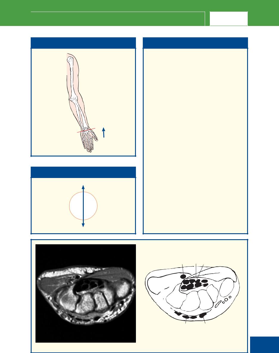
Coronal magnetic resonance image (MRI)
UPPER LIMB Selected images – Shoulder girdle
3D computed tomogram (CT)
A
3D computed tomogram (CT)
B
242

Selected images – Shoulder girdle |
|
|
UPPER LIMB |
|
|
|
|
|
|
6 |
|
|
14 |
|
|
|
|
|
|
|
|
|
|
|
|
|
|
|
|
|
5 |
|
|
|
|
1 |
5 |
|
|
|
|
22 |
21 |
14 |
|
|
||
|
7 |
|
1 |
|
7 |
||||
|
10 |
|
15 |
|
|||||
8 |
16 |
|
3 |
|
|
|
|
|
|
11 |
|
|
|
|
|
|
|||
|
|
|
|
|
|
|
|
||
|
17 |
|
4 |
|
|
|
|
16 |
|
|
12 |
|
|
|
|
11 |
|||
|
|
|
|
|
|
|
|
||
|
|
18 |
|
2 |
|
|
|
|
|
|
|
9 |
|
|
|
|
12 |
|
23 |
|
|
|
|
|
|
|
|
||
|
|
19 |
|
|
|
|
|
|
|
|
20 |
|
21 |
|
|
|
|
|
26 |
|
|
|
|
|
|
|
|
|
|
A |
|
|
|
B |
|
|
13 |
|
|
■ Orientation |
|
|
|
■ Orientation |
||||||||
|
|
|
|
|
|
|
|
|
|
|
|
|
|
|
Superior |
|
|
|
|
|
|
Superior |
|||
|
Lateral |
|
Medial |
|
|
Medial |
|
|
|
Lateral |
||
|
|
|
|
|
|
|
|
|||||
|
|
|
|
|
|
|
|
|||||
|
(right) |
|
|
|
|
|
|
|
|
|
(right) |
|
A |
|
|
|
|
|
|
B |
|
|
|
|
|
|
Inferior |
|
|
|
|
|
Inferior |
|||||
|
|
|
|
|
|
|
|
|
|
|
|
|
|
|
|
|
|
|
|
|
|
|
|
|
|
1 |
Shaft of clavicle |
11 |
Neck of scapula |
|
20 |
Shaft (proximal third) of |
||||||
2 |
Body of sternum |
12 |
Subscapular fossa of scapula |
|
humerus |
|||||||
3 |
Sternal end of clavicle |
13 |
Inferior angle of scapula |
|
21 |
Surgical neck of humerus |
||||||
4 |
Sternoclavicular joint |
14 |
Superior angle of scapula |
|
22 |
Costotransverse joint between |
||||||
5 |
Acromial end of clavicle |
15 |
Spine of scapula |
|
|
third rib and transverse process |
||||||
6 |
Acromioclavicular joint |
16 |
Head of humerus |
|
|
of third thoracic vertebra |
||||||
7 |
Subacromial space |
17 |
Greater tubercle of humerus |
23 |
Third rib |
|||||||
8 |
Acromion of scapula |
18 |
Lesser tubercle of humerus |
24 |
Infraspinous fossa |
|||||||
9 |
Lateral border of scapula |
19 |
Intertubercular sulcus of |
|
|
|
|
|
||||
10 Coracoid process of scapula |
|
humerus |
|
|
|
|
|
|||||
|
|
|
|
|
|
|
|
|
|
|
|
|
■ Notes
Surface-shaded three-dimensional volume-rendered |
important subacromial space (8) (normal in this |
|
|
CT images. Because bone attenuates the X-ray beam |
subject). The rotator cuff tendons (especially |
|
|
so much, its CT attenuation value (around +1000 HU) |
supraspinatus) have to pass though this limited space. |
|
|
is much greater than that of the surrounding soft |
Mild congenital variations in anatomy and the |
|
|
tissues. Thus, the bones can be ‘extracted’, with no |
inevitable degenerative changes in the |
|
|
overlying artefacts, to provide information equivalent |
acromioclavicular joint combine to impinge on this |
|
|
to that from a cadaveric skeleton. This subject is |
tendon. A high percentage of elderly people have |
|
|
holding the upper arm in mild internal rotation, which |
damaged rotator cuffs – one of the design flaws |
|
|
means that the bicipital groove (the groove for the |
associated with man’s evolution to a biped. |
|
|
tendon of the long head of biceps – also known as |
Note the thinness of the scapula (12), which is |
|
|
the intertubercular suclus) (19) is directed medially |
translucent in places. The strength of the scapula lies |
|
|
rather than anteriorly. |
in the border and processes; the lateral border (9) is |
|
|
The relationship of the acromioclavicular joint (6) to |
especially thick and strong for the attachment of |
|
243 |
the humeral head is well appreciated, along with the |
muscles. |
|
|
|
|
||
|
|
|
|
UPPER LIMB
Arm left – Axial section 1 – Male
1 |
|
|
|
|
|
|
|
1 |
|
|
2 |
|
|
3 |
1817 |
|
4 |
5 |
|
16 |
|
|||
14 |
|
|||
17 |
|
|
|
|
|
15 |
|
6 |
|
|
|
|
7 |
|
|
13 |
|
||
12 |
|
|
||
10 |
11 |
|
8 |
|
|
|
|
9 |
|
1 |
Deep fascia of arm |
10 |
Medial |
2 |
Biceps |
|
intermuscular |
3 |
Cephalic vein |
|
septum |
4 |
Brachialis |
11 |
Ulnar nerve |
5 |
Lateral |
12 |
Basilic vein |
|
intermuscular |
13 |
Superior ulnar |
|
septum |
|
collateral artery and |
6 |
Radial nerve, with |
|
vein |
|
profunda brachii |
14 |
Humerus shaft |
|
artery and vein |
15 |
Median nerve |
7 |
Triceps – lateral |
16 |
Musculocutaneous |
|
head |
|
nerve |
8 |
Triceps – medial |
17 |
Venae comitantes of |
|
head |
|
brachial artery |
9 |
Triceps – long head |
18 |
Brachial artery |
|
|
|
|
3 |
|
|
|
|
|
|
|
|
|
|
|
|
2 |
|
|
|
|
|
|
|
4 |
|
|
|
|
|
|
7 |
|
|
|
|
1817} |
|
14 |
|
|
|
|
12 |
9 |
8 |
|
|
|
|
|
|
|
||
|
|
|
|
|
|
|
|
|
|
|
|
|
|
|
|
|
|
|
Axial computed tomogram (CT) |
|
|
|
|
|
|
|
|
|
|
|
|
|
|
|
|
|
■ Notes |
|
|
|
|
|
|
|
|
|
||
|
|
This section passes through the mid-shaft of the humerus (14). It gives |
||||
|
|
a clear view of the fascial arrangements of the upper arm – the |
||||
|
|
investing sheath of the deep fascia (1), with its lateral (5) and medial |
||||
|
|
(10) intermuscular septa, which attach to the humeral shaft. These |
||||
|
|
septa divide the extensor group of muscles, the triceps (7, 8, 9), from |
||||
|
|
the anterior flexor group. The medial septum is pierced by the ulnar |
||||
|
|
nerve (11) and its accompanying vessels (13); the lateral septum is |
||||
|
|
pierced by the radial nerve with its accompanying profunda brachii |
||||
|
|
artery and vein (6). |
||||
|
|
The median nerve (15) and brachial artery (18) bear a close |
||||
|
|
relationship to each other in the upper arm, as shown in this section. |
||||
|
|
Superiorly, the nerve lies on the lateral side of the artery. At the mid- |
||||
|
|
humerus level, the artery is crossed superficially (sometimes deeply) by |
||||
244 |
|
|||||
|
the nerve, which then descends on its medial side. |
|||||
|
|
|
|
|
|
|
■ Orientation
Anterior
Medial 
 Lateral
Posterior
■ Section level
View

Elbow left – Axial section 1 – Male
UPPER LIMB
|
|
1 |
1 |
Cephalic vein |
10 |
Ulnar nerve |
|
2 |
|
2 |
Biceps |
11 |
Medial |
15 |
|
|
3 |
Brachialis |
|
intermuscular |
16 |
3 |
4 |
4 |
Brachioradialis |
|
septum |
|
|
5 |
Extensor carpi |
12 |
Basilic vein |
|
15 |
|
|
||||
|
|
|
radialis longus |
13 |
Medial cutaneous |
|
13 |
14 |
|
|
|||
|
6 |
Lateral |
|
nerve of forearm |
||
|
|
5 |
|
|||
12 |
|
|
intermuscular |
14 |
Median nerve |
|
|
|
|
||||
|
|
|
septum |
15 |
Venae comitantes of |
|
|
9 |
6 |
|
|||
|
7 |
Triceps tendon |
|
brachial artery |
||
|
|
8 |
8 |
Triceps |
16 |
Brachial artery |
11 |
|
9 |
Humerus |
|
|
|
10 |
|
|
|
|
|
|
|
|
7 |
|
|
|
|
1 4
2
5
 1516 3 14
12
98
7
Axial computed tomogram (CT)
■ Notes
This section transects the lower end of the humeral shaft as it expands to form its medial and lateral supracondylar ridges.
The origin of extensor carpi radialis longus (5) is from the upper part of the lateral ridge, and this muscle arises superior to, and separate from, the remaining extensor muscles of the forearm, which originate from a common origin from the lateral epicondyle of the humerus.
The ulnar nerve (10), just distal to the line of this section, will pass behind the medial epicondyle of the humerus; pressure here will elicit discomfort and often paraesthesia.
■ Orientation
Anterior
Medial 
 Lateral
Posterior
■ Section level
View
245

UPPER LIMB
Elbow left – Axial section 2 – Male
|
|
1 |
|
1 |
Cephalic vein |
12 |
Articular cartilage |
|
|
|
2 |
Biceps tendon |
13 |
Medial collateral |
|
|
|
3 |
|
||||
22 |
23 |
|
3 |
Brachioradialis |
|
ligament of elbow |
|
|
2 |
|
4 |
Extensor carpi |
14 |
Trochlea of humerus |
|
|
21 |
20 |
4 |
||||
|
|
radialis longus |
15 |
Capitulum of |
|||
|
|
5 |
|
|
|||
20 |
|
6 |
5 |
Radial nerve with |
|
humerus |
|
|
16 |
|
|||||
|
|
|
|
profunda brachii |
16 |
Brachialis |
|
|
18 |
|
|
|
|||
|
|
|
|
artery and vein |
17 |
Common flexor |
|
|
|
8 |
|
|
|||
19 |
|
7 |
6 |
Common extensor |
|
origin |
|
|
15 |
|
|||||
|
17 |
|
|
origin |
18 |
Median nerve |
|
|
|
|
|
||||
|
|
|
7 |
Lateral collateral |
19 |
Basilic vein |
|
|
|
14 |
|
||||
|
|
|
|
ligament of elbow |
20 |
Venae comitantes of |
|
|
13 |
|
|
||||
|
8 |
8 |
Joint capsule of |
|
brachial artery |
||
|
|
12 |
|
|
elbow |
21 |
Brachial artery |
|
|
11 |
|
9 |
Olecranon bursa |
22 |
Median cubital vein |
|
|
10 |
|
10 Ulnar nerve |
23 |
Bicipital aponeurosis |
|
|
|
|
|
11 Olecranon process |
|
|
|
|
|
9 |
|
|
of ulna |
24 |
Anconeus |
1
23 2  3
3
22
16 |
6 |
{2021

19
15
14
17
24 11
Axial computed tomogram (CT)
■ Notes
This section transects the elbow joint. The cartilage (12) covering the articular surfaces of the lower end of the humerus (14, 15) and the olecranon process of the ulna (11), together with the joint cavity and collateral ligaments (8), are readily appreciated.
The posterior surface of the olecranon process of the ulna is separated from the skin by a bursa (9). This is a common site for bursitis (‘student’s elbow’, ‘miner’s elbow’).
The median nerve (18), here lying medial to the brachial artery (21) (see note on page 244) is well-named. It lies in the median position throughout its course in the upper arm, at the elbow, in the forearm and at the wrist as it passes into the carpal tunnel below the flexor retinaculum.
246
■ Orientation
Anterior
Medial 
 Lateral
Posterior
■ Section level
View

Elbow left – Axial section 3 – Male
UPPER LIMB
|
|
|
|
1 |
Brachioradialis |
12 |
Flexor carpi ulnaris |
|
19 |
|
|
2 |
Extensor carpi |
13 |
Ulnar nerve, with |
|
25 |
|
|
radialis longus |
|
posterior recurrent |
|
|
|
|
3 |
Extensor carpi |
|
ulnar artery and |
|
|
23 |
22 |
|
26 |
radialis brevis |
|
vein |
|
|
1 |
Radial nerve with |
14 |
Radial notch of ulna |
||
|
21 |
|
4 |
||||
17 |
|
24 |
|
radial recurrent |
15 |
Flexor digitorum |
|
18 |
22 |
|
artery |
|
superficialis |
||
|
|
|
|
|
|||
16 |
|
2 |
5 |
Supinator |
16 |
Palmaris longus |
|
20 |
|
||||||
|
|
4 |
6 |
Head of radius |
17 |
Flexor carpi radialis |
|
15 |
|
|
5 |
||||
|
|
7 |
Common extensor |
18 |
Pronator teres |
||
|
|
|
|
|
origin |
19 |
Basilic vein |
13 |
14 |
6 |
3 |
8 |
Annular ligament of |
20 |
Brachialis |
|
|
superior radio-ulnar |
21 |
Median nerve |
|||
|
|
|
|||||
12 |
|
|
|
|
joint |
22 |
Venae comitantes of |
11 |
|
8 |
7 |
9 |
Anconeus |
|
brachial artery |
|
|
9 |
10 Deep fascia of the |
23 |
Brachial artery |
||
|
|
|
|||||
|
|
|
|
forearm |
24 |
Tendon of biceps |
|
|
10 |
|
|
|
|||
|
|
|
11 Flexor digitorum |
25 |
Median cubital vein |
||
|
|
|
|
||||
|
|
|
|
|
profundus |
26 |
Cephalic vein |
12
25 |
|
3 |
|
|
24 |
7 |
|
22 |
5 |
||
6 |
|||
23 |
|
|
18
20 14 9
17
16 15 12 11
Axial computed tomogram (CT)
■ Notes
This section passes through the superior radio-ulnar joint between the head of the radius (6) and the radial notch of the ulnar (14). The annular ligament (8), which maintains the congruity of this pivot joint, is shown well. In this CT image, the hand is in the neutral position alongside the body.
The median cubital vein (25) passes obliquely across the front of the elbow between the cephalic vein (26) and the basilic vein (19). It is separated from the underlying brachial artery (23) by a condensation of the deep fascia (10) termed the bicipital aponeurosis. Occasionally, in high division of the brachial artery, an abnormal ulnar artery may lie immediately below the median cubital vein in the superficial fascia. This vein is therefore best avoided for intravenous injections in order to protect against inadvertent intra-arterial injection.
■ Orientation
Anterior
Medial 
 Lateral
Posterior
■ Section level
View
247

UPPER LIMB
Elbow left – Coronal section 1 – Female
4 |
1 |
5
6
7
18
5
16
17
2
3
4
9
8
10
11
12
14
13
15
19
19 20
1 |
Shaft of humerus |
15 |
Extensor carpi radialis |
2 |
Lateral head of triceps |
|
longus |
3 |
Radial nerve |
16 |
Flexor carpi ulnaris |
4 |
Medial head of triceps |
17 |
Flexor digitorum |
5 |
Ulnar nerve |
|
profundus |
6 |
Medial epicondyle of |
18 |
Coronoid process of |
|
humerus |
|
ulna |
7 |
Trochlea of humerus |
19 |
Shaft of radius |
8 |
Capitulum of humerus |
20 |
Extensor carpi radialis |
9 |
Brachioradialis |
|
brevis |
10 Annular ligament |
|
|
|
11 Head of radius |
21 |
Olecranon fossa of |
|
12 Neck of radius |
|
humerus |
|
13 Tendon of biceps |
22 |
Lateral epicondyle of |
|
14 Supinator |
|
humerus |
|
|
|
|
|
248

Elbow left – Coronal section 1 – Female
UPPER LIMB
■ Section level |
■ Notes |
View
■ Orientation
Proximal
Medial 
 Lateral
Distal
The ulnar nerve (5) passes posterior to the medial epicondyle of the humerus (6), where it may be palpated. It may be injured at this site in fractures or dislocations around the elbow, or stretched in valgus deformity of this joint.
The tendon of biceps (13) inserts into the posterior lip of the tuberosity of the radius. It is a powerful supinator of the radio-ulnar joints and a flexor of the elbow joint.
The brachial vessels are in close anterior proximity to the elbow joint; the artery may be compromised in supracondylar fractures, which are relatively common in children.
The epicondyles have developed to provide attachment of the common extensor (lateral epicondyle) and flexor (medial epicondyle) muscle groups. Inflammation of the extensor origin on the lateral epicondyle (22) is known as ‘tennis elbow’. This section provides an excellent view of the superior radio-ulnar joint between the head of the radius (11) and the radial notch of the ulna (18). It communicates freely with the elbow joint. Together with the inferior radio-ulnar joint, it allows the movements of pronation and supination of the forearm, which are unique to the primate upper limb.
2
1
6
21
722
8
18
11
Coronal magnetic resonance image (MRI)
249

UPPER LIMB
Forearm left – Axial section 1 – Male
|
|
|
|
|
|
|
1 |
Palmaris longus |
15 |
Extensor digitorum |
|
|
|
|
|
|
|
2 |
Flexor carpi radialis |
16 |
Radius |
|
|
|
|
|
|
|
3 |
Flexor digitorum |
17 |
Posterior |
|
|
|
|
|
|
|
|
superficialis |
|
interosseous nerve |
|
|
1 |
|
|
|
|
4 |
Pronator teres – |
18 |
Posterior |
|
|
|
|
|
2 |
|
|
humeral head |
|
interosseous artery |
|
26 |
3 |
|
|
|
5 |
Ulnar artery |
|
and vein |
|
|
|
|
|
|
6 |
Ulnar vein |
19 |
Extensor carpi |
||
|
24 |
|
|
|
|
|||||
23 |
5 |
|
|
4 |
|
7 |
Median nerve, with |
|
ulnaris |
|
25 |
|
|
9 |
|
||||||
|
|
6 |
|
8 |
|
anterior |
20 |
Anconeus |
||
|
|
|
7 |
11 |
|
|
interosseous artery |
21 |
Ulna |
|
|
22 |
|
|
|
|
|||||
|
|
|
|
10 |
|
|
and vein |
22 |
Flexor digitorum |
|
|
|
|
|
16 |
12 |
|
8 |
Radial artery, with |
|
profundus |
|
|
|
|
|
|
|
venae comitantes |
23 |
Basilic vein |
|
|
|
|
|
|
13 |
|
|
|||
|
21 |
|
18 |
|
|
9 |
Cephalic vein |
24 |
Flexor carpi ulnaris |
|
|
|
|
|
17 |
14 |
|
10 Brachioradialis |
25 |
Ulnar nerve |
|
|
20 |
|
|
|
|
11 Radial nerve |
26 |
Deep fascia of |
||
|
|
|
|
|
|
|||||
|
|
19 |
|
15 |
|
|
12 Supinator |
|
forearm |
|
|
|
|
|
|
|
13 Extensor carpi |
|
|
||
|
|
|
|
|
|
|
|
|
||
|
|
|
|
|
|
|
|
radialis longus |
27 |
Pronator teres (ulnar |
|
|
|
|
|
|
|
14 Extensor carpi |
|
head) |
|
|
|
|
|
|
|
|
|
radialis brevis |
|
|
|
|
|
|
|
|
|
|
|
|
|
|
|
|
|
2 |
|
|
|
23 |
24 |
3 |
|
|
|
|
|
25 |
5/6 |
7 |
|
|
|||
|
|
4 |
|
||||
|
|
|
|
|
|||
|
|
|
|
|
|
|
|
|
22 |
|
|
|
27 |
9 |
|
|
|
|
|
|
|
||
|
|
21 |
|
|
|
8/11 |
10 |
|
|
|
16 |
|
|
||
|
|
|
|
|
12 |
|
|
|
20 19 |
|
|
|
|
|
|
13
14
15 
Axial computed tomogram (CT)
■ Notes
This section passes through the mid forearm. In both the section and the CT image, the forearm is viewed in the supinated position. Note how the median nerve (7) characteristically hugs the deep aspect of flexor digitorum superficialis (3). The ulnar nerve (25) lies sandwiched between flexor carpi ulnaris (24) and flexor digitorum profundus (22), and the radial nerve (11) lies beneath brachioradialis (10).
■ Orientation
Anterior
Medial 
 Lateral
Posterior
■ Section level
View
250

Forearm left – Axial section 2 – Male
UPPER LIMB
|
|
|
|
|
|
|
|
1 |
Palmaris longus |
17 |
Extensor pollicis |
|
|
|
|
|
|
|
|
|
tendon |
|
longus |
|
|
|
|
|
|
|
|
2 |
Flexor digitorum |
18 |
Extensor carpi ulnaris |
|
|
|
|
1 |
|
|
|
|
superficialis |
19 |
Ulna |
|
|
|
|
|
|
|
3 |
Flexor carpi radialis |
20 |
Interosseous |
|
|
|
|
|
|
|
|
|
||||
|
26 |
|
27 |
2 |
|
|
|
4 |
Median nerve |
|
membrane |
|
|
|
|
|
|
5 |
Radial artery |
21 |
Anterior interosseous |
||
|
|
|
|
3 |
|
|
|||||
|
25 |
24 |
|
|
|
|
6 |
Brachioradialis |
|
artery, vein and nerve |
|
|
|
|
|
5 |
6 |
|
|
||||
|
|
|
4 |
|
7 |
Radial nerve |
22 |
Flexor digitorum |
|||
|
|
|
|
|
|||||||
23 |
|
|
22 |
|
|
7 |
8 |
8 |
Cephalic vein |
|
profundus |
|
|
|
|
|
9 |
Pronator teres tendon |
23 |
Basilic vein |
|||
|
|
|
12 |
|
|
||||||
|
|
|
|
|
|
|
|||||
|
|
|
|
|
|
|
10 Extensor carpi radialis |
24 |
Ulnar nerve |
||
|
|
|
|
21 |
9 |
|
|
||||
|
|
|
|
|
|
|
longus and brevis |
25 |
Deep fascia of |
||
|
|
|
|
11 |
|
|
|
||||
|
|
|
19 |
|
|
|
11 Radius |
|
forearm |
||
|
|
|
20 |
|
10 |
|
|
||||
|
|
|
|
|
12 Flexor pollicis longus |
26 |
Flexor carpi ulnaris |
||||
|
|
|
|
|
|
||||||
|
|
|
|
|
|
|
|
||||
|
|
18 |
17 15 16 |
|
|
|
13 Extensor digitorum |
27 |
Ulnar artery with |
||
|
|
|
|
|
|
|
14 Extensor digiti minimi |
|
venae comitantes |
||
|
|
|
|
|
|
|
|
|
|||
|
|
|
14 |
13 |
|
|
|
15 Posterior interosseous |
|
|
|
|
|
|
|
|
|
|
|
||||
|
|
|
|
|
|
|
nerve, with artery |
28 |
Superficial flexor |
||
|
|
|
|
|
|
|
|
||||
|
|
|
|
|
|
|
|
|
and vein |
|
group of muscles |
|
|
|
|
|
|
|
|
16 Abductor pollicis |
29 |
Extensor group of |
|
|
|
|
|
|
|
|
|
|
longus |
|
muscles |
|
|
|
|
|
|
|
|
|
|
|
|
|
|
28 |
|
|
|
27 |
|
4 |
6 |
26 |
|
|
||
|
|
|
||
22 |
|
|
|
8 |
|
|
|
|
5 |
|
|
20 |
12 |
|
19 |
|
|
|
|
|
|
|
|
|
18 |
|
29 |
|
11 |
|
|
|
Axial computed tomogram (CT)
■ Notes
This section transects the supinated forearm at the junction of its upper two-thirds and lower one-third. Note that the very extensive origin of flexor digitorum profundus (22) is demonstrated clearly by this section. It arises from both the anterior and medial surfaces of the upper threequarters of the ulna (19), from the ulnar half of the interosseous membrane (20) and also from the superior three-quarters of the posterior border of the ulna by an aponeurosis that is in common with that of flexor carpi ulnaris (26) and extensor carpi ulnaris (18).
■ Orientation
Anterior
Medial 
 Lateral
Posterior
■ Section level
View
251
UPPER LIMB
Wrist left – Axial section 1 – Male
|
|
|
1 |
|
|
|
10 |
2 |
|
|
4 |
|
|
|
|
3 |
|
9 |
8 |
7 |
6 |
5 |
12 |
|
|
||||
|
|
|
|
||
|
|
|
11 |
|
|
|
|
|
|
|
15 |
|
|
|
20 |
|
18 |
|
|
|
|
|
|
|
26 |
|
|
|
|
|
25 |
24 |
23 |
|
|
|
|
|
|
|
21 |
|
|
|
22 |
|
|
|
1 |
Palmaris longus tendon |
15 |
Extensor pollicis brevis |
|
|
2 |
Flexor digitorum |
|
tendon |
|
|
|
superficialis tendons |
16 |
Radial nerve |
|
|
3 |
Median nerve |
17 |
Cephalic vein |
|
|
4 |
Flexor carpi radialis |
18 |
Extensor carpi radialis |
|
|
|
tendon |
|
longus tendon |
|
|
5 |
Flexor pollicis longus |
19 |
Extensor carpi radialis |
|
|
|
tendon |
|
brevis tendon |
|
|
6 |
Flexor digitorum |
20 |
Radius |
|
13 |
|
profundus tendon to |
21 |
Extensor pollicis longus |
|
|
index finger |
|
tendon |
||
14 |
|
|
|||
7 |
Flexor digitorum |
22 |
Extensor digitorum |
||
|
|||||
16 |
|
profundus tendon to |
|
tendon |
|
|
|
remaining fingers |
23 |
Extensor indicis |
|
17 |
8 |
Ulnar nerve |
24 |
Extensor digiti minimi |
|
9 |
Flexor carpi ulnaris |
|
tendon |
||
19 |
|
tendon |
25 |
Extensor carpi ulnaris |
|
|
10 Ulnar artery |
|
tendon |
||
|
11 Pronator quadratus |
26 |
Ulna |
||
|
12 Radial artery |
|
|
||
|
13 Brachioradialis insertion |
27 |
Superficial vein |
||
|
14 Abductor pollicis longus |
|
|
||
|
|
|
|||
|
|
tendon |
|
|
|
|
2 |
1 |
|
3 |
|
12 |
|
|
|
|
4 |
|
|
||
|
|
|
|
|
|
|
|
9 |
|
6 |
|
5 |
|
13 |
|
|
7 |
|
|
|
|
||
|
|
|
|
|
14 |
|
|
|
|
11 |
|
20 |
15 |
17 |
|
|
|
|
|
|
|
|
|
|
|
|
|
|
|
18 |
|
|
|
|
|
|
|
19 |
|
|
26 |
|
|
|
|
27 |
|
|
|
25 |
|
|
|
|
|
|
|
|
23 |
|
|
|
|
|
|
24 |
|
21 |
|
|
|
|
|
|
22 |
|
|
|
|
Axial magnetic resonance image (MRI)
■ Orientation
Anterior
Medial 
 Lateral
Posterior
■ Notes |
|
■ Section level |
|
|
|
This section transects the forearm immediately proximal to the wrist joint. The arrangement of the extensor tendons on the posterior and radial aspects of the wrist can be appreciated clearly. Note that extensor carpi ulnaris tendon (25) grooves the dorsal aspect of the distal ulna (26).
At this level, flexor digitorum profundus has given off a separate tendon to the index finger (6), while those for the remaining three fingers are still closely applied to each other (7).
Usually the cephalic vein (17) is easily visible at this site; here, it is a common locus for venous cannulation.
View
252
Wrist left – Axial section 2 – Male
UPPER LIMB
|
|
|
|
|
|
1 |
Flexor pollicis longus |
15 |
Extensor pollicis longus |
|
|
|
|
|
|
|
tendon |
|
tendon |
|
|
|
|
|
|
2 |
Median nerve |
16 |
Extensor indicis tendon |
|
|
|
|
|
|
3 |
Flexor digitorum |
17 |
Extensor digitorum |
|
|
|
|
|
|
|
superficialis tendons |
|
tendon |
|
24 |
25 |
2 |
|
|
4 |
Flexor digitorum |
18 |
Extensor digiti minimi |
23 |
|
5 |
|
|
profundus tendons |
|
tendon |
||
|
3 |
|
|
|
5 |
Flexor carpi radialis |
19 |
Extensor carpi ulnaris |
|
|
|
|
|
|
|||||
21 |
22 |
4 |
|
1 |
6 |
|
tendon |
|
tendon |
|
6 |
Abductor pollicis longus |
20 |
Pisiform |
|||||
|
|
|
7 |
|
|||||
20 |
|
|
|
|
|
tendon |
21 |
Basilic vein |
|
|
|
|
|
8 |
|
||||
|
|
|
|
|
7 |
Radial artery |
22 |
Ulnar nerve |
|
|
|
11 |
|
|
|
8 |
Extensor pollicis brevis |
23 |
Flexor carpi ulnaris |
|
|
10 |
9 |
|
|
tendon |
|
tendon |
|
|
12 |
|
|
9 |
24 |
||||
|
|
|
|
|
13 |
Styloid process of radius |
Ulnar artery |
||
|
|
|
|
14 |
10 Scaphoid |
25 |
Flexor retinaculum |
||
|
|
|
|
|
|||||
19 |
|
|
|
|
|
11 Lunate |
|
|
|
18 |
17 |
16 |
15 |
|
12 Triquetral |
26 |
Capitate |
||
|
|
||||||||
|
|
|
|
13 Extensor carpi radialis |
27 |
Hamate |
|||
|
|
|
|
|
|
|
longus tendon |
28 |
Trapezoid |
|
|
|
|
|
|
14 Extensor carpi radialis |
29 |
Trapezium |
|
|
|
|
|
|
|
|
brevis tendon |
|
|
|
|
|
|
|
|
|
|
|
|
|
|
24 |
25 |
|
|
|
|
|
|
2 |
|
|
6 |
|
|
|
|
29 |
8 |
|
|
22 |
|
3 |
10 |
|
|
23 |
3 |
|
|
|||
|
4 |
|
|
|
||
|
|
|
|
|
|
20 |
4 |
|
13
|
|
26 |
28 |
14 |
|
|
|
||
12 |
27 |
|
15 |
|
|
|
|
||
|
|
|
16 |
|
|
|
|
17 |
|
Axial computed tomogram (CT)
■ Notes
This section passes through the proximal row of carpal bones and the radial styloid process. The CT image is at a more distal level.
The radius (9) extends more distally than the ulna; thus, abduction of the wrist is more limited than adduction.
The pisiform bone (20) can be considered as a sesamoid within the termination of the tendon of flexor carpi ulnaris (23), which anchors via the pisohamate ligament to the hook of the hamate and via the pisometacarpal ligament to the base of the fifth metacarpal bone.
The flexor retinaculum (25) is a tough fibrous band across the front of the carpus, which converts its concavity into the carpal tunnel, transmitting the flexor tendons of the digits together with the median nerve (2). Its attachments can be seen in this section and on page 254, medially to the pisiform (20) and to the hook of the hamate (27), laterally as two laminae, the more superficial one being attached to the tubercles of the scaphoid (10) and the trapezium (29) and the deep lamina to the medial lip of the groove on the latter.
■ Orientation
Anterior
Medial 
 Lateral
Posterior
■ Section level


 View
View
253

UPPER LIMB
Wrist left – Axial section 3 – Male
|
|
29 |
|
30 |
|
|
25 |
27 |
22 |
|
|
|
|
|
|
|
|
|
||
|
|
|
28 |
18 |
|
|
26 |
|
|
|
|
1 |
|
|
21 |
|
17 |
|
||
|
|
|
|
|
||
|
22 |
20 |
19 |
|
|
2 |
23 |
|
|
|
|
||
|
|
|
|
|
|
|
24 |
|
|
16 |
15 |
|
3 |
|
|
|
4 |
|||
|
12 |
|
|
|||
|
|
|
|
|
|
|
11 |
13 |
14 |
|
|
5 |
|
|
|
|
|
|||
|
|
|
|
|
|
|
|
|
|
|
|
6 |
|
|
|
|
8 |
|
7 |
|
10 |
|
9 |
|
|
|
|
1 |
Abductor pollicis longus tendon |
18 |
Flexor carpi radialis tendon |
2 |
Extensor pollicis brevis tendon |
19 |
Flexor pollicis longus tendon |
3 |
Cephalic vein |
20 |
Flexor digitorum profundus |
4 |
Radial artery |
|
tendons |
5 |
Extensor pollicis longus tendon |
21 |
Flexor digitorum superficialis |
6 |
Extensor carpi radialis longus |
|
tendons |
|
tendon |
22 |
Flexor retinaculum |
7 |
Extensor carpi radialis brevis |
23 |
Muscles of hypothenar |
|
tendon |
|
eminence |
8 |
Extensor indicis tendon |
24 |
Pisometacarpal ligament |
9 |
Extensor digitorum tendons |
25 |
Palmaris brevis |
10 Extensor digiti minimi tendon |
26 |
Ulnar nerve |
|
11 Extensor carpi ulnaris tendon |
27 |
Ulnar artery |
|
12 Triquetral |
28 |
Median nerve |
|
13 Hamate |
29 |
Palmaris longus tendon |
|
14 Capitate |
30 |
Muscles of thenar eminence |
|
15 Trapezoid |
|
|
|
16 Scaphoid |
31 |
Base of thumb metacarpal |
|
17 Trapezium |
|
|
|
|
|
||
254
Wrist left – Axial section 3 – Male
UPPER LIMB
■ Section level |
■ Notes |
View
■ Orientation
Anterior
Medial 
 Lateral
Posterior
This section passes through the distal part of the carpus. The bony arch is seen well. The flexor retinaculum (22) has already been described (see page 253). Here, its distal attachment to the trapezium (17) and the hook of the hamate (13) can be seen. Note the tendon of flexor carpi radialis (18) lying in the tunnel formed by the groove on the trapezium and the two laminae of the lateral attachment of the retinaculum.
Swelling or deformity within the carpal tunnel compresses the median nerve (28) and produces carpal tunnel syndrome. The ulnar nerve (26) – part of a neurovascular bundle with the ulnar artery and its venae commitantes (27) – passes superficially to the flexor retinaculum and is, therefore, not implicated in this syndrome.
27 |
22 |
28 |
|
|
30
23
31 17
13 |
14 |
15 |
|
||
|
|
9 |
8 |
255
Axial magnetic resonance image (MRI)

UPPER LIMB
Wrist/hand left – Coronal section 1 – Female
34
|
|
|
|
33 |
|
|
|
|
|
|
|
29 |
28 |
|
|
|
|
|
|
|
|
|
27 |
|
|
|
|
|
|
|
32 |
|
|
26 |
|
|
24 |
|
|
|
|
|
|
|
|
|
|
|
|
|
|
|
|
25 |
|
|
|
|
|
|
|
|
|
|
|
23 |
|
|
|
|
|
|
|
|
|
|
22 |
|
|
|
|
31 |
30 |
|
|
|
|
|
|
|
|
|
20 |
21 |
|
18 |
|
|
|
|
|
|
|
17 |
19 |
|
|
||
|
|
|
|
|
|
|
|||
|
|
|
|
15 |
16 |
|
|
|
|
|
|
|
12 |
|
14 |
|
|
|
|
|
|
|
|
|
|
|
|
||
|
|
|
11 |
13 |
|
|
10 |
|
|
|
|
|
|
|
|
9 |
|
|
|
|
|
|
|
|
|
7 |
|
|
|
|
|
|
6 |
|
|
8 |
|
|
|
|
|
|
|
|
|
|
|
|
|
|
|
|
|
5 |
|
|
|
|
|
|
|
|
|
|
|
4 |
|
|
|
|
|
|
|
3 |
|
|
|
|
|
|
|
|
1 |
|
|
|
|
|
|
|
|
|
|
|
|
2 |
|
|
|
1 |
Shaft of ulna |
15 |
Hamate |
|
|
|
28 |
Common digital artery, vein and |
|
2 |
Shaft of radius |
16 |
Capitate |
|
|
|
|
|
nerve |
3 |
Flexor digitorum profundus (see |
17 |
Trapezoid |
|
|
|
29 |
Digital fibrous sheath of ring |
|
|
also 33) |
18 |
Trapezium |
|
|
|
|
|
finger |
4 |
Flexor pollicis longus |
19 |
Radial artery in anatomical |
30 |
Flexor digiti minimi |
||||
5 |
Pronator quadratus |
|
snuffbox |
|
|
|
31 |
Abductor digiti minimi |
|
6 |
Head of ulna |
20 |
Base of little finger bone |
32 |
Base of proximal phalanx of |
||||
7 |
Distal end of radius |
21 |
Distal opening of carpal tunnel |
little finger |
|||||
8 |
Abductor pollicis longus |
|
(arrowed) |
|
|
|
33 |
Tendon of flexor digitorum |
|
9 |
Extensor pollicis brevis |
22 |
Extensor pollicis longus |
|
|
profundus of index finger (see |
|||
10 Radial styloid process |
23 |
Abductor pollicis |
|
|
|
|
also 3) |
||
11 Articular disc (triangular |
24 |
Head of first metacarpal |
34 |
Tendon of flexor digitorum |
|||||
|
fibrocartilaginous complex, |
25 |
Second lumbrical |
|
|
|
|
superficialis of index finger |
|
|
TFCC) |
26 |
Tendon of flexor digitorum |
|
|
|
|||
12 Triquetral |
|
profundus |
|
|
|
|
35 |
Ulnar styloid |
|
13 Lunate |
27 |
Tendon of flexor digitorum |
|
36 |
Base of index metacarpal bone |
||||
14 Scaphoid |
|
superficialis |
|
|
|
|
|
|
|
|
|
|
|
|
|
|
|||
256

Wrist/hand left – Coronal section 1 – Female
UPPER LIMB
■ Section level
View
■ Orientation
Distal
Medial 
 Lateral
Proximal
■ Notes
Note that in the anatomical position of the wrist joint, the scaphoid (14) and lunate (13) are in contact with the distal end of the radius (7). The triquetral (12) articulates against the articular disc (11) only when the hand is adducted. The triquetral is, therefore, almost never injured in falls on the hand.
The pulse of the radial artery (19) is readily palpated in the anatomical snuffbox as the artery lies against the underlying scaphoid (14).
The distal end of the ulna is fractionally shorter than that of the radius. Thus, an articular disc (the triangular fibrocartilaginous complex, TFCC) runs from the ulnar styloid to the radius to complete the proximal part of the ellipsoid wrist joint. An articular disc implies two types of movement: the radius supinates and pronates around the ulna proximal to the disc. Minor variance in ulnar length probably contributes to damage to the TFCC in later life.
20 |
|
36 |
|
31 |
20 |
36 |
18 |
|||
|
|
|
|
|
||||||
|
|
15 |
17 |
|
|
|
|
15 |
17 |
|
|
|
16 |
|
|
|
|
|
16 |
|
|
11 12 |
|
|
|
|
12 |
|
|
|||
|
|
|
|
|
14 |
|
||||
14 |
|
|
|
|
13 |
|
||||
35 |
13 |
|
|
|
|
10 |
|
|||
|
10 |
|
|
|
|
6 |
|
|||
|
|
6 |
|
|
|
|
|
|||
|
|
|
|
|
|
7 |
|
|||
|
|
|
|
|
|
|
|
|||
|
|
|
7 |
|
|
|
|
5 |
|
|
|
|
|
|
|
|
|
|
|
|
|
5 |
|
|
|
|
|
|
|
|
|
|
|
1 |
|
|
|
|
|
|
|
|
|
A |
|
|
A |
|
|
|
|
|
|
|
|
|
|
B |
1 |
|
|
|
|||
|
|
|
|
|
|
|
||||
|
|
|
|
|
|
|
|
|
|
|
Coronal magnetic resonance image (MRI)
B
Coronal magnetic resonance image (MRI)
257

UPPER LIMB
Wrist/hand left – Sagittal section 1 – Female
19
17 18
16
15
14
8 13 11
12
10
7 9
65
4
3
2
1
1 |
Extensor digitorum |
12 |
Tendon of flexor |
2 |
Pronator quadratus |
|
digitorum profundus |
3 |
Distal end of radius |
13 |
Adductor pollicis |
4 |
Wrist joint |
14 |
Extensor expansion |
5 |
Capsule of wrist joint |
15 |
Proximal phalanx of |
6 |
Lunate |
|
middle finger |
7 |
Capitate |
16 |
Middle phalanx of |
8 |
Metacarpal bone of |
|
middle finger |
|
middle finger |
17 |
Distal phalanx of |
9 |
Flexor retinaculum |
|
middle finger |
10 Palmar aponeurosis |
18 |
Pulp space of distal |
|
11 Tendon of flexor |
|
phalanx |
|
|
digitorum superficialis |
19 |
Nail bed |
258

Wrist/hand left – Sagittal section 1 – Female
UPPER LIMB
■ Section level
8
11
12
7
6
View
3
2
Sagittal magnetic resonance image (MRI)
■ Orientation
Distal
Dorsal 
 Palmar
Proximal
■ Notes
The ‘half-moon’ of the lunate (6) is demonstrated well in this sagittal section. This characteristic appearance enables it to be identified readily in a lateral radiograph of the hand. Lateral radiographs are needed to assess lunate or perilunate dislocations, which are often missed on anteroposterior radiographs. Note the continuous alignment of the radius, lunate, capitate and metacarpal bones.
259

UPPER LIMB
Hand left – Axial section 1 – Male
|
|
|
|
|
|
|
|
|
1 |
Abductor pollicis brevis |
17 |
Extensor digitorum |
|
|
|
|
|
|
|
|
|
2 |
Flexor pollicis brevis |
|
tendon |
|
|
|
|
|
|
|
|
|
3 |
Palmar aponeurosis |
18 |
Third metacarpal |
|
|
|
|
|
|
|
|
|
4 |
Oponens pollicis brevis |
19 |
Fourth metacarpal |
|
|
|
|
|
|
|
|
|
5 |
First metacarpal |
20 |
Extensor digiti minimi |
|
|
|
|
|
|
|
|
|
6 |
Extensor pollicis brevis |
|
tendon |
|
|
|
|
|
|
2 |
|
1 |
|
tendon |
21 |
Fifth metacarpal |
|
|
|
|
|
|
|
|
7 |
Extensor pollicis longus |
22 |
Flexor digitorum |
|
|
|
|
|
3 |
|
|
4 |
|
||||
|
|
28 |
26 |
|
|
|
|
tendon |
|
profundus tendons |
||
|
|
|
25 |
9 |
|
|
|
|||||
|
|
|
|
|
|
|
||||||
|
|
|
|
|
5 |
8 |
Cephalic vein |
23 |
Lumbrical |
|||
|
|
|
|
|
|
|
|
|||||
|
|
|
|
24 |
|
|
|
9 |
Flexor pollicis longus |
24 |
Flexor digitorum |
|
29 |
|
27 |
|
|
10 |
11 |
6 |
|||||
|
|
|
23 |
7 |
|
tendon |
|
superficialis tendons |
||||
|
|
22 |
|
|
|
|||||||
|
|
|
19 |
|
|
14 |
|
8 |
10 Adductor pollicis |
25 |
Median nerve |
|
|
|
|
|
|
12 |
|
||||||
|
21 |
|
|
|
|
|
11 Radial artery |
26 |
Ulnar artery and nerve |
|||
|
20 |
|
|
|
13 |
|
|
|||||
|
|
|
18 |
|
|
|
12 First dorsal |
27 |
Opponens digiti minimi |
|||
|
|
|
|
|
|
|
|
|||||
|
|
|
|
17 |
16 |
15 |
|
|
|
interosseous |
28 |
Flexor digiti minimi |
|
|
|
|
|
|
|
|
|
13 Second metacarpal |
29 |
Abductor digiti minimi |
|
|
|
|
|
|
|
|
|
|
14 Second palmar |
|
|
|
|
|
|
|
|
|
|
|
|
|
interosseous |
30 |
Muscles of thenar |
|
|
|
|
|
|
|
|
|
15 Second dorsal |
|
eminence |
|
|
|
|
|
|
|
|
|
|
|
interosseous |
31 |
Muscles of hypothenar |
|
|
|
|
|
|
|
|
|
16 Extensor indicis tendon |
|
eminence |
|
|
|
|
|
|
|
|
|
|
|
|
|
|
Axial computed tomogram (CT)
|
3 |
|
|
9 |
30 |
|
22 |
25 |
|
|
5 |
|
|
|
|
||
31 |
24 |
|
|
10 |
|
|
|
|
|
|
|
|
21 |
|
|
|
12 |
|
19 |
|
|
|
|
|
18 |
15 |
13 |
|
|
|
|
|
|
|
■ Notes
This section passes through the proximal shafts of the metacarpals. The dense central part of the palmar aponeurosis (3) is triangular, its
apex being continuous with the distal margin of the flexor retinaculum (see pages 253 and 254). The expanded tendon of palmaris longus (see page 252) is attached to it. It is bound strongly to the overlying skin by dense fibro-areolar tissue. Compare this with the loose superficial fascia over the extensor aspect of the hand. Oedema of the hand thus occurs only on its dorsal aspect. The lateral and medial extensions of the palmar aponeurosis are the thin superficial coverings of the thenar and hypothenar muscles, respectively.
260
■ Orientation
Anterior
Medial 
 Lateral
Posterior
■ Section level
View

Hand left – Axial section 2 – Male
UPPER LIMB
|
|
|
|
|
|
|
|
|
1 |
Proximal phalanx of |
9 Interosseous muscles |
|
|
|
|
|
|
|
|
|
|
|
thumb |
10 |
Third metacarpal head |
|
|
|
|
|
|
|
|
|
2 |
Flexor pollicis longus |
11 |
Extensor digitorum |
|
|
|
|
|
|
|
|
|
1 |
tendon |
|
tendon |
|
|
|
|
|
|
|
|
|
2 |
|
||
|
|
|
|
|
|
|
|
|
First lumbrical |
12 |
Fourth metacarpal |
|
|
|
|
|
|
|
|
|
|
3 |
|||
|
|
|
|
|
|
|
|
|
4 |
Neurovascular bundle |
|
head |
|
|
|
4 5 |
4 |
5 |
|
|
3 |
5 |
Flexor tendons within |
13 |
Fifth metacarpal head |
5 |
|
|
|
|
|
sheath |
14 |
Extensor digitorum |
||||
4 |
5 |
|
|
|
|
|
|
|||||
|
|
|
|
|
|
|
6 |
Second metacarpal |
|
tendon to little finger |
||
|
|
|
|
|
|
|
|
|
|
|||
15 13 |
|
12 |
|
|
9 |
|
6 |
|
|
head |
15 |
Extensor indicis |
|
|
|
10 |
|
|
7 |
Extensor digitorum |
|
tendon |
|||
|
|
|
|
|
|
|
|
|
||||
14 |
|
11 |
|
|
|
|
|
|
7 |
tendon to index finger |
|
|
|
|
|
|
8 |
|
|
|
|
|
|||
|
|
11 |
|
|
|
|
8 |
Extensor indicis |
|
|
||
|
|
|
|
|
|
|
|
|
|
|||
tendon
Axial computed tomogram (CT)
1
 2
2
|
|
5 |
|
|
5 |
|
6 |
13 |
|
|
|
12 |
10 |
|
|
|
|
||
|
|
9 |
7 |
■ Notes
This section passes through the heads of the metacarpals of the fingers and through the proximal phalanx of the thumb (1). In the distal part of the palm, the digital arteries pass deeply between the divisions of the digital nerves so that, on the sides of the digits, the neurovascular bundle (4) has the digital nerve lying anterior to the digital artery and vein. The bundles lie adjacent to the tendon sheaths anterior to the metacarpal heads; this relationship is also maintained in the fingers. Thus, an incision along the anterior border of the bone will avoid these important structures.
■ Orientation
Anterior
Medial 
 Lateral
Posterior
■ Section level
261
View