


|
|
Axial section 1 – Male |
|
HEAD |
|
|
|
|
1
6 |
2 |
8
7
3
4
5
6
1 Frontal bone
2 Parietal bone
3Dura mater
4 Arachnoid mater
5Pia mater
6 Superior sagittal sinus
7 Superior cerebral vein
8Arachnoid granulation
10

■ Section level
View
■ Orientation
Anterior
Right 
 Left
Left
Posterior
Axial section 1 – Male |
HEAD |
|
|
■ Notes
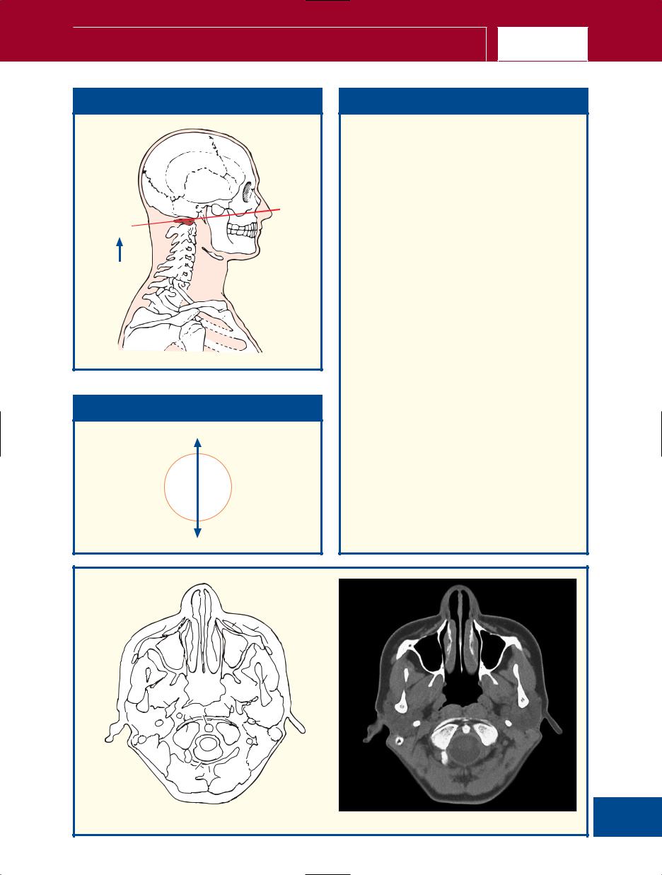
This section passes through the apex of the skull vault and traverses the parietal bones (2) and the superior portion of the frontal bone (1).
Between the inner and outer tables of the bones of the skull vault lie trabecular bone, termed diploe, which contains red bone marrow. This is highly vascular and a common site for blood-borne metastatic tumour deposits and multiple myeloma. Diploic veins (see (8) on page 18) occupy channels in this trabecular bone. These are absent at birth but begin to appear at about 2 years of age. They are large and thin-walled, being merely endothelium supported by elastic tissue, and they communicate with meningeal veins, dural sinuses and the pericranial veins. Radiographically they may appear as relatively transparent bands 3–4 mm in diameter.
The dura mater, which lines the inner aspect of the skull, comprises an outer, or endosteal, layer, or endocranium (3) (which is, in fact, the periosteum, which lines the inner aspect of the skull) and an inner, or meningeal, layer (4). Most of the intracranial venous sinuses are formed as clefts between these two layers, as demonstrated in this section by the superior sagittal sinus (6). The exceptions to this rule are the inferior sagittal sinus and the straight sinus, which are clefts within the meningeal layer.
6
2
 6
6
11
Axial computed tomogram (CT)
|
|
Axial section 2 – Male |
|
HEAD |
|
|
|
|
1
6
7
8
9
2
5
4
6
3
1 Frontal bone
2 Parietal bone
3 Sagittal suture
4Dura mater
5Arachnoid mater
6 Superior sagittal sinus
7Falx cerebri
8 Subarachnoid space
9Pia mater
12

■ Section level
View
■ Orientation
Anterior
Right 
 Left
Left
Posterior
Axial section 2 – Male |
HEAD |
|
|
■ Notes
This section, at a deeper plane through the skull vault, demonstrates the falx cerebri (7), which is formed as a double fold of the inner, meningeal, layer of the dura mater (5) and which forms the dural septum between the cerebral hemispheres.
The inner layer of the dura is lined by the delicate arachnoid mater. The pia mater (9) is vascular and invests the brain, spinal cord, cranial nerves and spinal nerve roots. It remains in close contact with the surface of the brain, including the depths of the cerebral sulci and fissures.
Over the convexities of the brain, the pia and arachnoid are in close contact. Over the cerebral sulci and the cisterns of the brain base, the pia and arachnoid are separated by the subarachnoid space (8), which contains cerebrospinal fluid. This space is traversed by a fine spider’s web of fibres (arachnoid: pertaining to the spider).
The total volume of cerebrospinal fluid in the adult is approximately 150 mL, of which some
25 mL is contained in the ventricular system, 25 mL in the spinal theca and the remaining 100 mL in the cerebral subarachnoid space.
1
8
7
2
13
Axial computed tomogram (CT)

|
|
Axial section 3 – Male |
|
HEAD |
|
|
|
|
10
1
12 |
|
|
13 |
11 |
6 |
|
|
3
2
4
5
9 |
6 |
8
10
7
1 |
Frontal bone |
7 |
Sagittal suture |
2 |
Parietal bone |
8 |
Dura mater |
3 |
Skin and dense subcutaneous |
9 |
Arachnoid mater |
|
tissue |
10 |
Superior sagittal sinus |
4 |
Epicranial aponeurosis (galea |
11 |
Falx cerebri |
|
aponeurotica) |
12 |
Grey matter |
5 |
Pericranium |
13 |
White matter |
6Branches of superficial temporal artery
14

■ Section level
View
■ Orientation
Anterior
Right 
 Left
Left
Posterior
Axial section 3 – Male |
HEAD |
|
|
■ Notes
This section, through the upper parts of the cerebral hemispheres, gives a clear picture of the distinction between the outer grey matter (12), which contains nerve cells, and the inner white matter (13), made up of nerve fibres. This is in contradistinction to the arrangement of the spinal cord, with the central grey and surrounding white matter.
Note the five layers of the scalp – skin, underlying dense connective tissue (3), dense epicranial aponeurosis, or galea aponeurotica (4), which is separated by a film of loose areolar connective tissue from the outer periosteum of the skull, the pericranium (5). The pericranium is densely adherent to the surface of the skull and passes through the various foramina, where it becomes continuous with the outer endosteal layer of the dura (8) and is also continuous with the sutural ligaments that occupy the cranial sutures.
Each of these layers is of clinical significance. The scalp is richly supplied with sebaceous glands and is the commonest site of epidermoid cysts. The connective tissue is made up of lobules of fat bound in tough fibrous septa. The blood vessels of the scalp lie in this layer; when the scalp is lacerated, the divided vessels retract between these septa and cannot be picked up with artery forceps in the usual way – they can be controlled by firm digital pressure against the skull on either side of the laceration. The aponeurotic layer is the occipitofrontalis, which is fibrous over the dome of the scalp but muscular in the occipital and frontal regions (see (2) on p. 24 and (2) on p. 26). The underlying loose areolar connective tissue accounts for the mobility of the scalp on the underlying bone. It is in this plane that surgical mobilization of scalp flaps is performed. Blood in this layer tracks forward into the orbits to produce periorbital haematomas. The periosteum adheres to the suture lines of the skull, so that a collection of blood or pus beneath this layer outlines the affected bone. This may produce the cephalohaematoma seen in birth injuries involving the skull.
1
11
2 
15
Axial computed tomogram (CT)

|
|
Axial section 4 – Male |
|
HEAD |
|
|
|
|
11 1
16
13
6
15 |
5 |
4
18 17
3
2
14 |
7 |
12
10
9 |
11 |
8
1 |
Frontal bone |
9 Dura mater |
|
2 |
Parietal bone |
10 |
Arachnoid mater |
3 |
Skin and dense |
11 |
Superior sagittal sinus |
|
subcutaneous tissue |
12 |
Falx cerebri |
4 |
Epicranial aponeurosis |
13 |
Cingulate gyrus |
|
(galea aponeurotica) |
14 |
Parieto-occipital sulcus |
5 |
Temporalis |
15 |
Corona radiata |
6 |
Pericranium |
16 |
Anterior cerebral artery |
7 |
Branch of superficial |
|
(branches) |
|
temporal artery |
17 |
Postcentral gyrus |
8 |
Sagittal suture |
18 |
Central sulcus |
16

■ Section level
View
■ Orientation
Anterior
Right 
 Left
Left
Posterior
Axial section 4 – Male |
HEAD |
|
|
■ Notes
This section allows some of the main gyri and sulci of the cerebrum to be identified. Cross-reference should be made to the photographs of the external aspects and sagittal section of the brain for orientation.
The corona radiata (15) comprises a fan-shaped arrangement of afferent and efferent projection fibres, which join the grey matter to lower centres. On the computed tomography (CT) image, it appears as a curved linear area of low attenuation termed the centrum semiovale.
The superficial temporal artery, of which the parietal branch can be seen at (7), is the smaller terminal branch of the external carotid artery, the other being the maxillary artery. The middle terminal branch can be seen immediately in front of (4). The blood supply to the scalp is the richest of all areas of the skin and there are free anastomoses between its various branches. It is for this reason that a partially avulsed scalp flap is usually viable.
1
12
15
2
17
Axial computed tomogram (CT)

|
|
Axial section 5 – Male |
|
HEAD |
|
|
|
|
12
1
14
25
24
|
22 |
21 |
23 |
|
20
18
19
15
14
12 |
13 |
|
|
11 |
|
7
6
5
4
17
2
16
3
8
10
9
1 |
Frontal bone |
9 Dura mater |
19 |
Corona radiata |
|
2 |
Parietal bone |
10 |
Arachnoid mater |
20 |
Corpus callosum |
3 |
Skin and dense subcutaneous |
11 |
Sagittal suture |
21 |
Longitudinal fasciculus |
|
tissue |
12 |
Superior sagittal sinus |
|
(corticocortical fibres) |
4 |
Temporal fascia |
13 |
Lunate sulcus |
22 |
Anterior cerebral artery |
5 |
Temporalis |
14 |
Falx cerebri |
|
(branches) |
6 |
Pericranium |
15 |
Cingulate gyrus |
23 |
Forceps minor |
7 |
Branches of superficial temporal |
16 |
Postcentral sulcus |
24 |
Cingulate sulcus |
|
artery |
17 |
Central sulcus |
25 |
Inferior sagittal sinus |
8 |
Diploic vein |
18 |
Roof of body of lateral ventricle |
|
|
18

■ Section level
View
■ Orientation
Anterior
Right 
 Left
Left
Posterior
Axial section 5 – Male |
HEAD |
|
|
■ Notes
This section passes through the roof of the lateral ventricle (18).
The central sulcus, or fissure of Rolando (17), is the most important of the sulcal landmarks, since it separates the precentral (motor) gyrus from the postcentral (sensory) gyrus. It also helps demarcate the frontal and parietal lobes of the cerebrum.
Again, the corona radiata (19), or centrum semiovale, is well seen in both the section and the CT image.
The corpus callosum (20) – and seen also on p. 7, in (5), (6) and (7) – is the largest fibre pathway of the brain. It links the cortex of the two cerebral hemispheres and roofs much of the lateral ventricles. Its anterior portion is termed the genu; its body is termed the trunk, which is arched and convex superiorly. It ends posteriorly as the splenium, which is its thickest part – see p. 20 (17). Congenital absence of the corpus callosum, or its surgical division, results in surprisingly little disturbance of function.
1
19
 2
2
12
19
Axial computed tomogram (CT)

|
|
Axial section 6 – Male |
|
HEAD |
|
|
|
|
39
1
10
11
38
37
36
|
35 |
34 |
|
|
32 |
31 |
33 |
|
|
30 |
29 |
|
|
|
26 |
|
19 |
18 |
|
16 |
17 |
|
12
14 |
13 |
10
3
5
4
28 |
6 |
27
7
23
24
22
25 |
8 |
21
2
20
8
16
15
9
|
1 |
Frontal bone |
13 |
Parieto-occipital sulcus |
27 |
Body of caudate nucleus |
|
2 |
Parietal bone |
14 |
Optic radiation |
28 |
Frontal horn of lateral |
|
3 |
Sutural bone |
15 |
Choroid plexus |
|
ventricle |
|
4 |
Skin and dense subcutaneous |
16 |
Posterior horn lateral ventricle |
29 |
Septum pellucidum |
|
|
tissue |
17 |
Splenium of corpus callosum |
30 |
Insula |
|
5 |
Epicranial aponeurosis (galea |
18 |
Lateral sulcus (Sylvian fissure) |
31 |
Claustrum |
|
|
aponeurotica) |
19 |
Third ventricle |
32 |
Putamen |
|
6 |
Temporalis |
20 |
Middle cerebral artery |
33 |
Internal capsule |
|
7 |
Pericranium |
|
(branches) |
34 |
Circular sulcus |
|
8 |
Branches of superficial |
21 |
Postcentral sulcus |
35 |
Genu of corpus callosum |
|
|
temporal artery |
22 |
Central sulcus |
36 |
Anterior cerebral artery |
|
9 |
Occipital vein |
23 |
Arachnoid mater |
|
(branches) |
20 |
10 Superior sagittal sinus |
24 |
Dura mater |
37 |
Forceps minor |
|
11 Falx cerebri |
25 |
Thalamostriate vein |
38 |
Cingulate sulcus |
||
|
12 Straight sinus |
26 |
Body of lateral ventricle |
39 |
Supra-orbital artery |
|

Axial section 6 – Male |
|
|
HEAD |
|
|
|
|
|
■ Section level |
|
|
■ Notes |
||
|
|
|
|
|
|
|
|
|
|
|
This section passes through the bodies of the lateral |
|
|
|
|
|
ventricles (26) and the third ventricle (19). |
|
|
|
|
|
The lateral ventricles comprise a frontal horn (28) |
|
|
|
|
|
and body (26), which continues with the posterior |
|
|
|
|
|
or occipital horn (16), which, in turn, enters the |
|
|
|
|
|
inferior horn within the temporal lobe. This will be |
|
|
|
|
|
seen in later sections. The lateral ventricles are |
View |
|
|
separated almost completely from each other by the |
||
|
|
|
|
|
septum pellucidum (29) but communicate indirectly |
|
|
|
|
|
via the third ventricle (19), a narrow slit-like cavity. |
|
|
|
|
|
The choroid plexuses of the lateral ventricles (15), |
|
|
|
|
|
which are responsible for the production of most of |
|
|
|
|
|
the cerebrospinal fluid extend from the inferior |
|
|
|
|
|
horn, through the body to the interventricular |
|
|
|
|
|
foramen, where they become continuous with the |
|
|
|
|
|
plexus of the third ventricle. |
|
|
|
|
|
In addition to the centres of ossification of the |
|
|
|
|
|
named bones of the skull, other centres may occur |
|
|
|
|
|
in the course of the sutures, which give rise to |
|
|
|
|
|
irregular sutural (Wormian) bones (3). They occur |
|
|
|
|
|
most frequently in the region of the lambdoid |
|
|
|
|
|
suture, as here, but sometimes they may be seen at |
■ Orientation |
|
|
the anterior, or more especially, the posterior |
||
|
|
fontanelle. They are usually limited to two or three |
|||
|
|
|
|
|
|
|
|
|
|
|
in number, but they may occur in greater numbers |
|
Anterior |
|
|||
|
|
|
in congenital hydrocephalic skulls and other |
||
|
|
|
|
|
|
|
|
|
|
|
congenital anomalies. |
|
|
|
|
||
Right |
|
|
Left |
||
|
|
||||
|
|
|
|
|
|
Posterior
1
28 35 15


 6 6
6 6 


2
16 17
12
11
10
21
Axial magnetic resonance image (MRI)
|
|
Axial section 7 – Male |
|
HEAD |
|
|
|
|
1
2
3
4
19
43
|
|
42 |
|
|
|
41 |
|
|
40 |
39 |
|
|
|
38 |
37 |
|
|
36 |
35 |
|
|
32 |
34 |
|
|
33 |
|
|
27 |
31 |
|
|
28 |
30 |
|
|
|
||
|
|
29 |
|
26 |
|
23 |
|
25 |
24 |
|
|
|
|
||
|
|
22 |
|
21
19
20
|
18 |
17 |
15 |
|
16 |
5
6 7
8
9
11
10
12
14
13
5
1 |
Supra-orbital artery |
15 |
Occipital artery |
31 |
Globus pallidus – internal |
2 |
Frontal belly of occipitofrontalis |
16 |
Squamous part of occipital |
|
segment |
3 |
Frontal sinus |
|
bone |
32 |
Globus pallidus – external |
4 |
Frontal bone |
17 |
Superior sagittal sinus |
|
segment |
5 |
Parietal bone |
18 |
Occipital lobe |
33 |
Choroid plexus in |
6 |
Middle meningeal artery and |
19 |
Falx cerebri |
|
interventricular foramen |
|
vein |
20 |
Calcarine sulcus |
|
(Monro) |
7 |
Branch of temporal artery |
21 |
Straight sinus |
34 |
Claustrum |
8 |
Sliver of squamous part of |
22 |
Great cerebral vein |
35 |
Insula |
|
temporal bone |
23 |
Fornix |
36 |
Putamen |
9 |
Skin and dense subcutaneous |
24 |
Internal cerebral vein (branches) |
37 |
Middle cerebral artery |
|
tissue |
25 |
Pulvinar of thalamus |
|
(branches) |
10 Epicranial aponeurosis (galea |
26 |
Optic radiation |
38 |
Anterior limb of internal capsule |
|
|
aponeurotica) |
27 |
Medial nucleus of thalamus |
39 |
Caudate nucleus – head |
11 Temporalis |
28 |
Third ventricle |
40 |
Corpus callosum |
|
22 12 Pericranium |
29 |
Ventroposterior thalamic |
41 |
Anterior cerebral artery |
|
13 Dura mater |
|
nucleus |
42 |
Frontal horn of lateral ventricle |
|
14 Arachnoid mater |
30 |
Circular sulcus |
43 |
Frontal lobe |
|

Axial section 7 – Male |
|
|
HEAD |
|
|
|
|
|
■ Section level |
|
■ Notes |
|
|
|
|
|
|
|
This section passes through the apex of the |
|
|
|
squamous part of the occipital bone (16) and the |
|
|
|
frontal sinus (3). These are paired but are rarely |
|
|
|
symmetrical, while the septum between them is |
|
|
|
usually deviated from the midline. They vary greatly |
|
|
|
in size, as may be appreciated from viewing a |
|
|
|
number of skull radiographs. Each lies posterior to |
|
View |
|
the supercilliary arch and extends upwards above |
|
|
the medial part of the eyebrow and back on to the |
||
|
|
||
|
|
medial part of the orbital roof. Sometimes they are |
|
|
|
divided by incomplete bony septa; rarely, one or |
|
|
|
both may be absent. Each drains into the anterior |
|
|
|
part of the middle meatus on the lateral wall of the |
|
|
|
nasal cavity via the frontonasal duct. |
|
|
|
The interventricular foramen of Monro (33) is well |
|
|
|
demonstrated and drains the lateral ventricle on |
|
|
|
both sides into the third ventricle (28), thus |
|
|
|
providing a linkage between the ventricular systems |
|
|
|
within the two cerebral hemispheres. |
|
|
|
This section also demonstrates the components of |
|
|
|
the basal ganglia, the claustrum (34), and the |
|
|
|
lentiform nucleus, made up of the globus pallidus |
|
■ Orientation |
|
(31, 32) and putamen (36). The latter is largely |
|
|
separated from the head of the caudate nucleus |
||
|
|
||
|
|
(39) by the anterior limb of the internal capsule |
|
Anterior |
|||
(38). |
|||
|
|||
Right 
 Left
Left
Posterior
4
11
37 |
42 39 |
31
32
27
29 
 36
36
28
26

 21
21  19
19
17
23
Axial magnetic resonance image (MRI)

|
|
Axial section 8 – Male |
|
HEAD |
|
|
|
|
45
30
29
28
24
1
2
43
5
19 47
46 |
|
|
|
|
|
|
|
6 |
|
|
|
41 |
7 |
|
44 |
|
9 |
||
43 |
42 |
|
||
|
8 |
|||
|
|
40 |
||
|
|
10 |
||
36 |
37 |
39 |
||
11 |
||||
|
|
38 |
||
32 |
35 |
|
||
|
|
|||
31 |
34 |
|
13 |
|
33 |
|
14 |
||
27 |
|
24 |
12 |
|
|
|
|||
26 |
|
|
||
|
|
|
||
|
25 |
|
|
23 |
22 |
|
|
|
21 |
|
15 |
|
20 |
19
16
18
17
|
1 |
Supra-orbital artery |
12 |
Superficial temporal |
26 |
Superior colliculus |
40 |
Claustrum |
|
2 |
Orbital part of |
|
artery |
27 |
Aqueduct of Sylvius |
41 |
Lateral sulcus (Sylvius) |
|
|
occipitofrontalis |
13 |
Dura mater |
28 |
Posterior cerebral |
42 |
Insula |
|
3 |
Frontal belly of |
14 |
Arachnoid mater |
|
artery |
43 |
Nucleus accumbens |
|
|
occipitofrontalis |
15 |
Parietal bone |
29 |
Tail of caudate nucleus |
|
septi |
|
4 |
Frontal sinus |
16 |
Occipital artery |
30 |
Cerebral peduncle |
44 |
Anterior cerebral |
|
5 |
Frontal bone |
17 |
Squamous part of |
31 |
Red nucleus |
|
artery |
|
6 |
Middle meningeal |
|
occipital bone |
32 |
Third ventricle |
45 |
Anterior perforated |
|
|
artery and vein |
18 |
Superior sagittal sinus |
33 |
Substantia nigra |
|
substance |
|
7 |
Skin and dense |
19 |
Falx cerebri |
34 |
Cornu ammonis |
46 |
Cingulate gyrus |
|
|
subcutaneous tissue |
20 |
Straight sinus |
|
(hippocampus) |
47 |
Orbitofrontal cortex |
|
8 |
Epicranial aponeurosis |
21 |
Tentorium cerebelli |
35 |
Mamillary body |
|
|
|
|
(galea aponeurotica) |
22 |
Collateral sulcus |
36 |
Hypothalamus |
48 |
Cisterna ambiens |
|
9 |
Temporalis |
23 |
Vermis of cerebellum |
37 |
Optic tract |
49 |
Temporal lobe |
24 |
10 Pericranium |
24 |
Lateral ventricle |
38 |
Amygdala |
50 |
Interpeduncular |
|
|
11 Squamous part of |
25 |
Parahippocampal |
39 |
Middle cerebral artery |
|
cistern |
|
|
|
temporal bone |
|
gyrus |
|
(branches) |
|
|

Axial section 8 – Male |
|
|
HEAD |
|
|
|
|
|
■ Section level |
|
|
■ Notes |
||
|
|
|
|
|
|
|
|
|
|
|
This section passes through the upper part of the |
|
|
|
|
|
squamous temporal bone (11) and traverses the |
|
|
|
|
|
midbrain at the level of the cerebral peduncle (30) |
|
|
|
|
|
and the red nucleus (31). |
|
|
|
|
|
The red nucleus (31) has a pinkish tinge, which is |
|
|
|
|
|
visible only in fresh tissue. The colour is produced by |
|
|
|
|
|
a ferric iron pigment present in the neurons of the |
|
|
|
|
|
red nucleus. |
View |
|
|
The aqueduct of Sylvius (27) is the |
||
|
|
|
|
|
communication between the third ventricle (see |
|
|
|
|
|
Axial section 7) and the fourth ventricle (see Axial |
|
|
|
|
|
section 10). |
|
|
|
|
|
The colliculi, two superior (26) and two inferior, |
|
|
|
|
|
blend to form the tectum over the aqueduct (27). |
|
|
|
|
|
This is sometimes termed the quadrigeminal plate, |
|
|
|
|
|
hence an alternative name for the cisterna ambiens |
|
|
|
|
|
(48) is the quadrigeminal cistern. Other names for |
|
|
|
|
|
this include superior cistern and cistern of the great |
|
|
|
|
|
cerebral vein. As this cistern contains the great |
|
|
|
|
|
cerebral vein and the pineal body, it is an important |
|
|
|
|
|
anatomical landmark. |
|
|
|
|
|
The squamous part of the temporal bone (11) is |
■ Orientation |
|
|
the thinnest bone of the calvarium (although, in |
||
|
|
contrast, its petrous part is the densest). It is, |
|||
|
|
|
|
|
|
|
|
|
|
|
however, ‘protected’ by the thick overlying |
|
Anterior |
|
|||
|
|
|
temporalis muscle (9). |
||
|
|
|
|
|
|
|
|
|
|
|
The middle meningeal artery (6) is a branch of the |
|
|
|
|
||
|
|
|
|
|
maxillary artery, and its accompanying vein, and |
|
|
|
|
|
may be torn, either together or individually, in |
Right |
|
|
Left |
|
fractures of the temporal bone. This constitutes the |
|
|
||||
|
|
|
|
|
commonest cause of a traumatic extradural |
|
|
|
|
|
haematoma. |
|
|
|
|
|
|
|
Posterior |
|
|
|
|
|
|
|
|
|
|
|
|
5 |
|
47 |
|
|
|
|
|
41 |
|
42 |
32 |
|
|
9 |
11 |
||
|
|||
|
|
49 |
|
50 |
|
|
|
30 |
26 |
|
|
|
|
||
23 |
48 |
15 |
18
17
25
Axial computed tomogram (CT)

|
|
Axial section 9 – Male |
|
HEAD |
|
|
|
|
36
35
21
|
3 |
|
6 |
|
5 |
4 |
|
|
|
|
|
|
||
|
7 |
|
12 |
|
7 |
|
|
13 |
|
|
|
7 |
8 |
|
|
|
11 |
|
|
|
|
22 |
9 10 |
|
|
|
|
14 |
15 |
|
|
|
|
|
|
38 |
|
40 |
44 |
|
39 |
|
||
|
|
|
|
|
|
37 |
|
|
|
|
|
41 |
43 |
|
|
34 |
33 |
42 |
16 |
|
|
|
|
|
|
32 |
31 |
|
|
|
|
|
|
|
30 |
28 |
29 |
|
|
|
|
|
|
|
27 |
|
25 |
|
|
|
|
|
|
|
26 |
|
23 |
|
17 |
|
|
|
|
22 |
|
|
18 |
|
|
|
|
21 |
|
|
|
|
|
24 |
19 |
|
|
|
|
20 |
|
|
|
1 |
Supra-orbital artery |
14 |
Lesser wing of |
27 |
Anterior lobe of |
40 |
Orbitofrontal cortex |
2 |
Frontal belly of |
|
sphenoid bone |
|
cerebellum |
41 |
Uncus of |
|
occipitofrontalis |
15 |
Temporalis |
28 |
Cerebellar vermis |
|
parahippocampal |
3 |
Frontal bone |
16 |
Temporal bone |
29 |
Inferior colliculus |
|
gyrus |
4 |
Frontal crest |
17 |
Parietal bone |
30 |
Aqueduct of Sylvius |
42 |
Hippocampus |
5 |
Frontal sinus |
18 |
Posterior belly of |
31 |
Locus coeruleus |
43 |
Temporal horn of |
6 |
Trochlea |
|
occipitofrontalis |
32 |
Decussation of |
|
lateral ventricle |
7 |
Ethmoidal air cells |
19 |
Occipital artery |
|
superior cerebellar |
44 |
Temporal pole |
8 |
Superior oblique |
20 |
Occipital bone |
|
peduncle |
|
|
9 |
Orbital plate of |
21 |
Superior sagittal sinus |
33 |
Basilar artery |
45 |
Vitreous humour |
|
ethmoid bone |
22 |
Falx cerebri |
34 |
Superior cerebellar |
46 |
Lens |
10 Superior rectus |
23 |
Straight sinus |
|
artery |
47 |
Middle cerebral artery |
|
|
underlying levator |
24 |
Occipital pole |
35 |
Posterior cerebral |
|
|
|
palpebri superioris |
25 |
Floor of lateral |
|
artery |
|
|
11 Orbital fat |
|
ventricle (occipital |
36 |
Internal carotid artery |
|
|
|
26 12 Lacrimal gland |
26 |
horn) |
37 |
Pituitary infundibulum |
|
|
|
13 Zygomatic process of |
Tentorium cerebelli |
38 |
Optic chiasma |
|
|
||
|
frontal bone |
|
(outer edge) |
39 |
Optic nerve (II) |
|
|

Axial section 9 – Male |
|
|
HEAD |
|
|
|
|
|
■ Section level |
■ Notes |
View
■ Orientation
Anterior
Right 
 Left
Left
Posterior
This section traverses the upper part of the orbits, the midbrain at the level of the inferior colliculus (29) and the anterior lobe of the cerebellum (27).
The straight sinus (23) lies in the sagittal plane of the tentorium cerebelli (26) at its attachment to the falx cerebri (22). It receives both the inferior sagittal sinus and the great cerebral vein, and drains posteriorly, usually into the left, but occasionally into the right, transverse sinus.
The optic nerves (39) have an intracranial course of about 10 mm. They unite at the optic chiasma (38), which lies immediately anterior to the infundibulum of the hypophysis cerebri, or pituitary gland (37). See also Coronal section 8 on p. 64.
46
45
|
47 |
|
35 |
32 |
30 |
|
|
28 |
27 |
|
23 |
21 |
24 |
|
16
27
Axial magnetic resonance image (MRI)

|
|
Axial section 10 – Male |
|
HEAD |
|
|
|
|
1
2
3
5
40
39
38
28
27
26
4 15
6
7
8
37
36
35
33
32
31 30
29
25
10
|
9 |
11 |
12 |
13 |
14
16
19
17
18
21
20
22
34
23
24
23
|
1 |
Nasal bone |
12 |
Orbicularis oculi – |
22 |
Mastoid air cells |
34 |
Hemisphere of |
|
2 |
Frontal process of |
|
orbital part |
23 |
Transverse sinus |
|
cerebellum |
|
|
maxilla |
13 |
Orbicularis oculi – |
24 |
Parietal bone |
35 |
Pontine tegmentum |
|
3 |
Nasolacrimal duct |
|
palpebral part |
25 |
Squamous part of |
36 |
Pontine nuclei |
|
4 |
Perpendicular plate of |
14 |
Frontal process of |
|
occipital bone |
37 |
Basilar artery |
|
|
ethmoid bone |
|
zygomatic bone |
26 |
Falx cerebelli |
38 |
Trigeminal nerve (V) |
|
5 |
Orbital plate of |
15 |
Medial rectus |
27 |
Superior sagittal |
39 |
Temporal lobe |
|
|
ethmoid bone |
16 |
Optic nerve (II) |
|
sinus |
40 |
Sclera |
|
6 |
Posterior ethmoidal |
17 |
Lateral rectus |
28 |
Posterolateral fissure |
|
|
|
|
air cells |
18 |
Greater wing of |
29 |
Emboliform |
41 |
Crista galli of ethmoid |
|
7 |
Sphenoidal sinus |
|
sphenoid bone |
|
(interposed) nucleus |
42 |
Petrous part of |
|
8 |
Internal carotid artery |
19 |
Temporalis |
30 |
Dentate nucleus |
|
temporal bone |
|
|
within cavernous sinus |
20 |
Squamous part of |
31 |
Vermis of cerebellum |
43 |
Internal auditory |
28 |
9 |
Cornea |
|
temporal bone |
32 |
Fourth ventricle |
|
meatus |
|
10 Lens |
21 |
Superficial temporal |
33 |
Middle cerebellar |
|
|
|
|
11 Vitreous humour |
|
artery and vein |
|
peduncle |
|
|
|

■ Section level
View
■ Orientation
Anterior
Right 
 Left
Left
Posterior
Axial section 10 – Male |
HEAD |
|
|
■ Notes
This section transects the eyeballs, the sphenoid sinus (7) and the pons (36) at the level of the middle cerebellar peduncles (33).
The structure of the orbit in horizontal section can be appreciated in this section. The eyeball with its cornea (9), lens (10) and vitreous humour (11) contained within the tough sclera (40), and the optic nerve (16) lie surrounded by the extrinsic muscles (15, 17). The slit-like nasolacrimal duct (3) drains downwards into the inferior meatus.
The fourth ventricle (32) lies above the tegmentum of the pons (35) and below the vermis of the cerebellum (31).
The ethmoidal air cells, or sinuses (6), are made up of some eight to ten loculi suspended from the outer extremity of the cribriform plate of the ethmoid bone and bounded laterally by its orbital plate. They thus occupy the upper lateral wall of the nasal cavity. The cells are divided into anterior, middle and posterior groups by bony septa. The middle group bulge into the middle meatus to form an elevation, the bulla ethmoidalis, into which they open. The anterior cells drain into the hiatus semilunaris, which is a groove below the bulla. The posterior cells drain into the superior meatus.
|
|
11 |
|
|
|
|
15 |
17 |
14 |
19 |
41 |
16 |
|
|
6 |
|
18 |
|
|
|
|
|
|
|
7
|
|
39 |
42 |
|
20 |
|
43 |
|
|
37 |
|
|
|
|
|
36 35 |
|
32 22
34
26
25
29
Axial computed tomogram (CT)

HEAD |
Axial section 11 – Male |
|
4 |
|
|
|
3 |
5 |
|
|
|
|
|
|
2 |
|
|
|
6 |
11 |
10 |
1 |
13 |
12 |
|
|
|
||
|
|
14 |
|
17
|
|
22 |
21 |
|
|
|
|
|
|
|
47 |
|
|
|
46 |
|
38 |
37 |
20 |
|
|
40 39 |
||
45 |
|
|
19 |
|
|
|
41 |
|
|
44 |
43 |
42 |
|
|
|
|
36 |
|
|
|
|
35 |
|
|
|
|
34 |
33 |
|
|
|
|
32 |
31 |
|
|
30 |
|
|
29
28
27
7
9
8
15
16
18
23
26
24
25
1 |
Inferior rectus |
14 |
Apex of maxillary |
25 |
Transverse sinus |
37 |
Basilar artery |
|
2 |
Nasolacrimal duct |
|
antrum |
26 |
Occipital artery and |
38 |
Abducent nerve (VI) |
|
3 |
Cartilage of nasal |
15 |
Frontal process of |
|
vein |
39 |
Trigeminal nerve (V) |
|
|
|
septum |
|
zygomatic bone |
27 |
Trapezius |
40 |
Labyrinthine artery |
4 |
Nasal bone |
16 |
Temporalis |
28 |
External occipital |
41 |
Facial nerve (VII) |
|
5 |
Frontal process of |
17 |
Greater wing of |
|
protuberance |
42 |
Vestibulocochlear |
|
|
|
maxilla |
|
sphenoid bone |
29 |
Falx cerebelli |
|
(auditory) nerve (VIII) |
6 |
Lacrimal bone |
18 |
Middle meningeal |
30 |
Vermis |
43 |
Cochlea |
|
7 |
Upper eyelid |
|
artery |
31 |
Middle cerebellar |
44 |
Stapes |
|
8 |
Orbicularis oculi |
19 |
Petrous part of |
|
peduncle |
45 |
External auditory |
|
9 |
Sclera |
|
temporal bone |
32 |
Fourth ventricle with |
|
meatus |
|
|
10 Vitreous humour |
20 |
Internal carotid artery |
|
choroid plexus |
46 |
Tympanic membrane |
|
|
11 Orbital plate of |
21 |
Sphenoidal sinus |
33 |
Medulla oblongata |
|
and handle of malleus |
|
|
|
ethmoid bone |
22 |
Septum of sphenoidal |
34 |
Inferior cerebellar |
47 |
Auditory tube |
30 |
|
|||||||
12 Ethmoidal air cells |
23 |
sinus |
35 |
peduncle |
|
(Eustachian) |
||
|
13 Perpendicular plate of |
Cerebellar hemisphere |
Flocculus |
|
|
|||
|
|
ethmoid bone |
24 |
Mastoid air cells |
36 |
Pyramidal tract |
|
|
|
|
|
|
|||||

■ Section level
View
■ Orientation
Anterior
Right 
 Left
Left
Posterior
Axial section 11 – Male |
HEAD |
■ Notes
This section passes through the upper part of the nasal cavity, the medulla oblongata (33) and, posteriorly, through the external occipital protuberance (28).
The sphenoidal sinus (21) is unusually large in this specimen. It is divided by a median septum (22) and drains anteriorly into the nasal cavity at the sphenoethmoidal recess.
Note the relations of the labyrinthine artery (40), a branch of the basilar artery (37), the facial nerve (41) and the vestibulocochlear (or auditory) nerve (42) as they enter the internal auditory meatus of the temporal bone together with the close relationships of the trigeminal nerve (V) (39) and the cerebellum (35). As an acoustic neuroma of the vestibulocochlear nerve enlarges, it stretches the adjacent cranial nerves V and VII anteriorly and also presses on the cerebellum and brain stem to produce the cerebello-pontine angle syndrome. Rather surprisingly, facial nerve weakness with unilateral taste loss is uncommon – occurring in less than five per cent of cases – although the facial nerve is at risk in surgical removal of the tumour.
3
15
14
|
37 |
41 |
43 |
42
32
23
28
31
Axial magnetic resonance image (MRI)
HEAD |
Axial section 12 – Male |
1
|
|
|
|
|
10 |
|
|
3 |
2 |
|
|
9 |
|
|
|
|
|
|
||
4 |
|
8 |
|
|
11 |
|
|
|
|
|
|
||
5 |
|
7 |
|
|
12 |
|
|
|
|
|
|
||
|
6 |
|
|
|
14 |
13 |
|
|
|
|
|
|
|
|
|
|
|
|
16 |
|
|
|
|
|
|
15 |
|
|
|
43 |
|
44 |
17 |
18 |
|
41 |
|
|
|||
|
42 |
|
|
|
||
|
|
|
|
|
||
|
40 |
|
|
|
19 |
|
|
|
32 |
|
|
||
39 |
37 |
36 |
|
|
|
|
|
|
|
20 |
|||
|
|
|
|
|
||
|
|
33 |
|
|
|
|
38 |
|
|
31 |
|
|
|
|
35 |
30 |
|
|
||
|
|
29 |
34 |
|
|
|
|
|
28 |
|
27 |
|
|
|
|
26 |
|
|
|
|
|
21 |
25 |
24 |
|
|
|
22 |
|
23 |
1 |
Cartilage of nasal |
15 |
Trigeminal nerve (V) |
26 |
Vermis |
39 |
Bulb of internal |
|
|
|
septum |
16 |
Articular disc of |
27 |
Cerebellar hemisphere |
|
jugular vein |
2 |
Nasolacrimal duct |
|
temporomandibular |
28 |
Tonsil of cerebellum |
40 |
Glossopharyngeal |
|
3 |
Orifice of maxillary |
|
joint |
29 |
Fourth ventricle |
|
nerve (IX), vagus nerve |
|
|
|
sinus |
17 |
Head of mandible |
|
(median aperture of |
|
(X) and accessory nerve |
4 |
Maxillary sinus |
18 |
Superficial temporal |
|
roof) |
|
(XI) |
|
5 |
Maxillary artery |
|
artery and vein |
30 |
Anterior inferior |
41 |
Internal carotid artery |
|
6 |
Sphenoidal sinus |
19 |
External auditory |
|
cerebellar artery |
42 |
Basi-occiput |
|
7 |
Vomer |
|
meatus |
31 |
Glossopharyngeal |
43 |
Longus capitis |
|
8 |
Middle nasal concha |
20 |
Mastoid air cells |
|
nerve (IX) |
44 |
Auditory (Eustachian) |
|
9 |
Maxilla |
21 |
Sternocleidomastoid |
32 |
Hypoglossal nerve (XII) |
|
tube |
|
|
10 Orbicularis oculi |
22 |
Occipital artery and |
33 |
Pyramidal tract |
|
|
|
|
11 Zygomatic bone |
|
vein |
34 |
Medulla |
45 |
Pterygopalatine fossa |
|
|
12 Temporalis and tendon |
23 |
Trapezius |
35 |
Inferior olive |
|
(apex) |
|
32 |
13 Zygomatic process of |
24 |
Occipital bone – |
36 |
Vertebral artery |
46 |
Foramen ovale |
|
|
temporal bone |
|
squamous part |
37 |
Vagus nerve (X) |
|
|
|
|
|
|
|
|
||||
|
14 Lateral pterygoid |
25 |
Falx cerebelli |
38 |
Sigmoid sinus |
|
|
|

Axial section 12 – Male |
HEAD |
■ Section level |
|
|
■ Notes |
||
|
|
|
|
|
|
|
|
|
|
|
This section transects the maxillary sinus (4) and the |
|
|
|
|
|
basi-occiput (42) and passes through the external |
|
|
|
|
|
auditory meatus (19). |
|
|
|
|
|
At this level, the vertebral arteries (36) are |
|
|
|
|
|
running cranially from their entry into the skull at |
|
|
|
|
|
the foramen magnum to form the basilar artery. |
|
|
|
|
|
The sigmoid sinus (38) runs forward to emerge |
|
|
|
|
|
from the skull at the jugular foramen, at which it |
|
|
|
|
|
becomes the bulb of the internal jugular vein (39). |
|
|
|
|
|
Exiting through the jugular foramen anterior to the |
View |
|
|
vein lie, from anterior to posterior, the |
||
|
|
glossopharyngeal, vagus and accessory cranial |
|||
|
|
|
|
|
|
|
|
|
|
|
nerves (40). |
|
|
|
|
|
The maxillary nerve (Vii) passes into the |
|
|
|
|
|
pterygopalatine fossa (45 on this CT image) having |
|
|
|
|
|
traversed the foramen rotundum. The mandibular |
|
|
|
|
|
nerve (Viii) leaves the skull via the foramen ovale |
|
|
|
|
(46). |
|
|
|
|
|
|
The maxillary sinus (4) is the largest of the air |
|
|
|
|
|
sinuses, is pyramidal in shape and occupies the body |
|
|
|
|
|
of the maxilla. Medially, the sinus drains through its |
|
|
|
|
|
orifice (3) into the middle meatus below the middle |
■ Orientation |
|
|
concha (8). The ostium is placed high up on this |
||
|
|
wall and is thus located inefficiently from a |
|||
|
|
|
|
|
|
|
|
|
|
|
mechanical point of view; drainage depends mainly |
|
Anterior |
|
|||
|
|
|
on the effectiveness of the cilia that line the walls of |
||
|
|
|
|
|
|
|
|
|
|
|
the sinus. |
|
|
|
|
||
Right |
|
|
Left |
||
|
|
||||
|
|
|
|
|
|
Posterior
1
|
2 |
|
9 |
|
|
|
|
|
11 |
|
|
|
4 |
|
|
|
45 |
7 |
12 |
|
|
||
|
|
|
|
13 |
46 |
|
|
|
|
|
|
|
17 |
42 |
41 |
|
44 |
|
|
|
19 |
34 |
39 |
|
|
||
|
20 |
|
38 |
|
27 |
|
|
24
23
33
Axial computed tomogram (CT)

HEAD |
Axial section 13 – Male |
1
|
|
8 |
7 |
|
|
|
|
|
|
|
|
|
|
|
|
|
|
|
|
6 |
|
3 |
|
|
|
|
|
|
|
4 |
|
|
|
|
|
|
|
|
|
|
|
|
|
|
|
|
5 |
|
|
|
|
|
9 |
|
|
|
|
2 |
|
|
|
|
|
|
|
|
|
|
|
|
|
|
|
|
|
|
|
|
|
10 |
|
|
|
|
|
15 |
14 |
13 |
12 |
|
|
|
|
46 |
17 |
|
16 |
11 |
|
|
|
|
|
18 |
|
||
|
|
|
|
|
|
|
||
|
|
21 |
|
|
|
|
|
|
|
|
45 |
|
20 |
|
|
19 |
|
|
|
22 |
|
|
|
|
|
|
39 |
41 |
|
44 |
|
|
21 |
|
23 |
|
40 |
42 |
27 |
|
|
|
|
|
|
|
|
|
|
22 |
24 |
||
|
|
43 |
28 |
|
|
|
||
|
|
|
|
|
|
|
|
|
|
|
|
|
29 |
|
26 |
|
|
|
|
|
|
|
|
25 |
||
|
|
|
|
|
30 |
|
|
|
|
38 |
|
|
|
|
|
|
|
|
|
|
|
|
|
|
|
|
|
37 |
|
|
|
|
|
|
|
|
|
36 |
|
|
|
|
|
|
|
35 |
|
32 |
31 |
|
|
|
|
|
|
|
|
|
|
|
|
|
|
|
|
33 |
|
|
|
|
|
|
|
|
34 |
|
|
|
|
|
1 |
Cartilage of nasal |
14 |
Lateral pterygoid |
26 |
Base of occipital |
38 |
Obliquus capitis |
|
septum |
|
plate of sphenoid |
|
condyle |
|
superior |
2 |
Vomer |
15 |
Medial pterygoid |
27 |
Basilar part of |
39 |
Parotid gland |
3 |
Inferior nasal concha |
16 |
Lateral pterygoid |
|
occipital bone |
40 |
Facial nerve (VII) |
4 |
Orifice of nasolacrimal |
17 |
Pterygoid artery and |
28 |
Vertebral artery |
41 |
Styloid process |
|
duct |
|
pterygoid venous |
29 |
Spinal cord |
42 |
Glossopharyngeal |
5 |
Maxillary sinus |
|
plexus |
30 |
Tonsil of cerebellum |
|
nerve (IX), vagus |
6 |
Maxilla |
18 |
Lingual nerve (Viii) |
31 |
External occipital |
|
nerve (X) and |
7 |
Levator labii |
19 |
Inferior alveolar nerve |
|
crest |
|
accessory nerve (XI) |
|
superioris |
|
(Viii) |
32 |
Rectus capitis |
43 |
Hypoglossal nerve (XII) |
8 |
Facial vein |
20 |
Chorda tympani |
|
posterior minor |
44 |
Rectus capitis anterior |
9 |
Zygomatic bone |
21 |
Internal carotid artery |
33 |
Semispinalis capitis |
45 |
Longus capitis |
10 Tendon of temporalis |
22 |
Internal jugular vein |
34 |
Trapezius |
46 |
Opening of auditory |
|
11 Masseter |
23 |
Neck of condylar |
35 |
Splenius capitis |
|
(Eustachian) tube |
|
12 Coronoid process of |
|
process of mandible |
36 |
Rectus capitis |
|
|
|
34 |
mandible |
24 |
Superficial temporal |
|
posterior major |
47 |
Nasopharynx |
|
|
artery |
37 |
Occipital artery and |
48 |
Parapharyngeal space |
|
13 Maxillary artery and |
|
||||||
|
vein |
25 |
Mastoid air cells |
|
vein |
49 |
Pharyngeal recess |

■ Section level
View
■ Orientation
Anterior
Right 
 Left
Left
Posterior
Axial section 13 – Male |
HEAD |
■ Notes
This section traverses the nasal septum (1) at the level of the inferior nasal concha (3), beneath which opens the nasolacrimal duct (4). This is the only structure that drains into the inferior meatus of the nasal cavity. Its termination is guarded by a mucosal valve, which prevents reflux from the nose. Posteriorly, the plane passes through the uppermost part of the spinal cord (29) and the cerebellar tonsil (30).
The internal jugular vein (22) in this specimen is small, especially on the left side. The chorda tympani (20) is seen here as it emerges from the petrotympanic fissure to join the lingual nerve (18) about 2 cm below the base of the skull. It subserves taste sensation to the anterior two-thirds of the tongue and supplies secretomotor fibres to the submandibular and sublingual salivary glands.
The tonsil of the cerebellum (30), on the inferior aspect of the cerebellar hemisphere, lies immediately above the foramen magnum. Withdrawal of cerebrospinal fluid at lumbar puncture in a patient with raised intracranial pressure is dangerous as it may result in potentially lethal herniation of the tonsils through this bony ring.
1 
9 5 


|
3 |
|
|
10 |
11 |
|
2 |
15 |
|
12 |
|
14 |
16 |
|
|||
46 47 |
|
|
|||
|
|
|
48 23 |
||
|
44/45 |
|
|
||
49 |
21 |
|
|
39 |
|
27 |
22 |
|
|||
|
41 |
||||
25 |
29 |
26 |
|||
|
|
|
|
||
|
|
|
|
|
|
 36/3835 32
33/34
35
Axial computed tomogram (CT)

HEAD |
Axial section 14 – Male |
1
|
|
|
|
|
6 |
|
|
2 |
|
|
|
|
|
|
|
|
|
3 |
|
|
5 |
|
|
|
|
|
|
|
|
|
7 |
|
|
|
|
|
|
8 |
9 |
|
|
|
4 |
|
|
|
|
|
|
|
|
|
|
|
|
|
|
50 |
52 |
|
|
10 |
12 |
|
|
|
|
|
|||
|
|
|
|
|
13 |
||
|
|
|
|
|
11 |
||
|
|
51 |
|
|
|
||
|
|
|
|
|
|
14 |
|
|
|
|
42 |
|
|
15 |
|
|
|
|
|
|
|
||
|
49 |
|
|
|
|
|
|
|
|
|
41 |
|
16 |
|
|
48 |
43 |
|
40 |
|
17 |
||
|
|
|
|
||||
47 |
44 |
|
39 |
37 |
35 |
18 |
20 |
18 |
46 |
38 |
|
|
19 |
||
45 |
|
|
36 |
|
|||
|
|
|
|
|
33 |
|
21 |
|
31 |
|
32 |
|
|
|
|
|
|
|
|
34 |
22 |
|
|
|
30 |
|
|
|
|
||
|
|
|
|
|
|
|
|
29
27
28
23
26 24
25
1 |
Cartilage of nasal |
17 |
Maxillary artery |
31 |
Spinal root of |
42 |
Nasopharynx |
|
septum |
18 |
Styloid process |
|
accessory nerve (XI) |
43 |
Internal carotid artery |
2 |
Facial vein |
19 |
External carotid artery |
32 |
Spinal cord within |
44 |
Glossopharyngeal |
3 |
Inferior nasal concha |
20 |
Retromandibular vein |
|
dural sheath |
|
nerve (IX) and vagus |
4 |
Horizontal plate of |
21 |
Posterior belly of |
33 |
Spinal dura mater |
|
nerve (X) |
|
palatine bone |
|
digastric |
34 |
Vertebral artery |
45 |
Sympathetic chain |
5 |
Maxillary sinus |
22 |
Anastomotic vertebral |
35 |
Atlanto-occipital joint |
46 |
Internal jugular vein |
6 |
Levator labii superioris |
|
vein |
36 |
Condyle of occipital |
47 |
Parotid gland |
7 |
Zygomaticus major |
23 |
Sternocleidomastoid |
|
bone |
48 |
Stylopharyngeus |
8 |
Maxilla |
24 |
Splenius capitis |
37 |
Alar ligament |
49 |
Accessory nerve (XI) |
9 |
Buccal pad of fat |
25 |
Trapezius |
38 |
Transverse ligament of |
50 |
Pterygoid venous |
10 Lateral pterygoid |
26 |
Semispinalis capitis |
|
atlas (first cervical |
|
plexus |
|
11 Medial pterygoid |
27 |
Rectus capitis |
|
vertebra) |
51 |
Tensor veli palatini |
|
12 Temporalis |
|
posterior major |
39 |
Dens of axis (odontoid |
52 |
Soft palate |
|
13 Masseter |
28 |
Ligamentum nuchae |
|
process of second |
|
|
|
14 Ramus of mandible |
29 |
Posterior atlanto- |
|
cervical vertebra) |
53 |
Pharyngeal recess |
|
36 15 Lingual nerve (Viii) |
30 |
occipital membrane |
40 |
Anterior arch of atlas |
54 |
Parapharyngeal space |
|
16 Inferior alveolar artery |
Posterior arch of |
|
(first cervical vertebra) |
|
|
||
|
vein and nerve (Viii) |
|
atlas |
41 |
Longus capitis |
|
|
■ Section level
C1
View
■ Orientation
Anterior
Right 
 Left
Left
Posterior
Axial section 14 – Male |
HEAD |
■ Notes
This section traverses the nasal cavity through its inferior meatus below the inferior concha (3), the hard palate at the horizontal plate of the palatine bone (4) and the tip of the dens of the axis, the second cervical vertebra (39).
The external carotid artery (19) divides at the neck of the mandible into the superficial temporal artery and the maxillary artery (17).
Note that the outer endosteal layer of the dura mater of the skull blends with the pericranium at the foramen magnum. The dural sheath surrounding the spinal cord (32) represents the continuation of the inner meningeal layer of the cerebral dura (see Axial section 1).
Note that the large vertebral canal of the atlas (first cervical vertebra), demonstrated well in this section between the posterior atlanto-occiptal membrane (29) and the anterior arch of the atlas (40), and seen well also in the Axial section 15, can be conveniently divided by the ‘rule of three’ into three roughly equal areas – that occupied by the cervical spinal cord (32), that occupied by the dens of the axis (39) and that occupied by the dural sheath and the extradural space.
1
|
|
|
|
6 |
7 |
|
|
|
|
|
|
||
|
5 |
|
|
8 |
|
|
|
|
3 |
|
|
|
|
|
|
|
|
|
|
|
|
|
|
|
|
12 |
14 |
|
|
42 |
11 10 |
13 |
||
54 |
|
|
||||
|
53 |
41 |
|
|
47 |
|
|
40 39 |
|
47 |
|||
|
43/46 |
18 |
||||
|
|
|
|
|
||
|
35 |
|
|
36 |
|
23 |
|
32 |
|
|
|
||
|
|
|
|
|
||
|
|
|
|
27 |
24 |
|
|
29 |
|
|
|
||
|
28 |
26 |
|
|
||
|
|
|
|
25 |
|
|
37
Axial computed tomogram (CT)

HEAD |
Axial section 15 – Male |
2
|
|
|
|
|
|
|
|
1 |
|
|
|
|
|
|
|
|
|
|
|
|
|
4 |
3 |
|
|
|
|
|
|
|
|
|
|
|
|
|
|
|
|
|
|
|
|
|
|
8 |
|
|
|
5 |
|
|
|
|
|
|
|
|
|
|
7 |
6 |
|
|
|
|
|
|
|
|
|
9 |
|
11 |
|
10 |
|
|
|
|
|
|
|
|
|
|
|
|
||
|
|
|
|
|
|
|
|
|
|
15 |
|
|
|
|
|
|
|
|
|
|
|
|
|
|
|
|
|
|
|
|
|
38 |
|
|
12 |
13 |
|
|
|
|
|
44 |
45 |
|
|
|
|
|
|
||
|
|
|
40 |
|
|
|
|
|
14 |
|
||
|
|
43 |
|
|
|
37 |
|
|
|
|
||
|
|
|
|
|
|
36 |
|
|
|
|
||
|
|
|
|
39 |
|
|
35 |
|
|
16 |
||
|
|
|
|
|
|
|
|
|
||||
|
|
|
|
42 |
34 |
|
33 |
|
|
|
|
17 |
|
|
|
|
|
|
32 |
|
|
||||
|
|
|
|
41 |
|
|
|
|
18 |
|
||
|
|
|
|
|
|
|
|
|
|
|
|
|
|
|
28 |
|
|
|
|
31 |
|
|
|
|
|
|
|
|
|
|
|
|
|
30 |
|
|
|
|
|
|
26 |
|
|
|
|
|
|
|
|
||
|
|
25 |
|
|
29 |
|
|
|
||||
|
|
|
|
|
|
|
|
|
||||
|
|
|
|
|
|
|
|
|
|
|
||
|
27 |
|
|
|
|
|
|
24 |
|
|
|
|
|
|
|
|
|
|
|
|
|
|
|
|
|
|
|
|
|
22 |
|
|
|
|
|
|
|
|
|
|
|
21 |
|
|
|
|
|
|
|
|
|
|
|
|
|
|
|
|
23 |
|
|
|
|
|
|
|
|
|
20 |
|
|
|
|
|
|
|
|
|
|
|
|
|
19 |
|
|
|
|
|
|
|
|
1 Nasopalatine nerve |
16 |
Retromandibular vein |
|
30 Dorsal root ganglion |
40 Internal carotid artery |
||||||
|
(Vii) within incisive |
17 |
Parotid gland |
|
|
|
of second cervical |
41 Vertebral artery |
||||
|
canal |
18 |
External carotid |
|
|
|
nerve |
|
|
42 Transverse process of |
||
|
2 Orbicularis oris |
|
artery |
|
|
|
31 Spinal cord within |
atlas (first cervical |
||||
|
3 Levator angulis oris |
19 |
Dermoid cyst of scalp |
|
|
dural sheath |
|
vertebra) |
||||
|
4 Maxillary antrum |
20 |
Trapezius |
|
|
|
32 Transverse ligament of |
43 Internal jugular vein |
||||
|
5 Zygomaticus major |
21 |
Splenius capitis |
|
|
|
atlas |
|
|
44 Styloid process, with |
||
|
6 Buccinator |
22 |
Semispinalis capitis |
|
|
33 Dens of axis (odontoid |
origins of styloglossus |
|||||
|
7 Alveolar process of |
23 |
Ligamentum nuchae |
|
|
|
process of second |
and stylohyoid and |
||||
|
maxilla |
24 |
Spine of axis |
|
|
|
|
cervical vertebra) |
glossopharyngeal |
|||
|
8 Hard palate |
25 |
Obliquus capitis |
|
|
34 Lateral mass of atlas |
nerve (IX) |
|||||
|
9 Soft palate |
|
inferior |
|
|
|
|
(first cervical vertebra) |
45 Stylopharyngeus |
|||
|
10 Temporalis |
26 |
Longissimus capitis |
|
|
35 Longus capitis |
|
|||||
|
11 Medial pterygoid |
27 |
Sternocleidomastoid |
|
|
36 Longus colli |
|
46 Levator and tensor |
||||
|
12 Lingual nerve (Viii) |
28 |
Posterior belly of |
|
|
37 Anterior arch of atlas |
veli palatini |
|||||
|
13 Ramus of mandible |
|
digastric |
|
|
|
|
(first cervical vertebra) |
47 Parapharyngeal space |
|||
38 |
14 Inferior alveolar artery |
29 |
Posterior arch of |
|
|
38 Nasopharynx |
|
48 Inferior nasal concha |
||||
|
vein and nerve |
|
atlas (first cervical |
|
|
39 Vagus nerve (X) and |
49 Cartilage of nasal |
|||||
|
15 Masseter |
|
vertebra) |
|
|
|
|
hypoglossal nerve (XII) |
septum |
|||
|
|
|
|
|
|
|
|
|
|
|
|
|

Axial section 15 – Male |
HEAD |
■ Section level |
|
|
■ Notes |
||||
|
|
|
|
|
|
|
|
|
|
|
|
|
|
This section traverses the hard (8) and soft (9) |
|
|
|
|
|
|
|
palate, the nasopharynx (38), the dens (33) and the |
|
|
|
|
|
|
|
spine of the axis (24). The CT image is rather more |
|
|
|
|
|
|
|
cranial. |
|
|
|
|
|
|
|
Flexion and extension of the skull (nodding |
|
|
|
|
|
|
|
movements of the head) take place at the atlanto- |
|
|
|
|
|
|
|
occipital joint between the upper facet of the lateral |
|
|
|
|
|
|
|
mass of the atlas (34) and the corresponding facet |
|
|
|
|
|
|
|
on the occipital bone. Rotation of the skull (looking |
|
|
C1–2 |
|
|
to the left and right) takes place at the atlanto-axial |
|||
|
|
|
|
|
|
articulation between the dens (33) and the facet on |
|
|
|
|
|
|
|
the anterior arch of the atlas (37). The transverse |
|
|
|
|
|
||||
|
|
|
|
|
|
ligament of the atlas (32) is dense and is the |
|
View |
|||||||
|
|
principal structure in preventing posterior dislocation |
|||||
|
|
|
|
|
|
||
|
|
|
|
|
|
of the dens. |
|
|
|
|
|
|
|
Obliquus capitis inferior (25) forms the lower |
|
|
|
|
|
|
|
outer limb of the suboccipital triangle. The vertebral |
|
|
|
|
|
|
|
artery (41), on emerging from the foramen |
|
|
|
|
|
|
|
transversarium of the atlas, enters this triangle on its |
|
|
|
|
|
|
|
ascending course to the foramen magnum. |
|
|
|
|
|
|
|
The maxillary antrum, or sinus (4), may be |
|
|
|
|
|
|
|
somewhat asymmetrical between the two sides – |
|
■ Orientation |
|
|
here it projects more inferiorly on the left side. The |
||||
|
|
floor of the sinus relates to the roots of the upper |
|||||
|
|
|
|
|
|
||
|
|
|
|
|
|
teeth – at least the upper second premolar and first |
|
|
|
Anterior |
|
||||
|
|
|
|
molar. The sinus may extend forwards, however, as |
|||
|
|
|
|
|
|
||
|
|
|
|
|
|
far as the canine and behind to the third molar. |
|
|
|
|
|
|
|||
|
|
|
|
|
|
Note that this subject has a large dermoid cyst of |
|
|
|
|
|
|
|
the scalp (19). |
|
|
Right |
|
|
Left |
|||
|
|
|
|||||
|
|
|
|
|
|
|
|
Posterior
49
4
48
|
|
47 |
|
10 |
15 |
|
|
|
|
13 |
|
46 |
|
38 |
11 |
||
|
|
||||
|
|
|
|
17 |
|
|
|
36 |
47 |
44 |
|
|
|
37 |
35 |
|
17 |
|
|
40 |
|
||
|
34 |
33 |
28 |
||
|
43 |
||||
|
|
||||
|
|
31 |
32 |
|
27 |
|
|
|
|
|
|
25 |
21 |
|
|
22 |
|
20 |
|
39
Axial computed tomogram (CT)

HEAD |
Axial section 16 – Male |
41
40
39
37
36
35
33 |
32 |
|
31 |
30 |
|
|
|
|
34 |
|
29 |
27 |
26 |
|
|
|
25 |
23 |
24 |
|
|
|
5 |
21
22
20
18
19
17
16
38
28
10 9
1
8
6 |
7 |
2 |
11 |
|
3 |
5
4
12
13
14
15
1 |
Retromandibular vein |
15 |
Semispinalis cervicis |
27 |
Styloglossus and |
40 |
Orbicularis oris |
2 |
Parotid gland |
16 |
Dermoid cyst of scalp |
|
stylohyoid |
41 |
Mucous gland of lip |
3 |
External carotid artery |
17 |
Trapezius |
|
(posteriorly) |
|
|
4 |
Vertebral vein |
18 |
Splenius capitis |
28 |
Base of uvula |
42 |
Hard palate |
5 |
Vertebral artery |
19 |
Semispinalis capitis |
29 |
Nasopharynx |
43 |
Soft palate |
6 |
Scalenus medius |
20 |
Sternocleidomastoid |
30 |
Palatine tonsil |
44 |
Styloid process |
7 |
Internal jugular vein |
21 |
Longissimus capitis |
31 |
Medial pterygoid |
45 |
Parapharyngeal space |
8 |
Internal carotid artery |
22 |
Obliquus capitis |
32 |
Lingual nerve (Viii) |
46 |
Anterior arch of atlas |
9 |
Longus capitis |
|
inferior |
33 |
Ramus of mandible |
47 |
Dens of axis |
10 Longus colli |
23 |
Posterior belly of |
34 |
Inferior alveolar |
|
(odontoid process of |
|
11 Body of axis (second |
|
digastric |
|
artery, vein and nerve |
|
second cervical |
|
|
cervical vertebra) |
24 |
Vagus nerve (X) and |
|
(Viii) within |
|
vertebra) |
12 Spinal cord within |
|
hypoglossal nerve |
|
mandibular canal |
48 |
Posterior arch of atlas |
|
|
dural sheath |
|
(XII) |
35 |
Masseter |
|
(first cervical vertebra) |
13 Lamina of axis (second |
25 |
Sympathetic chain |
36 |
Buccinator |
49 |
Foramen |
|
|
cervical vertebra) |
26 |
Stylopharyngeus and |
37 |
Levator anguli oris |
|
transversarium |
40 14 Spine of axis (second |
|
glossopharyngeal |
38 |
Mouth |
|
|
|
|
cervical vertebra) |
|
nerve (IX) |
39 |
Alveolar margin |
|
|

Axial section 16 – Male |
HEAD |
■ Section level |
|
|
■ Notes |
|||
|
|
|
|
|
|
|
|
|
|
|
|
|
This section passes through the alveolar margin (39) |
|
|
|
|
|
|
of the upper jaw and through the body of the axis |
|
|
|
|
|
|
(11). The CT image is at a more cranial level. |
|
|
|
|
|
|
The vertebral artery (5) on the right side of this |
|
|
|
|
|
|
specimen is tortuous and bulges laterally between |
|
|
|
|
|
|
the transverse processes of the atlas and axis, a not |
|
|
|
|
|
|
uncommon feature in arteriosclerotic subjects. Each |
|
|
|
|
|
|
cervical vertebra bears its characteristic foramen |
|
C2 |
|
|
transversarium (49) within its transverse process. |
||
|
|
|
The vertebral artery, with its accompanying vein, |
|||
|
|
|
|
|
|
ascends through the foramina of C6 to C1 to gain |
|
|
|
|
|
|
access to the foramen magnum. Not uncommonly, |
|
|
|
|
|
|
the foramen transversarium is bifid, the larger |
View |
||||||
|
|
|
|
|
|
opening of the two being for the vertebral artery |
|
|
|
|
|
|
and the smaller for the vein. Sympathetic fibres |
|
|
|
|
|
|
from the superior cervical ganglion (C1, 2, 3, 4) |
|
|
|
|
|
|
accompany the artery. |
|
|
|
|
|
|
The lips are lined by mucous membrane enclosing |
|
|
|
|
|
|
orbicularis oris (40), the labial vessels and nerves, |
|
|
|
|
|
|
fibrofatty connective tissue and the labial mucous |
|
|
|
|
|
|
glands (41). These lie between the mucosa and |
|
|
|
|
|
|
underlying muscle, are about 0.5 cm in diameter |
■ Orientation |
|
|
and resemble mucous salivary glands. Their ducts |
|||
|
|
drain into the vestibule of the mouth. These glands, |
||||
|
|
|
|
|
|
|
|
|
|
|
|
|
like those studded over the oral aspect of the |
|
|
Anterior |
|
|||
|
|
|
|
palate, are occasional sites of pleomorphic |
||
|
|
|
|
|
|
|
|
|
|
|
|
|
adenomas, which are similar to those seen more |
|
|
|
|
|
||
|
|
|
|
|
|
commonly in the parotid gland. |
|
Right |
|
|
Left |
||
|
|
|
||||
|
|
|
|
|
|
|
Posterior
42
39
43
|
31 |
33 35 |
|
29 |
|
||
45 |
|
|
|
|
44 |
|
|
46 |
10 9 |
|
2 |
47 |
8 7 |
23 |
|
49 |
|
20 |
|
12 |
|
|
22 |
|
|
48 |
|
|
|
|
|
19 |
18 |
|
|
|
|
17 |
|
41
Axial computed tomogram (CT)
HEAD |
Axial section 17 – Male |
1
2
3
|
|
|
|
|
|
|
|
|
|
4 |
|
|
|
|
|
|
|
|
|
|
5 |
|
|
|
|
32 |
|
|
|
|
|
|
|
|
|
|
|
|
30 |
|
6 |
7 |
|
|
|
|
|
|
|
|
|
|
||
|
|
|
|
|
|
|
|
|
|
|
|
42 |
41 |
|
33 |
|
|
29 |
|
10 |
8 |
45 |
|
|
|
|
|
|
|
|||
|
40 |
|
|
|
|
|
|
9 |
||
43 |
|
34 |
31 |
|
|
|
|
|||
44 |
|
|
|
|
|
|
|
|||
|
|
|
|
|
|
|
|
|||
|
|
|
|
|
|
|
|
|
|
|
|
39 |
|
35 |
|
28 |
27 |
|
|
|
|
|
|
|
36 |
|
|
|
|
|
||
|
38 |
|
|
24 |
|
|
26 |
|
|
|
|
37 |
|
|
|
|
|
|
23 |
|
|
|
|
|
|
|
|
|
|
|
25 |
|
|
|
|
|
|
21 |
|
|
22 |
|
11 |
|
|
|
|
|
|
|
|
|
||
|
|
|
|
|
|
|
20 |
|
|
12 |
|
|
|
|
|
|
|
|
|
|
|
19
16 |
13 |
|
|
18 |
|
14 |
|
|
15 |
17
1 |
Upper lip |
15 |
Splenius capitis |
25 |
Scalenus medius |
39 |
Digastric (posterior |
|
2 |
Orbicularis oris |
16 |
Deep cervical vein |
26 |
Longus capitis |
|
belly) |
|
3 |
Tongue |
17 |
Trapezius |
27 |
Longus colli |
40 |
External carotid artery |
|
4 |
Buccinator |
18 |
Semispinalis cervicis |
28 |
Constrictor of pharynx |
41 |
Styloglossus |
|
5 |
Facial artery and vein |
19 |
Spine of axis (second |
29 |
Uvula |
42 |
Stylohyoid |
|
6 |
Lingual nerve (Viii) |
|
cervical vertebra) |
30 |
Oropharynx |
43 |
Posterior auricular |
|
7 |
Ramus of mandible |
20 |
Lamina of axis (second |
31 |
Palatopharyngeal arch |
|
artery |
|
8 |
Masseter |
|
cervical vertebra) |
|
with |
44 |
Retromandibular vein |
|
9 |
Inferior alveolar artery |
21 |
Spinal cord within |
|
palatopharyngeal |
45 |
Parotid gland |
|
|
|
vein and nerve (Viii) |
|
dural sheath |
32 |
Palatoglossal arch |
|
|
|
|
|
|
|
||||
|
|
within mandibular |
22 |
Inferior articular |
|
with palatoglossus |
46 |
Nasopharynx |
|
|
canal |
|
process of axis (second |
33 |
Palatine tonsil |
47 |
Parapharyngeal space |
10 |
Medial pterygoid |
|
cervical vertebra) |
34 |
Stylopharyngeus |
48 |
Alveolar process of |
|
|
11 |
Sternocleidomastoid |
23 |
Vertebral artery and |
35 |
Internal carotid artery |
|
maxilla |
42 |
|
|||||||
12 |
Levator scapulae |
24 |
vein |
36 |
Vagus nerve (X) |
|
|
|
|
|
|||||||
|
13 |
Longissimus capitis |
Body of axis (second |
37 |
Internal jugular vein |
|
|
|
|
14 |
Semispinalis capitis |
|
cervical vertebra) |
38 |
Accessory nerve (XI) |
|
|
|
|
|
|
|||||

Axial section 17 – Male |
HEAD |
■ Section level |
|
|
■ Notes |
||||
|
|
|
|
|
|
|
|
|
|
|
|
|
|
This section traverses the upper lip (1), the tongue |
|
|
|
|
|
|
|
(3), the uvula (29) and the axis (19, 20, 22, 24). The |
|
|
|
|
|
|
|
plane of the CT image is slightly more cranial. |
|
|
|
|
|
|
|
The palatine tonsil (33) lies in the tonsillar fossa |
|
|
|
|
|
|
|
between the anterior and posterior pillars of the |
|
|
|
|
|
|
|
fauces. The anterior pillar, or palatoglossal arch (32), |
|
|
|
|
|
|
|
forms the boundary between the buccal cavity and |
|
|
|
|
|
|
|
the oropharynx (30); it fuses with the lateral wall of |
|
|
|
|
|
|
|
the tongue and contains the palatoglossus muscle. |
|
|
C2 |
|
|
The posterior pillar, or palatopharyngeal arch (31), |
|||
|
|
|
|
|
|
blends with the wall of the pharynx and contains |
|
|
|
|
|
|
|
the palatopharyngeus muscle. |
|
|
|
|
|
||||
|
|
|
|
|
|
The tonsil consists of a collection of lymphoid |
|
View |
|||||||
|
|
tissue covered by a squamous epithelium, a unique |
|||||
|
|
|
|
|
|
||
|
|
|
|
|
|
histological combination that makes it easy to |
|
|
|
|
|
|
|
identify it under the microscope. From late puberty |
|
|
|
|
|
|
|
onwards, the lymphoid tissue undergoes progressive |
|
|
|
|
|
|
|
atrophy. |
|
|
|
|
|
|
|
The prominent deep cervical vein (16) is a useful |
|
|
|
|
|
|
|
landmark in separating the deeply placed |
|
|
|
|
|
|
|
semispinalis cervicis muscle (18) from the more |
|
|
|
|
|
|
|
superficially placed semispinalis capitis (14); this is |
|
■ Orientation |
|
|
seen again in Axial section 18. |
||||
|
|
The intrinsic muscles of the tongue (3) are well |
|||||
|
|
|
|
|
|
||
|
|
|
|
|
|
shown and comprise longitudinal transverse and |
|
|
|
Anterior |
|
||||
|
|
|
|
cervical bands. Acting alone or in combination, they |
|||
|
|
|
|
|
|
||
|
|
|
|
|
|
give the tongue its precise and highly varied |
|
|
|
|
|
|
|||
|
|
|
|
|
|
mobility, which is important in both speech and |
|
|
|
|
|
|
|
swallowing. |
|
|
Right |
|
|
Left |
|||
|
|
|
|||||
|
|
|
|
|
|
|
|
Posterior
|
|
3 |
48 |
4 |
|
|
|
|
|
|
|
|
|
30 |
|
|
8 |
|
|
29 |
|
10 |
7 |
47 |
40 |
28 |
|
||
46 |
|
45 |
|||
|
|
|
39 |
||
|
|
27 |
26 |
||
|
|
|
|
||
|
|
24 |
35 |
37 |
|
|
|
|
|
11 |
|
|
|
|
|
|
21 |
12 |
|
14 |
||
|

 15
15
17
43
Axial computed tomogram (CT)

HEAD |
Axial section 18 – Male |
1
2
2
3
5
4
6
|
|
|
41 |
|
|
|
8 |
9 |
|
|
|
|
|
10 |
7 |
||
|
|
40 |
|
|
|
|
||
|
|
|
42 |
|
|
|
|
|
39 |
|
|
37 |
|
|
|
|
|
|
38 |
|
|
|
|
11 |
||
|
|
|
|
|
|
|||
|
|
36 |
|
|
|
|
||
|
|
|
|
|
|
|
|
|
|
|
34 |
35 |
29 |
28 |
27 |
|
|
|
|
|
|
|
||||
|
31 |
|
30 |
24 |
|
26 |
|
|
|
|
|
|
|
|
|||
33 |
|
|
|
|
25 |
|
|
|
|
32 |
|
|
|
|
12 |
|
|
|
|
|
23 |
|
|
|
|
|
|
|
|
|
|
|
|
|
|
|
|
|
|
|
|
|
15 |
13 |
|
|
|
|
|
|
|
|
|
|
|
|
|
22 |
|
16 |
|
14 |
17
18
21 |
20 |
19
1 |
Upper lip |
14 |
Splenius cervicis |
26 |
Anterior primary |
37 |
Stylohyoid |
|
2 |
Lower lip |
15 |
Longissimus capitis |
|
ramus of third cervical |
38 |
Tendon of digastric |
|
3 |
Orbicularis oris |
16 |
Deep cervical vein |
|
nerve |
39 |
Parotid gland |
|
4 |
Buccinator |
17 |
Semispinalis cervicis |
27 |
Longus capitis |
40 |
Submandibular |
|
5 |
Transverse intrinsic |
18 |
Semispinalis capitis |
28 |
Longus colli |
|
salivary gland |
|
|
|
muscle of tongue |
19 |
Splenius capitis |
29 |
Part of intervertebral |
41 |
Styloglossus entering |
6 |
Facial artery and vein |
20 |
Ligamentum nuchae |
|
disc between second |
|
tongue |
|
7 |
Ramus of mandible |
21 |
Trapezius |
|
and third cervical |
42 |
Oropharynx |
|
8 |
Inferior alveolar artery |
22 |
Spine of third cervical |
|
vertebrae |
|
|
|
|
|
vein and nerve (Viii) |
|
vertebra |
30 |
Vagus nerve (X) |
43 |
Genioglossus |
|
|
within mandibular |
23 |
Spinal cord within |
31 |
Accessory nerve (XI) |
44 |
Constrictor of pharynx |
|
|
canal |
|
dural sheath |
32 |
Deep cervical lymph |
45 |
Base of tongue |
9 |
Masseter |
24 |
Body of third cervical |
|
node |
46 |
Mylohyoid |
|
|
10 |
Medial pterygoid |
|
vertebra |
33 |
Sternocleidomastoid |
47 |
Hyoglossus |
44 |
|
|||||||
11 |
Retromandibular vein |
25 |
Vertebral artery and |
34 |
Internal jugular vein |
48 |
External jugular vein |
|
|
12 |
Scalenus medius |
|
vein within foramen |
35 |
Internal carotid artery |
|
|
|
|
|
|
|||||
|
13 |
Levator scapulae |
|
transversarium |
36 |
External carotid artery |
|
|
|
|
|
|
|||||

Axial section 18 – Male |
HEAD |
■ Section level |
|
|
■ Notes |
|||
|
|
|
|
|
|
|
|
|
|
|
|
|
This section passes between the lips (1 and 2), the |
|
|
|
|
|
|
body of the third cervical vertebra (24) and the |
|
|
|
|
|
|
spine of the third cervical vertebra (22). The CT |
|
|
|
|
|
|
image is from a different subject and comes from |
|
|
|
|
|
|
the series that traverse the neck. This is because few |
|
|
|
|
|
|
cranial CT runs extend as caudal as this level. |
|
|
|
|
|
|
Moreover, artefacts from the amalgam of dental |
|
|
|
|
|
|
fillings often obscure this region. Bolus |
|
|
|
|
|
|
enhancement with intravenous iodinated contrast |
|
C3 |
|
|
medium has opacified the major vessels (34–36) and |
||
|
|
|
assists in their identification. |
|||
|
|
|
|
|
|
|
|
|
|
|
|
|
The submandibular salivary gland (40) lies against |
|
|
|
|
|
|
the ramus of the mandible (7) at its angle, |
View |
|
|||||
|
|
separated by the medial pterygoid muscle (10). Its |
||||
|
|
|
|
|
|
|
|
|
|
|
|
|
close relationship to the parotid gland (39) is well |
|
|
|
|
|
|
demonstrated; it is separated from the latter only by |
|
|
|
|
|
|
the fascial sheet of the stylomandibular ligament. |
|
|
|
|
|
|
The foramen transversarium (25), lying within the |
|
|
|
|
|
|
transverse process, is the characteristic feature of all |
|
|
|
|
|
|
seven of the cervical vertebrae. That of the seventh |
|
|
|
|
|
|
cervical vertebra, the vertebra prominens, is often of |
|
|
|
|
|
|
small size because it transmits only small accessory |
■ Orientation |
|
|
vertebral veins and not the vertebral artery, which |
|||
|
|
usually enters at the sixth cervical vertebra. The |
||||
|
|
|
|
|
|
|
|
|
|
|
|
|
artery is surrounded by a plexus of sympathetic |
|
|
Anterior |
|
|||
|
|
|
|
nerve fibres and is accompanied by the smaller |
||
|
|
|
|
|
|
|
|
|
|
|
|
|
vertebral vein. Not infrequently, the foramen |
|
|
|
|
|
||
|
|
|
|
|
|
transversarium will be seen to be double – the |
|
|
|
|
|
|
smaller compartment in such examples conveys the |
|
Right |
|
|
Left |
|
vein. |
|
|
|
||||
|
|
|
|
|
|
|
Posterior
|
2 |
|
|
4 |
|
|
|
9 |
43 |
46 |
7 |
|
|
47 |
|
40 |
45 |
|
36 |
11 |
42 |
44 |
|
48 |
|
|
|
27 28 |
25 |
|
34 |
33 |
|
35 |
|
13 |
23 |
|
|
14 |
|
|
|
15 |
|
|
|
|
17 |
18 |
|
|
|
19 |
|
21 |
|
|
|
45
Axial computed tomogram (CT)

HEAD |
Axial section 19 – Male |
|
48 |
|
|
|
|
|
47 |
|
|
|
45 |
|
43 |
44 |
46 |
|
|
||
|
|
|
|
42 |
|
|
41 |
|
|
|
40 |
|
36 |
39 |
|
|
|
|
37 |
35 |
38 |
|
|
|
|
20
1
2
3
4
6
7
8
10
33
32
34 |
|
|
|
31 |
30 |
|
|
|
25 |
|
29 |
|
|
|
24 |
|
|
23 |
|
26 |
|
|
|
22 |
|
|
21
18
19
5
9
11
12
28
27 13
14
15
17 16
1 |
Lower lip |
16 |
Splenius capitis |
|
of fourth cervical |
40 |
External carotid artery |
|
2 |
Orbicularis oris |
17 |
Semispinalis capitis |
|
nerve |
41 |
Origin of facial artery |
|
3 |
Under surface of |
18 |
Semispinalis cervicis |
27 |
Scalenus medius |
42 |
Parotid gland |
|
|
|
tongue |
19 |
Ligamentum nuchae |
28 |
Scalenus anterior |
43 |
Retromandibular vein |
4 |
Body of mandible |
20 |
Deep cervical vein |
|
origin |
44 |
Submandibular |
|
5 |
Depressor anguli oris |
21 |
Spine of fourth |
29 |
Vertebral artery and |
|
salivary gland – |
|
6 |
Genioglossus |
|
cervical vertebra |
|
vein within foramen |
|
superficial lobe |
|
7 |
Lingual artery and |
22 |
Spinal cord within |
|
transversarium |
45 |
Tendon of digastric |
|
|
|
vein |
|
dural sheath |
30 |
Longus capitis |
46 |
Styloglossus |
8 |
Hyoglossus |
23 |
Part of body of |
31 |
Longus colli |
47 |
Deep lobe of |
|
9 |
Mylohyoid |
|
fourth cervical |
32 |
Prevertebral fascia |
|
submandibular |
|
|
10 Lingual nerve (Viii) |
|
vertebra |
33 |
Oropharynx |
|
salivary gland |
|
|
11 Inferior alveolar nerve |
24 |
Part of intervertebral |
34 |
Constrictor muscles of |
48 |
Facial artery and vein |
|
|
|
(Viii) within |
|
disc between third |
|
pharynx |
|
|
|
|
mandibular canal |
|
and fourth cervical |
35 |
Vagus nerve (X) |
49 |
Platysma |
|
12 |
Ramus of mandible |
|
vertebrae |
36 |
Internal jugular vein |
50 |
Hyoid bone |
46 |
|
|||||||
13 |
Cervical lymph nodes |
25 |
Part of body of third |
37 |
Accessory nerve (XI) |
51 |
External jugular vein |
|
|
14 |
Levator scapulae |
|
cervical vertebra |
38 |
Sternocleidomastoid |
|
|
|
|
|
|
|||||
|
15 |
Splenius cervicis |
26 |
Dorsal root ganglion |
39 |
Internal carotid artery |
|
|
|
|
|
||||||

■ Section level
C3–4
View
■ Orientation
Anterior
Right 
 Left
Left
Posterior
Axial section 19 – Male |
HEAD |
■ Notes
This section passes through the upper border of the lower lip (1), genioglossus at the base of the tongue (6) and the cartilaginous disc between the third and fourth cervical vertebrae (24).
The prevertebral fascia (32) invests the front of the bodies of the cervical vertebrae, the prevertebral muscles (30, 31) and the scalene muscles (27, 28). It forms an almost avascular transverse plane behind the pharynx (33) and the great vessels (36, 39). It extends from the skull base above to the superior mediastinum below, where it blends with the anterior longitudinal vertebral ligament. It provides an avascular plane for the anterior surgical approach to the cervical vertebrae. It is deep to this fascia that tuberculous pus will track from an infected cervical vertebra.
The facial artery at its origin from the external carotid artery (40) is seen at (41). It arches over the submandibular salivary gland (44) to cross the lower border of the mandible (4), where its pulse is palpable (48).
|
|
4 |
|
|
49 |
6 |
8 9 |
|
|
|
|
|
||
|
50 |
47 |
|
|
|
44 |
|
||
43 |
33 |
40 |
48 |
51 |
|
|
|
||
|
34 |
39 |
36 |
|
|
31 30 |
|
||
|
|
28 |
|
38 |
|
29 |
27 |
|
|
|
|
14 |
|
|
|
|
|
|
|
18 |
15 |
|
16 |
||
17 |
||
|
47
Axial computed tomogram (CT)

HEAD |
Selected images – Axial MRIs |
1 |
4 |
|
|
5 |
|
2 |
6 |
|
7 |
||
9 |
||
8 |
3
12 |
11 |
|
20 |
||
30 |
||
|
||
18 |
21 |
|
|
||
|
16 |
|
|
22 |
|
|
17 |
A A
Axial magnetic resonance image (MRI)
1 |
|
2 |
|
3 |
14 |
|
|
11 |
|
30 |
20 |
|
|
18 |
19 |
|
|
23 |
16 |
|
|
|
27 |
B B
Axial magnetic resonance image (MRI)
|
1 |
|
10 |
30 |
13 |
24/25 |
20 |
|
28 |
23 |
26 |
15 |
|
29 |
27 |
|
48 |
C |
C |
|
|
Axial magnetic resonance image (MRI)

■ Section level
A
B
C
View
■ Orientation
Anterior
Selected images – Axial MRIs |
HEAD |
■ Notes
These three axial T2-weighted images show many important structures at the base of the brain, along with the orbits and sinuses. The water content of the globe provides good contrast with the lens. On this sequence, the fluid in the globe and cerebrospinal spaces yields similar signal intensity to the fat within the orbit. The T2 weighting also demonstrates the emerging nerves within the cerebrospinal fluid to good effect. Demonstration of a normal VIII (vestibulocochlear) nerve and fluid entering the internal auditory canal (meatus) effectively excludes a neuroma here. Possible lesions at this site (the cerebello-pontine angle) provide one of the commonest referrals for magnetic resonance imaging (MRI). On the spin-echo sequence used here, flowing arterial blood returns no signal and thus appears black; in this way, the internal carotid and basilar arteries are well visualized. Aircontaining structures, such as the sphenoidal sinus, also appear black.
The pons is seen well on these images, and areas of infarction and other lesions should be looked for on T2-weighted images. The fourth ventricle is another important landmark in this region; it should be central and symmetrical.
Right 
 Left
Left
Posterior
1 |
Nasal bone |
16 |
Pinna |
|
|
|
2 |
Ethmoidal air cells |
17 |
Straight sinus |
|
|
|
3 |
Sphenoidal sinus |
18 |
Pons |
|
|
|
4 |
Cornea |
19 |
Trigeminal nerve (V) |
|
|
|
5 |
Lens |
20 |
Temporal lobe of brain |
|
|
|
6 |
Vitreous humour |
21 |
Temporal horn of lateral ventricle |
|
|
|
7 |
Medial rectus |
22 |
Anterior lobe of cerebellum |
|
|
|
8 |
Optic nerve (II) |
23 |
Fourth ventricle |
|
|
|
9 |
Lateral rectus |
24 |
Facial nerve (VII) |
|
|
|
10 Maxillary sinus |
25 |
Vestibulocochlear (auditory) nerve (VIII) |
|
|
||
11 Internal carotid artery – within cavernous sinus |
26 |
Middle cerebral peduncle |
|
|
||
|
(b11) |
27 |
Cerebellar hemisphere |
|
|
|
12 Pituitary fossa |
28 |
Internal auditory meatus |
|
|
||
13 Greater wing of sphenoid bone |
29 |
Vermis of cerebellum |
|
|
||
49 |
||||||
14 Temporalis |
30 |
Basilar artery |
|
|||
15 Mastoid |
|
|
|
|
||
|
|
|
|
|
|
|

|
|
Coronal section 1 – Female |
|
HEAD |
|
|
|
|
11 |
|
|
|
|
16 |
7 |
|
|
8 |
1 |
|
|
|
9 |
|
|
|
|
|
12 |
10 |
6 |
2 |
|
|||
|
17 |
|
|
|
|
23 |
4 |
|
|
|
|
|
18 |
|
|
14 |
24 |
22 |
|
|
|
||
13 |
19 |
|
|
|
|
|
|
|
20 |
21 |
3 |
15 |
|
|
|
|
|
|
|
|
25 |
|
|
|
27 |
|
|
|
26 |
|
|
|
5 |
|
|
|
30 |
28 |
|
32 |
31 |
29 |
|
|
|
|
|
1 |
Skin and dense subcutaneous |
10 |
Pia mater |
22 |
Middle temporal gyrus |
|
|
|
tissue |
11 |
Superior sagittal sinus |
23 |
Inferior parietal lobule |
|
|
2 |
Epicranial aponeurosis (galea |
12 |
Falx cerebri |
24 |
Posterior horn of lateral |
|
|
|
aponeurotica) |
13 |
Tentorium cerebelli |
|
ventricle |
|
|
3 |
Occipital belly of |
14 |
Straight sinus |
25 |
Cerebellar hemisphere |
|
|
|
occipitofrontalis |
15 |
Transverse sinus |
26 |
Horizontal fissure of cerebellum |
|
|
4 |
Parietal bone |
16 |
Superior parietal lobule |
27 |
Internal occipital crest |
|
|
5 |
Occipital bone |
17 |
Precuneus |
28 |
Trapezius |
|
|
6 |
Dura mater |
18 |
Cuneus |
29 |
Splenius capitis |
|
|
7 |
Subdural space |
19 |
Lingual gyrus |
30 |
Semispinalis capitis |
|
50 |
|||||||
8 |
Arachnoid mater |
20 |
Medial occipitotemporal gyrus |
31 |
Ligamentum nuchae |
||
|
9 |
Subarachnoid space |
21 |
Lateral occipitotemporal gyrus |
32 |
Greater occipital nerve |
|
|
|
|
|
|
|
|

■ Section level
View
■ Orientation
Superior
Right 
 Left
Left
Inferior
Coronal section 1 – Female |
HEAD |
|
|
■ Notes
This coronal section passes through the posterior part of the occipital bone (5) immediately anterior to the external occipital protuberance. It passes through the posterior extremity of the posterior, or occipital, horn of the lateral ventricle (24).
In this, as in all subsequent sections, crossreference should be made between a coronal section with the photographs of the external aspects and sagittal section of the brain for orientation of the positions of the main sulci and gyri (see pages 2–7).
On this proton-density magnetic resonance image, flowing blood in the venous sinuses appears black (low signal intensity) because the protons that were excited have moved out of the slice before measurement (creating a flow void).
11
4 1
12
14
15 |
13 |
25
5
 30
30
51
Coronal magnetic resonance image (MRI)
|
|
Coronal section 2 – Female |
|
HEAD |
|
|
|
|
11
14
13 |
|
8 |
|
12 |
|
7 |
|
9 |
|
|
|
10 |
6 |
1 |
|
|
|
|
15
2
|
|
21 |
3 |
|
|
20 |
|
16 |
|
|
22 |
|
|
|
|
|
|
19 |
|
17 |
18 |
26 |
23 |
|
|
|
|
|
5 |
|
|
|
24 |
|
|
27 |
25 |
|
|
|
||
|
|
4 |
|
|
28 |
|
|
|
|
29 |
|
|
|
30 |
|
34 |
|
31 |
|
35 |
32 |
||
|
|||
|
|
||
|
|
33 |
36
|
1 |
Skin and dense subcutaneous |
12 |
Falx cerebri |
24 |
Transverse sinus |
|
|
|
tissue |
13 |
Precuneus |
25 |
Cerebellar hemisphere |
|
|
2 |
Pericranium |
14 |
Superior parietal lobule |
26 |
Superior vermis |
|
|
3 |
Parietal bone |
15 |
Inferior parietal lobule |
27 |
Inferior vermis |
|
|
4 |
Occipital bone |
16 |
Middle temporal gyrus |
28 |
Falx cerebelli |
|
|
5 |
Occipital belly of |
17 |
Lateral occipitotemporal gyrus |
29 |
Internal occipital crest |
|
|
|
occipitofrontalis |
18 |
Medial occipitotemporal gyrus |
30 |
Rectus capitis posterior minor |
|
|
6 |
Dura mater |
19 |
Lingual gyrus |
31 |
Semispinalis capitis |
|
|
7 |
Subdural space |
20 |
Cuneus |
32 |
Splenius capitis |
|
|
8 |
Arachnoid mater |
21 |
Cingulate gyrus |
33 |
Trapezius |
|
|
9 |
Subarachnoid space |
22 |
Posterior horn of lateral |
34 |
Greater occipital nerve |
|
52 |
|||||||
10 Pia mater |
|
ventricle |
35 |
Ligamentum nuchae |
|||
|
11 Superior sagittal sinus |
23 |
Tentorium cerebelli |
36 |
Semispinalis cervicis |
||
|
|
|
|
|
|
|
|

■ Section level
View
■ Orientation
Superior
Right 
 Left
Left
Inferior
Coronal section 2 – Female |
HEAD |
|
|
■ Notes
The makeup of the layers of the scalp can be appreciated in this and subsequent sections. It comprises hair-bearing skin, lying on dense, highly vascular connective tissue (1); note the large vessels seen in many of the sections. This, in turn, is adherent to a tough aponeurosis, which is the aponeurotic sheet joining the occipital belly of occipitofrontalis (5) to the frontalis muscle. The former arises from the superior nuchal line, while the latter inserts into the fascia above the eyebrows. The occipital part is supplied by the auricular, and the frontal part by the temporal, branch of the facial nerve (VII). Paralysis of the facial nerve is followed by inability to wrinkle the forehead on the affected side. Beneath the aponeurosis lies a layer of loose areolar tissue, which again can be appreciated in these sections. It is in this plane that avulsion of the scalp can take place in tearing injuries and in which a flap of scalp can be turned down during surgical exposure of the skull. The final layer, the periosteum, is closely adherent to the skull.
This T2-weighted image is through the same position as in the MRI image in coronal section 1. The cerebrospinal fluid in the subarachnoid space (9) now yields high signal intensity (white), providing contrast with the gyri.
The various layers of the meninges are demonstrated well (6, 8, 10). Haemorrhage around these layers is a serious event. An extradural haematoma develops between bone (3) and the dura mater (6) and usually arises soon after trauma that ruptures a meningeal vessel. A subdural haematoma collects in the subdural space (7), usually due to venous bleeding following minor trauma in the elderly. Subarachnoid haemorrhage develops suddenly in the subarachnoid space (9), usually following the spontaneous rupture of a cerebral artery, or berry aneurysm.
11
1 3
12


 23
23  24
24 
 25
25
4
31
53
Coronal magnetic resonance image (MRI)

|
|
Coronal section 3 – Female |
|
HEAD |
|
|
|
|
|
12 |
1 |
|
|
|
|
|
15 |
|
10 |
|
|
|
9 |
|
|
13 |
8 |
|
|
|
11 |
2 |
|
|
7 |
|
|
|
|
|
16 |
14 |
|
|
17 |
|
|
|
|
22 |
|
|
|
|
24 |
|
18 |
|
23 |
|
|
|
|
|
21 |
|
3 |
4 |
19 |
25 |
|
|
20 |
|
|
41 |
|
|
|
5 |
|
|
|
|
|
|
26 |
27 |
|
|
|
|
|
|
|
|
|
28 |
|
6 |
|
|
|
|
|
|
|
|
|
36 |
|
|
29 |
|
33 |
|
|
|
|
|
|
|
40 |
30 |
|
|
|
|
|
|
|
|
|
31 |
32 |
|
|
|
|
|
|
|
37 |
|
|
|
|
39 |
|
|
|
|
38 |
|
34 |
|
|
|
|
|
|
|
|
|
35 |
|
1 |
Skin and dense subcutaneous |
13 |
Falx cerebri |
28 |
Cerebello-medullary cistern |
|
|
tissue |
14 |
Precuneus |
29 |
Rectus capitis posterior minor |
|
2 |
Epicranial aponeurosis (galea |
15 |
Superior parietal lobule |
30 |
Rectus capitis posterior major |
|
|
aponeurotica) |
16 |
Inferior parietal lobule |
31 |
Semispinalis capitis |
|
3 |
Parietal bone |
17 |
Superior temporal gyrus |
32 |
Splenius capitis |
|
4 |
Occipital belly of |
18 |
Middle temporal gyrus |
33 |
Superior oblique |
|
|
occipitofrontalis |
19 |
Inferior temporal gyrus |
34 |
Trapezius |
|
5 |
Occipital margin of temporal |
20 |
Tentorium cerebelli |
35 |
Levator scapulae |
|
|
bone |
21 |
Lingual gyrus |
36 |
Occipital artery and vein |
|
6 |
Occipital bone |
22 |
Cingulate gyrus |
37 |
Bifid spine of axis |
|
7 |
Dura mater |
23 |
Calcarine sulcus |
38 |
Semispinalis cervicis |
|
8 |
Subdural space |
24 |
Posterior horn of lateral |
39 |
Occipital lymph nodes |
54 |
9 |
Arachnoid mater |
|
ventricle |
40 |
Ligamentum nuchae |
10 Subarachnoid space |
25 |
Superior vermis |
41 |
Transverse sinus |
||
11 Pia mater |
26 |
Cerebellar hemisphere |
|
|
||
|
|
|
||||
|
12 Superior sagittal sinus |
27 |
Inferior vermis |
42 |
Small infarct (see notes) |
|

■ Section level
View
■ Orientation
Superior
Right
 Left
Left
Inferior
Coronal section 3 – Female |
HEAD |
|
|
■ Notes
This and neighbouring sections give clear views of the structure of the superior sagittal sinus (12) and the transverse sinus (41), which are formed as clefts between the outer (endosteal) and inner (meningeal) layers of the dura mater.
The internal structure of the cerebellum (26) can be appreciated in this section, with its superficial highly convoluted cortex over a dense core of white matter, which contains the deep cerebellar nuclei. The highly branched appearance of the cerebellum in section is given the fanciful name of ‘arbor vitae’
– tree of life.
The bifid spine of the axis (37) gives attachment to semispinalis cervicis (38), rectus capitis posterior major (30) and the ligamentum nuchae (40), as well as the inferior oblique, which can be seen in the next section.
The small occipital lymph nodes (39) are of clinical significance in that they are classically enlarged in rubella (German measles) and some forms of cancer.
This T2-weighted magnetic resonance image shows cerebrospinal fluid (white) in the subarachnoid space (10) surrounding the gyri and within the posterior horn of the left lateral ventricle (24).
The magnetic resonance image shows a small area of abnormal high signal intensity (42) medially in the occipital lobe. The clinical features and radiological features were those of a small infarct.
12
|
10 |
1 |
|
|
|
|
|
3 |
|
13 |
|
|
24 |
|
42 |
|
|
|
25 |
20 |
41 |
|
|
|
|
|
26 |
27 |
|
|
6 |
|
|
30 |
|
|
31 |
32 |
|
|
34 |
55
Coronal magnetic resonance image (MRI)

|
|
Coronal section 4 – Female |
|
HEAD |
|
|
|
|
8
9
23
24
22
21
25
26
29
|
|
30 |
50 |
34 |
33 |
|
39
10 |
4 |
|
5 |
||
|
||
6 |
1 |
|
|
11 |
7 |
2 |
|
|
|
3 |
|
|
12 |
|
20 |
19 |
|
|
18 |
|
13 |
|
|
|
|
|
17 |
|
|
|
16 |
|
|
|
|
15 |
14 |
35 |
|
|
|
|
31 |
27 |
|
|
|
|
|
|
28 |
|
36 |
|
|
|
|
|
32 |
|
|
|
|
37 |
|
|
|
|
41 |
|
38 |
42 |
45 |
|
|
|
|
|
43 |
44 |
40 |
|
|
|
|
|
|
|
48 |
|
|
47 |
49 |
|
46 |
|
|
1 |
Skin and dense |
15 |
Lateral |
27 |
Tentorium cerebelli |
39 |
Posterior tubercle of |
|
|
|
subcutaneous tissue |
|
occipitotemporal gyrus |
28 |
Cerebellar hemisphere |
|
atlas |
2 |
Epicranial aponeurosis |
16 |
Medial |
29 |
Superior vermis |
40 |
Bifid spinous process |
|
|
|
(galea aponeurotica) |
|
occipitotemporal gyrus |
30 |
Superior medullary |
|
of axis |
3 |
Dura mater |
17 |
Parahippocampal gyrus |
|
vellum |
41 |
Superior oblique |
|
4 |
Subdural space |
18 |
Fimbria of |
31 |
Fourth ventricle |
42 |
Rectus capitis posterior |
|
5 |
Arachnoid mater |
|
hippocampus |
32 |
Cerebello-medullary |
|
major |
|
6 |
Subarachnoid space |
19 |
Tapetum |
|
cistern |
43 |
Inferior oblique |
|
7 |
Pia mater |
20 |
Posterior horn of |
33 |
Tonsil of cerebellum |
44 |
Sternocleidomastoid |
|
8 |
Superior sagittal sinus |
|
lateral ventricle |
34 |
Dentate nucleus |
45 |
Splenius capitis |
|
9 |
Falx cerebri |
21 |
Splenium of corpus |
35 |
Parietal bone |
46 |
Levator scapulae |
|
|
10 Postcentral gyrus |
|
callosum |
36 |
Mastoid air cells within |
47 |
Longissimus capitis |
|
|
11 Inferior parietal lobule |
22 |
Cingulate gyrus |
|
petrous part of |
48 |
Semispinalis capitis |
|
|
12 Superior temporal |
23 |
Paracentral lobule |
|
temporal bone |
49 |
Semispinalis cervicis |
|
56 |
|
|||||||
|
gyrus |
24 |
Cingulate sulcus |
37 |
Occipital bone |
50 |
Transverse sinus |
|
13 Middle temporal gyrus |
25 |
Optic radiation |
38 |
Posterior atlanto- |
|
|
||
|
|
|
||||||
|
|
|
||||||
|
14 Inferior temporal gyrus |
26 |
Great cerebral vein |
|
occipital membrane |
51 |
Small infarct (see notes) |
|
|
|
|||||||
|
|
|
|
|
|
|
|
|

Coronal section 4 – Female |
|
|
HEAD |
|
|
|
|
|
■ Section level |
■ Notes |
View
■ Orientation |
Superior |
||
|
|
|
|
|
|
|
|
Right |
|
|
Left |
|
|
||
|
|
|
|
|
Inferior |
||
This section passes through the posterior part of the opening in the tentorium cerebelli (27). Note the great cerebral vein (26), a short median vessel formed by the union of the two internal cerebral veins. It passes backwards to open into the anterior end of the straight sinus, which lies at the junction of the falx cerebri (9) with the tentorium cerebelli.
The dentate nucleus of the cerebellum (34) is the largest and most lateral of the four cerebellar nuclei. Fibres from this nucleus form the bulk of the superior cerebellar peduncle.
On the two T2-weighted images shown here, the extent of the cerebrospinal fluid is well demonstrated in image (A), especially in the subarachnoid space (6) around the gyri, but also in the cisterns around the base of the brain (32).
Also to be seen in image (A) is a small infarct (51); this is an area where there has been damage caused by interruption to the blood supply, most commonly due to a small embolus.
|
|
|
Coronal magnetic resonance |
8 |
1 |
|
image (MRI) |
|
|
||
|
6 |
|
35 |
|
|
|
|
9 |
|
|
|
51 |
20 |
|
|
|
|
|
|
29 |
27 |
|
|
|
|
|
|
50 |
28 |
|
|
33 |
|
|
|
32 |
|
|
|
39 |
|
|
|
A |
|
|
A |
|
|
1 |
Coronal magnetic resonance |
|
|
image (MRI) |
|
8 |
|
|
|
9 |
|
|
|
20 |
35 |
|

 27 29
27 29
 31
31
B |
B |
57 |
|

|
|
Coronal section 5 – Female |
|
HEAD |
|
|
|
|
|
|
12 |
|
|
|
|
|
|
|
|
|
|
3 |
|
|
|
|
|
|
17 |
|
|
|
|
14 |
|
|
|
|
5 |
|
15 |
|
|
16 |
|
|
|
|
|
13 |
|
6 |
|
|
||
|
|
|
|
7 |
4 |
||
|
|
|
|
|
|
||
|
|
|
|
28 |
|
|
|
|
|
|
27 |
|
|
|
|
|
|
|
|
|
|
|
|
|
|
|
26 |
|
|
18 |
|
|
|
|
24 |
|
19 |
|
|
|
|
|
|
|
|
||
|
|
|
|
|
|
|
|
|
|
|
|
25 |
|
|
20 |
|
|
29 |
|
|
|
23 |
|
|
|
|
|
|
|
|
|
|
|
30 |
|
|
|
|
21 |
|
|
31 |
|
|
|
||
|
|
|
|
|
|
||
|
|
|
|
|
|
|
22 |
|
|
|
|
35 |
|
|
|
|
|
|
32 |
34 |
33 |
|
9 |
58 |
|
|
|
|
|
38 |
|
|
|
|
|
|
|
|
|
|
|
|
36 |
11 |
|
10 |
50 |
|
|
|
|
39 |
|||
|
|
|
|
40 |
|
||
|
|
|
37 |
|
54 |
52 |
|
|
|
|
41 |
|
|
|
|
|
|
|
|
|
|
|
|
|
|
|
|
|
55 |
|
|
|
|
|
|
42 |
|
51 |
53 |
|
|
|
|
43 |
56 |
|
|
|
|
|
|
57 |
|
||
|
|
|
|
|
|
|
|
|
|
|
46 |
44 |
|
|
|
|
|
|
|
|
|
||
|
47 |
48 |
49 |
45 |
|
||
|
|
|
|||||
1
2
8
1 |
Skin and dense |
18 |
Supramarginal gyrus |
33 |
Cerebellar hemisphere |
46 |
Dorsal nerve root C5 |
||
|
|
subcutaneous tissue |
19 |
Lateral sulcus |
34 |
Middle cerebellar |
47 |
Dorsal root ganglion |
|
2 |
Epicranial aponeurosis |
20 |
Superior temporal |
|
peduncle |
|
C5 |
||
|
|
(galea aponeurotica) |
|
gyrus |
35 |
Tentorium cerebelli |
48 |
Ventral nerve root C5 |
|
3 |
Dura mater |
21 |
Middle temporal gyrus |
36 |
Termination of |
49 |
Body of fifth cervical |
||
4 |
Subdural space |
22 |
Inferior temporal gyrus |
|
medulla oblongata |
|
vertebra |
||
5 |
Arachnoid mater |
23 |
Choroid plexus within |
37 |
Commencement of |
50 |
Posterior belly of |
||
6 |
Subarachnoid space |
|
posterior horn of |
|
spinal cord |
|
digastric |
||
7 |
Pia mater |
|
lateral ventricle (see |
38 |
Sigmoid sinus |
51 |
Longissimus capitis |
||
8 |
Parietal bone |
|
25) |
39 |
Vertebral artery |
52 |
Splenius capitis |
||
9 |
Mastoid air cells within |
24 |
Body of lateral |
|
entering foramen |
53 |
Sternocleidomastoid |
||
|
|
petrous part of |
|
ventricle |
|
magnum |
54 |
Superior oblique |
|
|
|
temporal bone |
25 |
Choroid plexus within |
40 |
Atlanto-occipital |
55 |
Inferior oblique |
|
|
10 Occipital bone |
|
body of lateral |
|
joint |
56 |
Semispinalis capitis |
||
|
11 Margin of foramen |
|
ventricle (see 23) |
41 |
Posterior arch of atlas |
57 |
Levator scapulae |
||
|
|
magnum |
26 |
Corpus callosum |
42 |
Lamina of axis |
58 |
Auricular cartilage of |
|
|
12 Superior sagittal sinus |
27 |
Cingulate gyrus |
43 |
Facet joint between |
|
ear |
||
|
13 Falx cerebri |
28 |
Cingulate sulcus |
|
C2/3 vertebrae |
|
|
||
|
14 Precentral gyrus |
29 |
Thalamus |
44 |
Facet joint between |
59 |
Occipital condyle |
||
58 |
|||||||||
15 Postcentral gyrus |
30 |
Pineal gland |
45 |
C3/4 vertebrae |
60 |
Dens of axis (odontoid |
|||
|
16 Para central lobule |
31 |
Aqueduct (of Sylvius) |
Facet joint between |
|
peg of second cervical |
|||
|
17 Precentral gyrus |
32 |
Pons |
|
C4/5 vertebrae |
|
vertebra) |
||
|
|
|
|||||||
|
|
|
|
|
|
|
|
|
|

Coronal section 5 – Female |
|
|
HEAD |
|
|
|
|
|
■ Section level |
View |
■ Orientation |
Superior
Right 
 Left
Left
Inferior
■ Notes
This section provides an excellent view of the foramen magnum (11) in the coronal section. It can be appreciated that the medulla oblongata (36) terminates at its superior margin and the spinal cord (37) commences at its inferior margin.
The vertebral artery (39) passes over the posterior arch of the atlas (41) to enter the skull through the foramen magnum. The first cervical dorsal spinal ramus lies between the artery and posterior arch. Muscular branches of the artery supply the deep muscles of this region and anastomose with the occipital, ascending and deep cervical arteries.
Formation of the fifth cervical spinal nerve from its dorsal (46) and ventral (48) roots is seen clearly. Note that the dorsal root ganglion (47) lies within the intervertebral foramen between the fourth and fifth (49) cervical vertebrae.
The nerve roots in the cervical spine emerge cranial to their numbered vertebra (i.e. C5 roots emerge between C4 and C5; C8 emerges between C7 and T1).
In the thoracic, lumbar and sacral spine, roots emerge caudal to their numbered vertebra (i.e. L5 emerges between L5 and S1).
Note the close relationship between the pons (32), medulla oblongata (36) and the atlanto-occipital joints (40) and dens of the axis (odontoid peg) (60). This explains why injuries at the C1/C2 level are so serious and diseases that affect this region, e.g. rheumatoid arthritis eroding the dens of the axis (odontoid peg) and weakening ligaments, can be so disabling.
12
|
|
6 |
|
|
26 |
24 |
|
|
|
23 |
|
|
|
35 |
|
|
|
33 |
|
|
|
34 |
58 |
|
|
33 |
|
|
36 |
11 |
|
41 |
37 |
|
|
|
|
|
|
A |
|
|
A |
Coronal magnetic resonance image (MRI)
12
1
68
26
24
34
32
59
60 |
41 |
40 |
|
|
|
|
|
|
42 |
|
|
|
|
B |
|
|
B |
|
|
|
|
|
|
|
|
||
|
|
|
|
|
|
59 |
|
|
|
Coronal magnetic resonance |
|||
|
|
|
image (MRI) |
|
||
|
|
|
|
|
|
|

|
|
Coronal section 6 – Female |
|
HEAD |
|
|
|
|
|
|
|
11 |
|
|
|
|
|
|
|
|
8 |
|
|
|
46 |
12 |
9 |
|
7 |
|
|
|
|
|
||
|
|
45 |
13 |
|
10 |
6 |
|
|
|
|
|
|
|
|
44 |
14 |
|
|
|
1 |
|
|
|
|
|
|
2 |
|
|
|
15 |
21 |
|
|
|
28 |
|
|
|
|
|
|
27 |
16 |
17 |
23 |
22 |
3 |
29 |
|
|||||
|
18 |
|
|
|
||
|
|
19 |
20 |
24 |
|
|
30 |
|
47 |
25 |
|
34 |
5 |
|
|
26 |
|
33 |
|
|
|
|
32 |
|
|
||
|
31 |
|
38 |
|
4 |
|
|
|
37 |
|
|||
|
|
|
|
|
||
|
|
|
|
|
|
|
|
|
|
35 |
36 |
41 |
49 |
|
|
|
|
|||
|
|
|
|
|
||
|
|
|
39 |
|
42 |
58 |
|
|
|
|
|
||
|
|
|
43 |
|
|
48 |
|
|
|
40 |
|
|
|
|
|
|
50 |
|
|
|
|
|
|
|
51 |
52 |
53 |
|
|
|
|
|
|
|
59 |
60 |
|
|
|
56 |
|
55 |
|
57 |
54 |
|
|
1 |
Skin and dense |
22 |
Claustrum |
41 |
Facial nerve (VII) and |
49 |
Mastoid antrum within |
|
|
subcutaneous tissue |
23 |
Putamen |
|
vestibulocochlear nerve |
|
petrous part of temporal |
|
2 |
Epicranial aponeurosis |
24 |
Globus pallidus |
|
(VIII) entering internal |
|
bone |
|
|
(galea aponeurotica) |
25 |
Posterior limb of internal |
|
auditory meatus within |
50 |
Atlanto-occipital joint |
|
3 |
Parietal bone |
|
capsule |
|
petrous part of temporal |
51 |
Atlanto-axial joint |
|
4 |
Temporal bone |
26 |
Mamillary body |
|
bone |
52 |
Vertebral artery within |
|
5 |
Temporalis |
27 |
Insula |
42 |
Glossopharyngeal nerve |
|
foramen transversarium |
|
6 |
Dura mater |
28 |
Lateral cerebral fissure |
|
(IX), vagus nerve (X) and |
|
of axis (see 39) |
|
7 |
Subdural space |
|
and branches of middle |
|
cranial part of accessory |
53 |
Posterior belly of |
|
8 |
Arachnoid mater |
|
cerebral artery |
|
nerve (XI) entering jugular |
|
digastric |
|
9 |
Subarachnoid space |
29 |
Superior temporal gyrus |
|
foramen within petrous |
54 |
Body of C4 vertebra |
|
10 |
Pia mater |
30 |
Middle temporal gyrus |
|
part of temporal bone |
55 |
C4 dorsal root ganglion |
|
11 |
Superior sagittal sinus |
31 |
Inferior temporal gyrus |
43 |
Hypoglossal nerve (XII) |
56 |
Internal jugular vein |
|
12 |
Falx cerebri |
32 |
Hippocampus |
|
entering hypoglossal |
57 |
Sternocleidomastoid |
|
13 |
Medial frontal gyrus |
33 |
Inferior horn of lateral |
|
canal within petrous part |
58 |
Auricular cartilage of ear |
|
14 |
Cingulate gyrus |
|
ventricle |
|
of temporal bone |
59 |
Dura of spinal canal |
|
15 |
Body of corpus callosum |
34 |
Tail of caudate nucleus |
44 |
Postcentral gyrus |
60 |
Levator scapulae |
|
16 |
Choroid plexus within |
35 |
Pons |
45 |
Precentral gyrus |
|
|
|
|
lateral ventricle |
36 |
Trigeminal nerve (V) |
46 |
Superior frontal gyrus |
61 |
Lateral ventricle |
|
17 |
Septum pellucidum |
37 |
Trochlear nerve (IV) |
47 |
Substantia nigra |
62 |
Basilar artery |
|
18 |
Fornix |
38 |
Free margin of |
48 |
Mastoid air cells within |
63 |
Body of second cervical |
60 |
19 |
Third ventricle |
|
tentorium cerebelli |
|
mastoid process of the |
|
vertebra |
20 |
Thalamus |
39 |
Vertebral artery (see 52) |
|
petrous part of temporal |
|
|
|
|
|
|
|
|||||
|
21 |
Caudate nucleus |
40 |
Medulla oblongata |
|
bone |
|
|

Coronal section 6 – Female |
|
|
HEAD |
|
|
|
|
|
■ Section level |
View |
■ Orientation |
Superior
Right
 Left
Left
Inferior
■ Notes
The posterior limb of the internal capsule (25) is transected in this section and can be seen descending into the pons (35). Medial to the internal capsule can be seen the tail of the caudate nucleus (21) and the thalamus (20), while laterally lies the lentiform nucleus, made up of the putamen (23) and, more medially, the globus pallidus (24). Lateral to the lentiform nucleus lies the claustrum (22), sandwiching the narrow external capsule between the two.
The squamous part of the temporal bone (4) is the thinnest part of the calvarium. Contrast it with the densest part of the skull – the wellnamed petrous temporal bone (49).
The internal auditory meatus is cut along its length and demonstrates the facial nerve (VII) and vestibulocochlear, or auditory, nerve (VIII) lying within it (41).
MRI is an excellent method of demonstrating small acoustic neurinomata, which develop close to the internal auditory meatus. It is now possible to diagnose these benign tumours long before the bony meatus becomes enlarged.
These two magnetic resonance images, obtained at the same anatomical plane, graphically demonstrate the different information that can be obtained using different magnetic resonance parameters: image
(A) is the proton-density image whereas image (B) is the T2-weighted image from a dual-echo acquisition.
|
|
|
11 |
|
1 |
|
|
|
12 |
|
3 |
|
|
|
15 |
61 |
|
|
||
|
|
|
|
|
||
|
|
|
19 |
|
|
|
|
|
|
25 |
|
|
|
41 |
35 |
|
|
|
||
|
|
|
|
|||
|
62 |
|
|
|
|
|
|
|
|
51 |
|
|
|
|
63 |
|
|
|
|
|
A |
|
|
|
|
A |
|
|
|
|
|
|
Coronal magnetic resonance |
|
|
|
|
|
|
image (MRI) |
|
|
|
|
11 |
9 |
1 |
|
|
|
|
3 |
|
||
|
|
|
|
|
|
|
28 |
61 |
|
15 |
|
|
|
|
|
|
|
|
||
|
|
|
|
|
|
|
|
20 |
|
19 |
|
|
|
32 |
|
35 |
|
|
|
|
|
|
|
|
|
||
41 |
|
|
|
|
|
|
|
|
|
62 |
|
|
|
B |
|
|
|
|
B |
|
|
|
|
|
|
Coronal magnetic resonance |
61 |
|
|
|
|
|
image (MRI) |
|
|
|
Coronal section 7 – Female |
|
HEAD |
|
|
|
|
35
30
31
32
11
3812
13
37
36 |
|
|
|
14 |
|
|
|
|
|
|
|
|
|
15 |
|
|
24 |
16 |
17 |
27 |
|
|
|
19 |
25 |
|
23 |
18 |
|
|
|
26 |
22 |
20 |
|
|
21 |
||
|
|
|
||
|
|
34 |
40 |
39 |
|
|
|
|
|
|
|
|
42 |
41 |
33 |
|
|
|
|
|
|
|
43 |
|
|
|
|
|
|
|
|
|
|
53 |
61 |
54 |
55 |
||
|
|
|
|
|
|
|
62 |
|
|
|
|
|
56 |
57 |
63 |
64 |
|
|
|
|
|
|
60 |
58 |
|
|
|
|
|
|
|
|
|
59 |
68 |
|
|
67 |
66 |
|
65 |
||
|
1
8
|
|
7 |
|
|
2 |
10 |
9 |
6 |
|
||
|
|
3 |
28
29
4
45
5
44 |
47 |
46
48 49
50
51 52
1 |
Skin and dense |
19 |
Choroid plexus with |
35 |
Postcentral gyrus |
54 |
Rectus capitis anterior |
|
subcutaneous tissue |
|
floor of lateral ventricle |
36 |
Central sulcus |
55 |
Anterior atlanto- |
2 |
Epicranial aponeurosis |
20 |
Third ventricle |
37 |
Precentral gyrus |
|
occipital membrane |
|
(galea aponeurotica) |
21 |
Mamillary body |
38 |
Superior frontal gyrus |
56 |
Anterior arch of atlas |
3 |
Parietal bone |
22 |
Thalamus |
39 |
Oculomotor nerve (III) |
57 |
Dens of axis (odontoid |
4 |
Squamous part of |
23 |
Anterior limb of |
40 |
Posterior cerebral |
|
peg of second cervical |
|
temporal bone |
|
internal capsule |
|
artery |
|
vertebra) |
5 |
Temporalis |
24 |
Caudate nucleus |
41 |
Basilar artery |
58 |
Body of axis |
6 |
Dura mater |
25 |
Putamen |
42 |
Superior cerebral artery |
59 |
C2/3 intervertebral disc |
7 |
Subdural space |
26 |
Globus pallidus |
43 |
Pons |
60 |
Atlanto-axial joint |
8 |
Arachnoid mater |
27 |
Claustrum |
44 |
Trigeminal nerve (V) |
61 |
Glossopharyngeal |
9 |
Subarachnoid space |
28 |
Insula |
45 |
Free margin of |
|
nerve (IX) |
10 Pia mater |
29 |
Lateral cerebral fissure |
|
tentorium cerebelli |
62 |
Vagus nerve (X) |
|
11 Superior sagittal sinus |
|
and branches of middle |
46 |
External auditory |
63 |
Spinal accessory nerve |
|
12 Falx cerebri |
|
cerebral artery |
|
meatus |
|
(XI) |
|
13 Medial frontal gyrus |
30 |
Superior temporal gyrus |
47 |
Tympanic membrane |
64 |
Hypoglossal nerve (XII) |
|
14 Cingulate gyrus |
31 |
Middle temporal gyrus |
48 |
Internal carotid artery |
65 |
Posterior wall of |
|
15 Body of corpus |
32 |
Inferior temporal gyrus |
49 |
Styloid process |
|
pharynx |
|
|
callosum |
33 |
Lateral |
50 |
Parotid gland |
66 |
Thyroid cartilage |
16 Body of lateral |
|
occipitotemporal gyrus |
51 |
Internal jugular vein |
67 |
Sternocleidomastoid |
|
62 |
ventricle |
34 |
Parahippocampal gyrus |
52 |
Digastric |
68 |
Platysma |
17 Septum pellucidum |
|
adjacent (lateral) to |
53 |
Base of occipital bone |
|
|
|
18 Fornix |
|
hippocampus |
|
(clivus) |
|
|
|

■ Section level
View
■ Orientation
Superior
Right 
 Left
Left
Inferior
Coronal section 7 – Female |
HEAD |
|
|
■ Notes
This section passes through the external auditory meatus (46). This is about 37 mm long and has a peculiar S-shaped course, being directed first medially upwards and backwards, then medially and backwards, and finally medially forwards and downwards. The outer third of the canal is cartilaginous and somewhat wider than the medial osseous portion. It leads to the tympanic membrane, or eardrum (47), which faces laterally downwards and forwards.
This section provides a clear view of the dens (57) in coronal section and articulation with the anterior arch of the atlas (56). It also illustrates the importance of the transverse ligament of atlas keeping the dens of the axis (odontoid peg of second cervical vertebra) (57) in intimate contact with the atlas (first cervical vertebra).
1
35
3
30
9 |
|
15 |
|
16 |
29 |
20 |
28 |
41
53
57
56
50
58
63
Coronal magnetic resonance image (MRI)

|
|
Coronal section 8 – Female |
|
HEAD |
|
|
|
|
|
|
11 |
|
|
|
1 |
|
|
|
|
|
|
|
|
|
|
|
|
|
7 |
|
12 |
|
|
|
|
|
|
|
|
|
|
|
|
|
2 |
|
|
|
6 |
|
10 |
|
|
14 |
|
|
|
|
|
|
|
|
|
|
|
|||
8 |
9 |
13 |
|
|
|
|
|
|
|
|
|
|
17 |
|
|
15 |
|
|
|
|
|
|
|
|
|
|
|
|
|
|
|
18 |
|
|
|
|
|
|
|
|
|
19 |
|
|
16 |
|
|
|
|
|
|
|
|
|
|
|
3 |
|
|
|
|
20 |
21 22 |
|
|
|
|
||
|
|
23 |
27 |
37 |
|
|
|||
|
|
|
|
|
24 |
|
|
|
|
|
|
26 |
25 |
|
28 |
|
|
4 |
|
|
|
31 |
|
|
|||||
|
|
38 |
|
32 |
|
|
|||
|
|
|
|
|
|
||||
|
|
|
|
|
39 |
29 |
30 |
|
5 |
|
|
|
|
|
33 |
|
|||
|
|
41 |
|
|
|
|
|
||
|
|
|
|
36 |
|
|
|
|
|
|
|
|
|
|
|
|
|
|
|
|
|
|
42 |
|
40 |
35 |
34 |
|
|
|
|
|
44 |
|
|
|
45 |
||
|
|
43 |
|
|
|
|
|||
|
|
|
|
|
|
|
|||
|
|
|
|
|
|
|
|
|
|
|
|
|
|
|
|
47 |
46 |
|
|
|
|
|
48 |
|
|
|
|
|
|
|
|
50 |
|
|
|
51 |
61 |
|
62 |
|
|
|
|
|
|
|
|
|
|
|
|
|
69 |
|
|
|
53 |
|
|
|
|
|
49 |
|
52 |
57 |
60 |
59 |
|
|
|
|
|
|
|
|
|||
|
|
|
|
|
|
56 |
|
|
|
|
|
|
|
|
64 |
|
55 |
|
|
|
|
|
|
|
|
58 |
|
||
|
|
|
|
|
|
|
|
||
|
|
|
|
|
|
|
54 |
|
|
|
|
|
|
|
|
63 |
|
|
|
67 |
|
|
65 |
|
|
|
|
|
|
|
|
|
|
|
66 |
68 |
|
|
|
1 |
Skin and dense |
22 |
Head of caudate |
39 |
Oculomotor nerve (III) |
52 |
Internal carotid artery |
|
subcutaneous tissue |
|
nucleus |
40 |
Trigeminal ganglion |
53 |
Styloglossus |
2 |
Epicranial aponeurosis |
23 |
Lentiform nucleus |
41 |
Pituitary stalk |
54 |
Tendon of digastric |
|
(galea aponeurotica) |
24 |
Putamen |
42 |
Pituitary gland within |
55 |
Stylohyoid |
3 |
Parietal bone |
25 |
Nucleus accumbens |
|
pituitary fossa (sella |
56 |
Stylopharyngeus |
4 |
Squamous part of |
26 |
Anterior column of |
|
turcica) |
57 |
External carotid artery |
|
temporal bone |
|
fornix |
43 |
Internal carotid artery |
58 |
Hypoglossal nerve (XII) |
5 |
Temporalis |
27 |
Claustrum |
|
within cavernous sinus |
59 |
Parotid gland |
6 |
Dura mater |
28 |
Insula |
44 |
Body of sphenoid bone |
60 |
Retromandibular vein |
7 |
Subdural space |
29 |
Origin of middle |
|
and sphenoidal sinus |
61 |
Maxillary artery and |
8 |
Arachnoid mater |
|
cerebral artery (see 30) |
45 |
Intra-articular disc of |
|
vein |
9 |
Subarachnoid space |
30 |
Middle cerebral artery |
|
temporomandibular |
62 |
Superficial temporal |
10 |
Pia mater |
|
branches (see 29) |
|
joint |
|
vein |
11 |
Superior sagittal sinus |
31 |
Origin of anterior |
46 |
Head of mandible |
63 |
Greater horn of hyoid |
12 |
Falx cerebri |
|
cerebral artery (see 18) |
47 |
Middle meningeal |
|
bone |
13 |
Medial frontal gyrus |
32 |
Superior temporal |
|
artery within foramen |
64 |
Constrictor muscles of |
14 |
Superior frontal gyrus |
|
gyrus |
|
spinosum of sphenoid |
|
pharynx |
15 |
Middle frontal gyrus |
33 |
Middle temporal gyrus |
|
bone |
65 |
Cartilage of epiglottis |
16 |
Inferior frontal gyrus |
34 |
Inferior temporal gyrus |
48 |
Posterior wall of |
66 |
Superior margin of |
17 |
Cingulate gyrus |
35 |
Lateral |
|
nasopharynx |
|
lamina of thyroid |
18 |
Pericallosal artery |
|
occipitotemporal gyrus |
49 |
Posterior wall of |
|
cartilage |
19 |
Body of corpus |
36 |
Medial |
|
oropharynx |
67 |
Submandibular gland |
64 |
callosum |
|
occipitotemporal gyrus |
50 |
Auditory (Eustachian) |
68 |
Platysma |
Septum pellucidum |
37 |
Lateral sulcus |
|
tube |
69 |
Longus capitis |
|
20 |
|
||||||
21 |
Lateral ventricle |
38 |
Optic chiasma (II) |
51 |
Levator veli palatini |
|
|

Coronal section 8 – Female |
|
|
HEAD |
|
|
|
|
|
■ Section level
View
■ Orientation
Superior
Right 
 Left
Left
■ Notes
The plane of this section passes through the head of the mandible (46) and the temporomandibular joint. The articular surfaces of the joint are covered with fibrocartilage (not hyaline cartilage as is usual in a synovial joint). The joint contains a prominent fibrocartilaginous intra-articular disc (45), which divides it into an upper and lower compartment.
The parotid gland (59) and the submandibular salivary gland (67) are in contact with each other, separated only by a sheet of fascia, the stylomandibular ligament.
The anterior limb of the internal capsule relates medially to the head of the caudate nucleus (22) and laterally to the putamen (24). See also the note on the posterior limb of the internal capsule in Coronal section 6.
The pituitary gland (42) can be seen lying within its fossa, in close relationship to the optic chiasma. An enlarging tumour of the pituitary gland classically produces the visual disturbance of bitemporal hemianopia because of pressure on the medial aspect of the chiasma. The modern pernasal trans-sphenoidal fibre-optic approach for pituitary surgery via the sphenoid sinus (44) can be appreciated in this section.
On this T1-weighted magnetic resonance image, the sphenoid (44) is very bright because there is virtually no sinus aeration; this is very variable. The bright signal reflects a high narrow content of bone.
Inferior
11 9
22 21
38 29/31
28
41
42
44
3
37
46
59
65
Coronal magnetic resonance image (MRI)

|
|
Coronal section 9 – Female |
|
HEAD |
|
|
|
|
|
|
7 |
|
|
1 |
|
|
|
|
|
|
9 |
2 |
|
|
|
|
|
10 |
|
|
13 |
|
|
|
|
|
|
8 |
|
|
|
|
|
11 |
14 |
|
15 |
|
|
|
12 |
|
|
|
|||
|
|
|
|
|||
|
|
|
18 |
|
16 |
|
|
|
19 |
|
|
|
3 |
|
|
|
|
|
|
|
|
|
20 |
|
|
|
17 |
|
|
|
|
|
|
|
|
21 |
|
|
|
|
|
|
22 |
|
|
|
31 |
|
|
23 |
|
|
|
|
|
|
|
|
|
|
32 |
|
|
24 |
25 |
27 |
26 |
28 |
|
|
|
|
|
4 |
||
|
|
|
|
|
|
|
|
|
|
|
|
33 |
29 |
|
|
|
|
|
34 |
|
|
|
|
|
|
39 |
|
|
|
38 |
37 |
|
36 30 |
|
|
|
35 |
|
|||
|
|
6 |
|
|
5 |
|
|
|
|
|
|
||
|
60 |
|
|
42 |
|
|
|
|
59 |
|
|
|
|
|
|
|
|
|
|
|
|
|
|
61 |
|
|
41 |
|
|
|
62 |
|
||
|
|
|
|
43 |
||
|
|
|
|
|
|
|
|
|
65 |
|
64 |
|
46 |
|
|
|
|
44 |
||
|
|
|
|
|
45 |
|
|
|
|
|
|
63 |
|
|
|
57 |
|
|
56 |
|
|
|
|
|
|
|
|
|
58 |
|
55 |
|
48 |
|
|
|
52 |
|
|||
|
|
|
54 |
|
|
|
|
53 |
|
51 |
49 |
|
|
|
|
|
|
|||
|
|
|
|
|
50 |
|
40
47
1 |
Skin and dense |
17 |
Inferior frontal gyrus |
36 |
Ophthalmic nerve (III) |
53 |
Body of hyoid bone |
|
|
|
subcutaneous tissue |
18 |
Medial frontal gyrus |
|
with trochlear nerve |
54 |
Lesser horn of hyoid |
2 |
Epicranial aponeurosis |
19 |
Cingulate gyrus |
|
(IV) |
|
bone |
|
|
|
(galea aponeurotica) |
20 |
Body of corpus |
37 |
Internal carotid artery |
55 |
Stylohyoid ligament |
3 |
Parietal bone |
|
callosum |
|
in cavernous sinus |
56 |
Styloglossus |
|
4 |
Squamous part of |
21 |
Anterior horn of lateral |
38 |
Sphenoidal sinus |
57 |
Intrinsic muscle of |
|
|
|
temporal bone |
|
ventricle |
39 |
Temporalis |
|
tongue |
5 |
Zygomatic process of |
22 |
Head of caudate |
40 |
Intra-articular disc of |
58 |
Lingual artery |
|
|
|
temporal bone |
|
nucleus |
|
temporomandibular |
59 |
Nasopharynx |
6 |
Body of sphenoid bone |
23 |
Anterior cerebral artery |
|
joint |
60 |
Opening of auditory |
|
7 |
Superior sagittal sinus |
24 |
Olfactory sulcus |
41 |
Lateral pterygoid |
|
(Eustachian) tube |
|
8 |
Falx cerebri |
25 |
Olfactory tract (I) |
42 |
Maxillary artery |
|
(arrowed) |
|
9 |
Dura mater |
26 |
Orbital gyri |
43 |
Inferior alveolar nerve |
61 |
Levator veli palatini |
|
|
10 Subdural space |
27 |
Optic nerve (II) |
|
and artery |
62 |
Tensor veli palatini |
|
|
11 Arachnoid mater |
28 |
Superior temporal |
44 |
Medial pterygoid |
63 |
Palatoglossus |
|
|
12 Subarachnoid space |
|
gyrus |
45 |
Ramus of mandible |
64 |
Superior constrictor of |
|
|
13 Pia mater |
29 |
Middle temporal gyrus |
46 |
Masseter |
|
pharynx |
|
|
14 Callosomarginal branch |
30 |
Inferior temporal gyrus |
47 |
Parotid gland |
65 |
Soft palate |
|
|
|
of anterior cerebral |
31 |
Insula |
48 |
Facial artery and vein |
|
|
|
|
artery in longitudinal |
32 |
Middle cerebral artery |
49 |
Submandibular gland |
66 |
Internal carotid artery |
66 |
|
|||||||
|
fissure |
33 |
Oculomotor nerve (III) |
50 |
Platysma |
67 |
Anterior clinoid process |
|
|
15 Superior frontal gyrus |
34 |
Abducent nerve (VI) |
51 |
Hyoglossus |
|
of sphenoid bone |
|
|
16 Middle frontal gyrus |
35 |
Maxillary nerve (Vii) |
52 |
Tendon of digastric |
68 |
Temporal lobe |
|
|
||||||||

Coronal section 9 – Female |
|
|
HEAD |
|
|
|
|
|
■ Section level
View
■ Orientation
■ Notes
The line of this section passes through the zygomatic process of the temporal bone (5), the posterior part of the tongue (57) and the body of the hyoid bone (53). We peer into the nasopharynx (59) with the termination of the auditory, or Eustachian, tube (60) just visible.
The oculomotor nerve (III) (33) passes through the sharp edge of the tentorium cerebelli to enter the cavernous sinus (37). The cerebral hemisphere, compressed by an extradural or subdural clot, presses upon the nerve at the tentorial edge and produces dilation of the pupil; hence, the neurosurgical aphorism, ‘explore the side with the dilated pupil’. Damage to the internal carotid artery within the cavernous sinus (37), usually as a result of trauma, may produce a carotico-cavernous fistula and results in a pulsating exophthalmos.
The intrinsic muscles of the tongue (57) comprise longitudinal, transverse and vertical bands of muscle. These, acting alone or in combination, give the tongue its precise and highly variable mobility in speech and swallowing. Their nerve supply is the hypoglossal nerve (XII).
Superior
Right 
 Left
Left
Inferior
|
12 |
3 |
|
|
|
|
|
|
20 |
|
|
22 |
21 |
|
|
|
|
|
|
|
67 |
|
|
66 |
|
68 |
|
6 |
|
|
|
|
|
41 |
|
59 |
|
44 |
47 |
|
|
|
|
65 |
|
45 |
46 |
67
Coronal magnetic resonance image (MRI)

|
|
Coronal section 10 – Female |
|
HEAD |
|
|
|
|
10 |
|
1 |
|
|
|
2 |
|
11 |
|
7 |
6 |
|
12 |
|
|
|
|
5 |
|
|
13 |
8 |
|
|
|
||
|
|
|
|
|
|
9 |
4 |
|
|
14 |
|
16 |
3 |
|
|
|
|
|
|
|
17 |
|
|
|
|
|
|
|
|
15 |
|
18 |
|
|
|
|
|
|
19 |
|
|
|
|
24 |
|
|
|
|
|
|
|
|
|
|
|
|
||
|
|
22 |
|
|
|
|
23 |
|
|
|
|
|
|
20 21 |
26 |
|
32 |
|
|||
|
|
|
|
|
|
25 |
|
|
|
33 |
|
|
|
|
|
|
|
|
|
|
|
|
|
|
51 |
|
|
30 |
29 |
|
|
|
|
|
|
|
|
|
|
|
|
||
|
|
50 |
|
|
28 |
|
|
|
|
|
|
|
|
|
|
|
|
|
|
||
|
|
|
|
|
|
27 |
|
|
|
35 |
|
|
|
|
|
|
|
|
|
|
|
|
|
49 |
|
|
31 |
|
|
34 |
|
|
|
|
|
|
|
|
|
37 |
|
36 |
|
|
|
|
|
|
|
|
|
|
||
|
|
48 |
|
|
|
|
|
|
|
|
|
|
|
|
|
|
|
38 |
|
|
|
|
|
45 |
|
|
46 |
|
|
|
||
|
|
|
|
|
39 |
|
|
|
||
|
|
47 |
|
|
|
|
|
|
|
|
|
|
|
|
|
|
|
40 |
|
|
|
|
|
44 |
|
|
|
|
41 |
|
|
|
|
|
43 |
|
|
42 |
|
|
|
|
|
|
|
|
|
|
|
|
|
|
||
|
|
|
|
|
|
|
|
|
|
|
1 Skin and dense |
18 |
Olfactory tract (I) |
|
28 |
Lateral pterygoid plate |
43 |
Mylohyoid |
|||
subcutaneous tissue |
19 |
Lesser wing of |
|
|
of sphenoid bone |
|
44 |
Geniohyoid |
||
2 Epicranial aponeurosis |
|
sphenoid bone |
|
29 |
Lateral pterygoid |
|
45 |
Transverse fibres of |
||
(galea aponeurotica) |
20 |
Septum between |
|
30 |
Maxillary artery |
|
|
intrinsic muscle of |
||
3 Branch of superficial |
|
sphenoidal sinuses |
|
31 |
Medial pterygoid |
|
|
tongue |
||
temporal artery |
21 |
Sphenoidal sinus |
|
32 |
Temporalis |
|
|
46 |
Sublingual gland (deep |
|
4 Frontal bone |
22 |
Optic nerve (II) |
|
33 |
Zygomatic arch |
|
|
part) |
||
5 Dura mater |
23 |
Temporal lobe of brain |
|
34 |
Masseter |
|
|
47 |
Lingual artery |
|
6 Subdural space |
|
within middle cranial |
|
35 |
Accessory parotid |
|
48 |
Uvula |
||
7 Arachnoid mater |
|
fossa |
|
|
gland |
|
|
49 |
Palatine glands of soft |
|
8 Subarachnoid space |
24 |
Greater wing of |
|
36 |
Parotid duct |
|
|
|
palate |
|
9 Pia mater |
|
sphenoid bone |
|
37 |
Body of mandible |
|
50 |
Nasal cavity |
||
10 Superior sagittal sinus |
25 |
Maxillary nerve within |
|
38 |
Inferior alveolar artery |
|
51 |
Nasal septum (vomer) |
||
11 Falx cerebri |
|
foramen rotundum of |
|
|
and nerve within |
|
|
|
||
12 Superior frontal gyrus |
|
greater wing of |
|
|
mandibular canal |
|
52 |
Anterior clinoid process |
||
13 Middle frontal gyrus |
|
sphenoid bone |
|
39 |
Facial artery and nerve |
|
(lesser wing of |
|||
14 Inferior frontal gyrus |
26 |
Pterygopalatine |
|
40 |
Platysma |
|
|
|
sphenoid bone) |
|
68 15 Gyrus rectus |
27 |
ganglion |
|
41 |
Submandibular gland |
|
53 |
Ramus of mandible |
||
16 Cingulate gyrus |
Medial pterygoid plate |
|
42 |
Anterior belly of |
|
54 |
Nasopharynx |
|||
17 Medial orbital gyrus |
|
of sphenoid bone |
|
|
digastric |
|
|
|
|
|
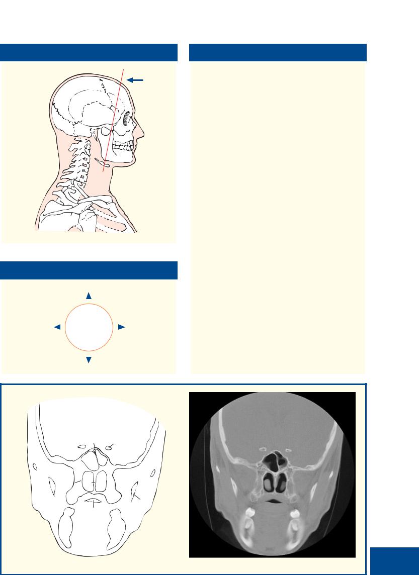
Coronal section 10 – Female |
|
|
HEAD |
|
|
|
|
|
■ Section level |
|
|
■ Notes |
||
|
|
|
|
|
|
|
|
|
View |
|
Do not be deceived! This section passes through the |
|
|
|
|
tip of the temporal lobe of the cerebrum (23), lying |
|
|
|
|
|
|
inferior to the lesser wing of the sphenoid (19) and |
|
|
|
|
|
not the orbit. Note that the plane of this section lies |
|
|
|
|
|
immediately anterior to the anterior horn of the |
|
|
|
|
|
lateral ventricle. |
|
|
|
|
|
The parotid duct (36) can be palpated easily in |
|
|
|
|
|
the living subject by tensing the masseter muscle |
|
|
|
|
|
(34) and feeling along the upper part of the anterior |
|
|
|
|
|
border of this muscle just inferior to the zygomatic |
|
|
|
|
|
arch (33). The accessory parotid gland, or pars |
|
|
|
|
|
accessoria (35), is usually completely detached from |
|
|
|
|
|
the main gland and lies between the parotid duct |
|
|
|
|
|
and the zygomatic arch. It accounts for an |
|
|
|
|
|
occasionally very anteriorly placed parotid tumour. |
|
|
|
|
|
The paired sphenoidal sinuses (21) lie within the |
|
|
|
|
|
body of the sphenoid bone and vary quite |
|
|
|
|
|
considerably in size and shape. They are rarely |
|
|
|
|
|
symmetrical, one often being much larger than the |
|
|
|
|
|
other and extending across the midline behind the |
|
|
|
|
|
other. Occasionally one overlaps the other sinus |
|
|
|
|
|
superiorly. Usually the septum (20) between the two |
■ Orientation |
|
|
sinuses is intact, although occasionally these |
||
|
|
communicate with each other. |
|||
|
|
|
|
|
|
|
|
|
|
|
As well as the main salivary glands, many other |
|
Superior |
|
|||
|
|
|
accessory salivary glands are found, some in the |
||
|
|
|
|
|
|
|
|
|
|
|
tongue, some between the crypts of the palatine |
|
|
|
|
||
|
|
|
|
|
tonsils and some on the inner aspects of the lip and |
|
|
|
|
|
cheeks. Large numbers are found in the posterior |
Right |
|
|
Left |
|
hard palate and the soft palate (49). They are mainly |
|
|
||||
|
|
|
|
|
mucous in type and are occasional sites for the |
|
|
|
|
|
development of a pleomorphic salivary tumour. |
|
|
|
|
|
The CT image is purposefully displayed at optimal |
|
Inferior |
|
|
setting for bony structure. |
|
|
|
|
|
||
|
|
|
|
|
|
21
2728 51 50
38
69
Coronal computed tomogram (CT)

|
|
Coronal section 11 – Female |
|
HEAD |
|
|
|
|
|
|
|
|
|
|
|
1 |
|
|
|
|
|
|
|
8 |
|
|
|
|
2 |
|
|
|
|
|
|
|
|
|
|
|
|
|
|
|
|
9 |
11 |
|
|
4 |
|
|
|
|
|
|
|
|
|
|
|
||
|
|
|
|
|
|
6 |
|
|
5 |
|
|
|
|
|
|
|
12 |
|
|
|
|
|
|
|
|
|
14 |
|
|
7 |
3 |
|
|
|
57 |
|
|
|
|
|
|
|
|
|
|
|
|
|
|
|
|
|
|
|
|
|
|
|
10 |
|
|
|
13 |
|
|
|
|
|
|
|
|
15 |
|
|
|
|
|
|
|
|
|
16 |
17 |
|
18 |
|
|
|
|
|
|
|
|
|
|
24 |
|
|
|
|
|
|
|
|
|
|
|
|
|
|
|
56 |
|
|
23 |
|
|
19 |
20 |
|
|
|
|
|
25 |
|
|
|
|
29 |
|
|
|
|
|
22 |
|
|
|
|
||
|
|
|
|
55 |
|
|
|
|
||
|
|
|
|
|
|
21 |
|
|
|
|
|
|
|
|
52 |
|
|
|
27 |
|
|
|
|
|
|
|
|
|
|
|
|
|
|
|
|
26 |
|
|
|
|
|
28 |
30 |
|
|
|
51 |
53 |
|
|
|
|
||
|
|
|
|
|
|
|
|
|
||
|
|
|
|
50 |
|
|
|
|
|
|
|
|
54 |
|
|
|
|
|
|
32 |
|
|
|
|
48 |
|
|
|
|
|
||
|
|
|
49 |
|
|
|
33 |
|
||
|
|
|
47 |
|
|
|
|
|||
|
|
|
|
|
|
|
31 |
|
||
|
|
|
|
|
|
|
|
|
|
|
|
|
|
|
|
|
|
|
|
34 |
|
|
|
|
|
43 |
|
|
|
|
|
|
|
|
46 |
|
|
|
|
|
|
|
|
|
|
|
|
|
|
|
|
|
35 |
|
|
|
45 |
|
42 |
|
|
|
|
36 |
|
|
|
|
|
|
|
|
|
|
||
|
|
|
44 |
|
41 |
|
|
|
|
|
|
|
|
|
|
|
|
|
|
|
|
|
|
|
|
|
40 |
|
|
|
37 |
|
|
|
|
|
39 |
38 |
|
|
|
|
|
|
1 Skin and dense |
11 Superior frontal |
23 |
Superior oblique |
|
37 |
Platysma |
50 Inferior nasal |
||
|
subcutaneous |
gyrus |
24 |
Branches of |
|
38 |
Anterior belly of |
concha |
||
|
tissue |
12 Middle frontal |
|
ophthalmic artery |
|
digastric |
51 Middle nasal |
|||
|
2 Epicranial |
gyrus |
|
and vein |
|
39 |
Mylohyoid |
concha |
||
|
aponeurosis |
13 Inferior frontal |
25 |
Ethmoidal air cells |
40 |
Geniohyoid |
52 Superior nasal |
|||
|
(galea |
gyrus |
26 |
Maxillary sinus |
|
41 |
Genioglossus |
concha |
||
|
aponeurotica) |
14 Longitudinal |
27 |
Maxillary nerve |
|
42 |
Lingual artery |
53 Nasal septum |
||
|
3 Dura mater |
fissure |
28 |
Maxillary artery |
|
43 |
Transverse fibres |
54 Parotid duct |
||
|
4 Subdural space |
15 Orbital gyri |
29 |
Temporalis |
|
|
of intrinsic |
55 Orbital part of |
||
|
5 Arachnoid mater |
16 Olfactory tract (I) |
30 |
Zygomatic arch |
|
|
muscle of tongue |
ethmoid bone |
||
|
6 Subarachnoid |
17 Levator palpebrae |
31 |
Masseter |
|
44 |
Sublingual gland |
56 Greater wing of |
||
|
space |
superioris |
32 |
Ramus of mandible |
45 |
Facial artery |
sphenoid bone – |
|||
|
7 Pia mater |
18 Superior rectus |
33 |
Buccal pad of fat |
|
46 |
Facial vein |
orbital surface |
||
|
8 Superior sagittal |
19 Optic nerve (II) in |
34 |
Buccinator |
|
47 |
Soft palate |
57 Frontal bone |
||
|
sinus |
dural sheath |
35 |
Body of mandible |
48 |
Horizontal plate |
|
|||
70 |
|
|
|
|
|
|
|
|
||
9 Falx cerebri |
20 Lateral rectus |
36 |
Inferior alveolar |
|
49 |
of palatine bone |
58 Zygoma |
|||
|
10 Medial frontal |
21 Inferior rectus |
|
nerve in |
|
Tuberosity of |
59 Dental artefacts |
|||
|
gyrus |
22 Medial rectus |
|
mandibular canal |
|
|
maxilla |
(see notes) |
||
|
|
|
|
|
|
|
|
|
|
|

Coronal section 11 – Female |
|
|
HEAD |
|
|
|
|
|
■ Section level
 View
View
■ Orientation
Superior
Right 
 Left
Left
Inferior
■ Notes
This section does indeed pass through the posterior part of the cavity of the orbit and demonstrates the close packing of the extrinsic muscles (17, 18, 20–23) and blood vessels (24) with the orbital fat and optic (II) nerve (19). Note that the optic nerve is surrounded by an extension of the dura mater and is, therefore, bathed in cerebrospinal fluid. Raised intracranial pressure is thus transmitted in the cerebrospinal fluid along the sheath and results in the changes of papilloedema.
The ethmoidal air cells, or sinuses (25), are small, thinwalled cavities in the ethmoidal labyrinth. They range in number from three large to 18 small cells on either side and are separated from the orbit by the paper-thin orbital plate of the ethmoid. Orbital cellulitis can thus easily result from ethmoid sinusitis (see (54) in Coronal section 13).
The three nasal conchae (still often referred to by ear, nose and throat (ENT) surgeons as the turbinate bones) project downwards like three scrolls from the lateral wall of the nasal cavity. The lowest, the inferior (50), is the largest and broadest. It is a separate bone, unlike the middle (51) and superior (52), which are part of the ethmoid bone (55). The middle and superior conchae are joined anteriorly, but diverge away from each other posteriorly so that the superior concha can be visualized only at posterior rhinoscopy and is invisible on viewing through the anterior nares. Beneath each concha is a space, termed the superior, middle and inferior meatus, respectively.
Metallic material used in dental fillings creates substantial problems for coronal CT. Even with careful positioning and gantry angulation, problems may be unavoidable: On the image, note the distortion created by the presence of a metallic dental filling.
57
1
57 |
|
19 |
|
|
|
|
|
56 |
|
|
|
|
|
25 |
|
|
51 |
53 |
58 |
26 |
|
||
50 |
|
|
|
|
|
|
49
59
35
71
Coronal computed tomogram (CT)
|
|
Coronal section 12 – Female |
|
HEAD |
|
|
|
|
|
|
2 |
|
1 |
|
|
|
|
|
|
|
|
|
|
|
|
|
|
|
|
|
|
|
|
|
|
|
5 |
|
|
9 |
|
|
|
|
|
|
|
|
|
|
|
|
|
|
|
|
|
|
|
|
|
|
4 |
6 |
|
|
10 |
|
11 |
|
|
|
|
|
3 |
|
|
|
|
|
|
|
||||
|
7 |
|
|
|
|
|
|
|
||||
|
|
|
|
|
|
|
|
|
|
|
||
|
|
|
|
|
|
17 |
|
|
|
|
|
|
|
|
8 |
|
|
|
|
|
|
|
|
|
|
|
|
|
|
|
|
|
|
12 |
|
|
||
|
|
|
|
|
|
|
|
|
|
|
||
|
|
|
|
|
|
|
16 |
|
13 |
|
|
|
|
58 |
|
|
|
|
|
|
|
|
|
||
|
|
|
|
|
|
|
14 |
|
|
|
||
|
|
57 |
|
|
|
|
15 |
|
|
|
||
|
|
|
|
|
|
|
19 |
|
20 |
21 |
|
|
|
|
|
|
|
|
|
|
|
27 |
22 |
|
|
|
|
|
|
|
|
|
|
|
|
|
|
|
|
|
|
|
54 |
|
|
18 |
|
|
|
||
|
|
|
|
|
|
|
|
25 |
26 |
23 |
|
|
|
|
|
|
|
|
|
|
|
|
|||
|
|
|
|
|
|
|
|
|
|
|
|
|
|
56 |
|
|
|
53 |
|
|
|
24 |
28 |
|
|
|
|
55 |
|
|
|
|
|
|
||||
|
|
|
|
|
|
|
|
|
|
|
29 |
|
|
|
|
|
|
|
|
|
|
|
59 |
|
|
|
|
|
|
51 |
|
|
|
|
|
|
|
|
|
|
52 |
|
|
|
|
|
|
|
|
|
|
|
|
|
|
|
50 |
|
|
|
|
|
30 |
|
|
|
|
|
49 |
|
|
|
|
|
|
|
|
|
|
|
|
|
|
|
|
|
|
|
|
|
|
|
|
|
|
|
58 |
|
|
|
|
31 |
32 |
|
|
|
|
48 |
|
|
|
|
|
|
|
|
|
|
|
|
|
|
|
|
|
|
|
|
|
|
|
47 |
|
|
|
|
|
|
|
33 |
|
33 |
|
|
|
|
|
|
|
|
|
|
|
|
|
|
|
|
|
|
|
|
|
|
45 |
34 |
35 |
|
|
|
|
|
|
|
|
|
|
|
|
||
|
|
|
|
|
|
|
|
|
44 |
|
36 |
|
|
|
|
|
|
43 |
|
|
|
|
|
||
|
|
|
|
|
|
|
|
|
|
|
||
|
|
46 |
|
42 |
|
|
|
41 |
37 |
|
|
|
|
|
|
|
|
|
|
|
|
|
|||
|
|
|
|
|
|
|
|
|
38 |
|
|
|
|
|
|
|
|
|
|
39 |
|
40 |
|
|
|
1 Skin and dense |
|
olfactory bulb (I) lying |
|
33 |
Buccal fat |
|
|
50 Nasal septum |
||||
subcutaneous tissue |
|
on cribriform plate of |
|
34 |
Buccinator |
|
|
51 Middle nasal concha |
||||
2 Epicranial aponeurosis |
|
ethmoid bone |
|
|
|
|
35 |
Facial vein |
|
|
52 Maxillary sinus |
|
(galea aponeurotica) |
19 |
Posterior part of crista |
|
36 |
Facial artery |
|
|
53 Ethmoidal air cells |
||||
3 Frontal belly of |
|
galli |
|
|
|
|
37 |
Inferior alveolar nerve |
54 Orbital part of ethmoid |
|||
occipitofrontalis |
20 |
Levator palpebrae |
|
|
|
in mandibular canal |
bone |
|||||
4 Dura mater |
|
superioris |
|
|
|
|
38 |
Body of mandible |
|
55 Orbital surface of |
||
5 Subdural space |
21 |
Superior rectus |
|
|
|
39 |
Platysma |
|
|
maxilla |
||
6 Arachnoid mater |
22 |
Branches of ophthalmic |
|
40 |
Anterior belly of |
|
56 Zygomatic bone |
|||||
7 Subarachnoid space |
|
artery and vein |
|
|
|
|
digastric |
|
|
57 Orbital part of frontal |
||
8 Pia mater |
23 |
Lateral rectus |
|
|
|
|
41 |
Mylohyoid |
|
|
bone |
|
9 Superior sagittal sinus |
24 |
Inferior rectus |
|
|
|
|
42 |
Geniohyoid |
|
|
58 Palatine process of |
|
10 Falx cerebri |
25 |
Medial rectus |
|
|
|
|
43 |
Genioglossus |
|
maxilla |
||
11 Superior frontal gyrus |
26 |
Optic nerve (II) in dural |
|
44 |
Sublingual gland |
|
59 Infra-orbital artery and |
|||||
12 Middle frontal gyrus |
|
sheath |
|
|
|
|
45 |
Intrinsic muscle of |
|
nerve within infra- |
||
13 Inferior frontal gyrus |
27 |
Superior oblique |
|
|
|
|
tongue |
|
|
orbital canal of maxilla |
||
14 Orbital gyri |
28 |
Temporalis |
|
|
|
|
46 |
Depressor anguli oris |
|
|||
15 Longitudinal fissure |
29 |
Zygomatic arch |
|
|
|
47 |
Buccal vestibule |
|
60 Teeth arising from |
|||
72 16 Cingulate gyrus |
30 |
Zygomaticus major |
|
|
48 |
Palatine glands of soft |
mandible |
|||||
17 Medial frontal gyrus |
31 |
Masseter |
|
|
|
|
|
palate |
|
|
61 Globe |
|
18 Posterior portion of |
32 |
Parotid duct |
|
|
|
|
49 |
Inferior nasal concha |
|
|||

Coronal section 12 – Female |
|
|
HEAD |
|
|
|
|
|
■ Section level
 View
View
■ Orientation
Superior
Right 
 Left
Left
■ Notes
From the roof of the nasal cavity, some 20 olfactory nerve (I) filaments on each side perforate the dura and arachnoid over the cribriform plate and pass upwards through the subarachnoid space to enter the olfactory bulb (18). From here, the olfactory tract passes posteriorly on the inferior surface of the frontal lobe. Fractures crossing the anterior cranial fossa, with tearing of the overlying dura, may result in cerebrospinal rhinorrhoea, watery fluid draining into the nose. Untreated, this communication between the nasal cavity and the subarachnoid space inevitably results in meningitis.
The maxillary sinus, or antrum (52), occupying most of the body of the maxilla, is the largest of the nasal accessory sinuses. In dentulous subjects, conical elevations, which correspond to the roots of the first and second molar teeth, project into the floor of the sinus, which they occasionally perforate. Less commonly, the roots of the two premolars, the third molar and, rarely, the canine may also project into the sinus. Upper dental infection may thus involve the sinus, and dental extraction may result in an oromaxillary fistula. The sinus opens into the nasal cavity in the lowest part of the hiatus semilunaris below the middle concha (51). A second orifice is often present in or just below the hiatus.
CT in the coronal plane is used to demonstrate the anatomy of the maxillary sinus and its drainage into the nasal cavity. Some ENT surgeons now perform flexible endoscopic sinus surgery (FESS) to improve matters. This CT projection is also useful for assessing fractures of the floor of the orbit.
Inferior
57 |
27 |
21 |
|
23 |
|||
19 |
|
||
53 |
|
61 |
56 |
|
25 |
|
|
|
|
55 |
52 |
|
|
|
|
|
49 |
|
|
58 |
|
|
60 |
73
Coronal computed tomogram (CT)

|
|
Coronal section 13 – Female |
|
HEAD |
|
|
|
|
2
1 |
|
|
|
|
|
4 |
|
|
|
|
|
3 |
|
|
|
|
|
5 |
|
|
|
|
|
6 |
|
9 |
|
|
|
|
|
10 |
11 |
12 |
|
|
|
|
|
||
7 |
23 |
|
|
|
|
8 |
|
|
|
||
33 |
|
|
|
|
13 |
|
22 |
20 |
|
14 |
|
|
|
|
21 |
|
15 |
32 |
|
|
|
16 |
|
|
|
|
34 |
||
31 |
|
|
|
|
|
|
|
|
|
|
|
30 |
24 |
|
19 |
|
17 |
|
|
18 |
|
||
|
26 |
|
|
25 |
35 |
53 |
|
|
|
|
|
27 |
|
|
|
36 |
|
|
|
|
|
|
|
|
28 |
|
|
37 |
|
47 |
|
|
|
||
|
|
|
|
38 |
|
|
|
|
29 |
|
|
|
|
|
|
39 |
|
|
|
|
|
|
|
46 |
45 |
|
|
40 |
|
|
|
|
|
||
|
|
|
|
|
|
|
|
|
|
|
41 |
52 |
|
|
|
42 |
|
50 |
44 |
|
|
|
|
|
|
|
|
||
51 |
|
|
|
|
|
49 |
43 |
|
48 |
||
|
1 |
Skin and dense |
12 |
Lacrimal gland (orbital |
26 |
Inferior nasal concha |
44 |
Sublingual gland |
|
|
|
subcutaneous tissue |
|
part) |
27 |
Palatine process of |
45 |
Genioglossus |
2 |
Epicranial aponeurosis |
13 |
Lacrimal gland |
|
maxilla |
46 |
Median septum of |
|
|
|
(galea aponeurotica) |
|
(palpebral part) |
28 |
Alveolar process of |
|
tongue |
3 |
Occipital belly of |
14 |
Lateral rectus |
|
maxilla |
47 |
Dorsum of tongue |
|
|
|
occipitofrontalis |
15 |
Inferior oblique |
29 |
Vestibule of mouth |
48 |
Platysma |
4 |
Frontal bone |
16 |
Inferior rectus |
30 |
Nasal septum |
49 |
Depressor anguli oris |
|
5 |
Dura mater |
17 |
Orbital margin of |
31 |
Middle nasal concha |
50 |
Depressor labii |
|
6 |
Frontal lobe of brain |
|
zygomatic bone |
32 |
Scleral layer of orbit |
|
inferioris |
|
|
|
covered with arachnoid |
18 |
Infra-orbital artery and |
33 |
Vitreous humour |
51 |
Mental nerve |
|
|
mater and blood vessels |
|
nerve within infra- |
34 |
Orbicularis oculi |
52 |
Sublingual papilla |
|
|
within the anterior |
|
orbital canal of maxilla |
35 |
Zygomaticus minor |
53 |
Hard palate |
|
|
cranial fossa |
19 |
Maxillary sinus |
36 |
Zygomaticus major |
|
|
7 |
Infundibulum draining |
20 |
Medial rectus |
37 |
Parotid duct |
54 |
Orbital margin of |
|
|
|
frontal sinus |
21 |
Orbital surface of |
38 |
Buccal fat pad |
|
ethmoid bone |
8 |
Roof of nasal cavity |
|
maxilla |
39 |
Facial vein |
55 |
Crista galli of ethmoid |
|
9 |
Orbital part of frontal |
22 |
Lacrimal bone |
40 |
Buccinator |
|
bone |
|
|
|
bone |
23 |
Tendon of superior |
41 |
Facial artery |
|
|
|
|
|
|
|||||
74 |
|
|
|
|||||
10 Superior rectus |
24 |
oblique |
42 |
Mucous membrane of |
|
|
||
|
11 Levator palpebrae |
Nasolacrimal duct |
|
mouth |
|
|
||
|
|
superioris |
25 |
Maxilla |
43 |
Mandible |
|
|
|
|
|
|
|||||

Coronal section 13 – Female |
|
|
HEAD |
|
|
|
|
|
■ Section level
 View
View
■ Orientation
Superior
Right 
 Left
Left
Inferior
■ Notes
The orbital part of the lacrimal gland (12) lies in the lacrimal fossa on the lateral part of the roof of the orbit supported by the lateral margin of levator palpebrae superioris (11). It projects around the lateral margin of this muscle and turns forward to form the palpebral part of the gland (13), which is visible through the superior fornix of the conjunctiva. Its dozen or so ducts drain into the superior fornix.
The nasolacrimal duct (24), about 2 cm in length, runs downwards and laterally to open into the inferior meatus below the inferior nasal concha (26), about
2 cm behind the nostril. Its mucosa is raised into several folds, which act as valves. These prevent air and nasal mucus being forced up the duct into the lacrimal sac when blowing the nose.
The buccal pad of fat (38) protrudes in front of the masseter to lie on the buccinator (40) immediately inferior to the parotid duct (37). Its estimated volume is 10 cm3. It is well developed in babies, where it forms a prominent elevation over the external surface of the face (the sucking pad). This helps to prevent collapse of the cheeks in vigorous sucking. It persists through life and is relatively ‘protected’, in that it does not decrease, even in emaciated subjects.
Note the paper-thin (lamina papyricea) orbital margin of the ethmoid bone (54). This portion and the relatively thin orbital margin of maxilla are liable to be damaged by a blow-out injury; the globe, being tougher, transmits injury to the walls of the orbit following trauma that is not absorbed by the bony margins. Squash balls are a particular culprit. The extrinsic eye muscles may get trapped between the fracture margins.
4
6
955
33 |
54 |
|
21
30
26
19
75
Coronal computed tomogram (CT)

|
|
|
Sagittal section 1 – Male |
|
|
|
|
|
|
|||||
|
HEAD |
|
|
|
|
|
|
|
||||||
|
|
|
|
|
|
|
|
|
|
|
|
|
|
|
|
|
|
|
|
|
|
4 |
|
|
|
|
|
|
|
|
|
|
|
3 |
|
|
|
|
|
|
5 |
|
|
|
|
|
|
|
|
|
|
|
|
|
|
|
|
|
|
|
|
|
|
|
|
|
10 |
|
9 |
13 |
|
6 |
|
|
|
|
|
|
|
|
|
|
|
|
|
|
|
||
|
|
|
|
|
|
|
12 |
|
|
|
|
|
|
|
|
|
1 |
|
|
|
|
|
|
7 |
|
|
|
||
|
|
|
|
|
14 |
|
|
|
|
|
|
|||
|
|
|
|
|
|
|
|
|
|
|
|
|
|
|
|
|
|
|
|
|
|
17 |
|
18 |
|
|
|
|
|
|
|
|
|
|
24 |
|
16 |
|
19 |
|
36 |
|
|
|
|
|
2 |
|
15 |
|
|
|
|
|
|||||
|
|
|
11 |
2523 |
|
|
8 |
|
|
|
|
|||
|
|
|
|
|
|
|
37 |
38 |
|
|
||||
|
|
|
|
|
|
21 |
20 |
|
|
|||||
|
|
|
|
|
26 |
22 |
|
|
|
|
||||
|
|
|
|
31 |
27 |
|
34 |
|
|
39 |
|
|
||
|
|
|
|
|
|
28 |
|
|
|
40 |
|
|
||
|
|
|
|
|
|
|
|
35 |
|
|
42 |
|
||
|
|
|
|
32 |
|
|
29 |
|
|
61 |
41 |
|
||
|
|
|
|
|
30 |
|
|
|
|
|
|
43 |
|
|
|
|
|
|
|
|
|
|
|
60 |
59 |
|
44 |
||
|
|
|
|
|
|
|
|
|
|
48 |
|
|||
|
|
|
|
|
|
|
|
|
62 |
57 |
|
|
|
|
|
|
|
|
33 |
|
|
66 |
|
|
|
|
|
||
|
|
|
|
|
|
|
56 |
|
58 |
|
|
44 |
||
|
|
|
|
|
|
|
|
|
|
|
|
|||
|
|
|
|
65 |
63 |
64 |
|
|
54 |
|
|
43 |
|
|
|
|
|
|
|
|
|
55 |
|
|
47 |
46 |
|
||
|
|
|
|
|
|
|
|
|
|
|
||||
|
|
|
|
|
|
|
|
|
|
|
|
|
|
|
|
45 |
|
53 |
49 |
|
|
||
52 |
50 |
|
51 |
||
|
1 |
Occipital bone |
21 |
Oculomotor nerve (III) |
38 |
Middle nasal concha |
56 |
Anterior arch of atlas |
|
2 |
Falx cerebri |
22 |
Posterior cerebral |
39 |
Middle meatus |
|
(first cervical vertebra) |
|
3 |
Superior sagittal sinus |
|
artery |
40 |
Inferior nasal concha |
57 |
Nasal part of pharynx |
|
4 |
Parietal bone |
23 |
Midbrain |
41 |
Inferior meatus |
|
(nasopharynx) |
|
5 |
Frontal bone |
24 |
Pineal body |
42 |
Hard palate |
58 |
Uvula |
|
6 |
Frontal sinus |
25 |
Superior colliculus |
43 |
Central incisor (upper |
59 |
Soft palate |
|
7 |
Crista galli of ethmoid |
26 |
Inferior colliculus |
|
and lower) |
60 |
Pharyngeal recess |
|
|
|
bone |
27 |
Aqueduct (of Sylvius) |
44 |
Lip (upper and lower) |
61 |
Opening of auditory |
8 |
Sphenoidal sinus |
|
connecting third and |
45 |
Body of mandible |
|
(Eustachian) tube |
|
9 |
Genu of corpus callosum |
|
fourth ventricles |
46 |
Sublingual gland |
62 |
Anterior margin of |
|
10 |
Body of corpus callosum |
28 |
Pons |
47 |
Genioglossus |
|
foramen magnum |
|
11 |
Splenium of corpus |
29 |
Fourth ventricle |
48 |
Dorsum of tongue |
63 |
Posterior margin of |
|
|
|
callosum |
30 |
Cerebellum |
49 |
Geniohyoid |
|
foramen magnum |
12 |
Septum pellucidum |
31 |
Tentorium cerebelli |
50 |
Mylohyoid |
64 |
Posterior arch of atlas |
|
13 |
Anterior lobe gyrus |
32 |
Straight sinus |
51 |
Body of hyoid bone |
65 |
External occipital |
|
14 |
Body of fornix |
33 |
Transverse sinus |
52 |
Epiglottis |
|
protuberance |
|
15 |
Third ventricle |
34 |
Basilar artery |
53 |
Valleculla |
66 |
Medulla oblongata |
|
16 |
Hypothalamus |
35 |
Clivus (basi-occipital |
54 |
Oral part of pharynx |
|
|
|
|
17 |
Mamillary body |
|
and basi-sphenoid |
|
(oropharynx) |
67 |
Lateral ventricle |
76 |
|
|
||||||
18 |
Optic chiasm |
36 |
bones) |
55 |
Dens of axis (odontoid |
|
|
|
|
|
|||||||
|
19 |
Pituitary stalk |
Superior nasal concha |
|
peg of second cervical |
|
|
|
|
20 |
Pituitary gland |
37 |
Superior meatus |
|
vertebra) |
|
|
|
|
|
|
|||||

Sagittal section 1 – Male |
|
|
HEAD |
|
|
|
|
|
■ Section level |
View |
■ Orientation |
Superior
Posterior 
 Anterior
Inferior
■ Notes
Note that the nasal septum has been removed from this section in order to display the nasal conchae on the lateral wall.
The roof of the hard palate (42) lies at the level of the atlas (first cervical vertebra). Note that a clear anteroposterior view of the dens of the axis (second cervical vertebra) can be obtained on radiological examination by asking the patient to open the mouth widely.
This section illustrates the approach to the pituitary gland (20) via the transnasal trans-sphenoidal sinus (8) route at fibre-optic endoscopic surgery.
The frontal sinuses (6) vary considerably in size and are rarely symmetrical, the septum between the two usually being deviated to one or the other side. Each may be divided further by incomplete bony septa. Occasionally, one or both may be absent.
These two sagittal T1-weighted magnetic resonance images are very close to the median sagittal plane. Indeed, image (A) is so midline that the falx cerebri has been traversed. Image (B) is minimally lateral to the median sagittal plane and thus cerebral gyri and sulci are visible. The anatomy of the cerebral aqueduct and fourth ventricle is demonstrated well. So too is the important close relationship of the pituitary gland to the optic chiasm.
|
|
|
|
|
|
|
|
|
|
Sagittal magnetic resonance |
|
|
|
|
|
|
|
|
|
|
|
|
image (MRI) |
|
|
4 |
|
|
|
|
2 |
5 |
|
|
|
|
||
|
|
|
|
10 |
|
|
|
|
|
|
|
|
|
|
|
|
|
|
|
9 |
|
|
|
|
|
|
|
|
11 |
15 |
|
|
|
6 |
|
|
|
|
|
|
|
|
|
|
18 |
|
|
|
|
||
|
|
|
|
|
19 |
|
|
|
|
|
||
3 32 |
|
27 |
8 |
|
|
|
|
|
||||
|
|
|
|
28 |
20 |
|
|
|
|
|
||
|
|
|
30 |
29 |
|
35 |
|
42 |
|
|
|
|
|
|
|
|
|
57 |
|
|
|
|
|||
|
|
|
|
34 |
|
|
|
|
|
|
||
|
|
|
|
|
|
|
|
|
|
|
||
|
|
|
|
66 |
|
|
59 |
47 |
|
|
|
|
|
|
|
1 |
|
|
|
|
|
|
|
|
|
|
|
|
|
|
|
|
|
|
|
|
|
|
|
|
|
63 |
|
|
55 |
54 |
|
|
|
|
|
|
|
|
64 |
|
|
|
|
|
|
|
||
|
A |
|
|
|
|
|
|
A |
|
|
|
|
|
|
|
|
|
|
|
|
|
|
|
||
|
|
|
|
|
|
|
|
|
|
Sagittal magnetic resonance |
|
|
|
|
|
|
|
|
|
|
|
|
|
|
|
|
|
|
4 |
|
|
|
|
|
|
image (MRI) |
|
|
|
|
|
|
10 |
|
|
|
5 |
|
|
|
|
|
|
|
|
67 |
|
|
|
|
|
|
||
|
|
|
|
|
9 |
13 |
|
|
|
|
||
|
|
|
11 |
|
|
|
|
6 |
|
|
|
|
|
|
|
|
|
|
|
|
|
|
|
|
|
|
|
|
27 |
|
|
|
|
|
|
|
|
|
|
|
|
|
28 |
|
|
36 |
|
|
|
|
|
|
|
|
|
35 |
|
|
|
|
|
|||
33 |
30 |
29 |
|
42 |
|
|
|
|
||||
|
|
57 |
|
|
|
|
||||||
|
|
|
|
66 |
|
34 |
|
|
|
|
|
|
|
|
|
|
|
|
|
59 |
|
|
|
|
|
|
|
|
|
|
|
|
54 |
47 |
|
|
|
|
|
|
|
|
|
55 |
|
|
77 |
||||
|
|
|
|
|
|
|
|
|
|
|||
|
B |
|
|
|
|
|
B |
|
|
|
||
|
|
|
|
|
|
|
|
|
|
|
||
|
|
|
|
|
|
|
|
|
|
|
|
|
|
|
|
|
|
|
|
|
|
|
|
|
|

|
|
Coronal sections 1–2 – Temporal bone/inner ear – Male |
|
HEAD |
|
|
|
|
20
2
11
29
19
18
17
1
29
17
18
28
27
2
1
2 |
4 |
|
3 |
5
|
|
15 |
|
|
12 |
|
16 |
|
|
2 |
|
|
|
|
14 |
10 |
13 |
8 |
6 |
|
|||
9 |
|
||
|
|
|
2 |
7 |
|
|
22 |
6 |
|
|
|
6 |
|
|
|
|
|
2 |
4 |
5 |
|
|
|
|
1 |
|
||
|
|
|
|
3 |
|
||
|
|
|
|
|
|
|
|
21 |
|
|
|
|
|
|
|
25 |
2 |
15 |
|
|
|
|
|
13 |
14 |
2 |
|
|
|
||
|
|
|
|
|
|||
2 |
|
|
26 |
|
|
|
|
10 |
9 |
|
|
|
|
|
|
|
|
|
7 |
|
|
||
|
|
|
|
|
|
||
|
|
2 |
8 |
|
|
|
|
|
|
|
|
|
|
||
22 |
|
|
24 |
6 |
|
|
|
|
|
|
|
|
|||
23
|
1 |
Dura mater |
12 |
Tendon of tensor tympani |
23 |
Styloid process |
|
2 |
Temporal bone |
13 |
Stapes |
24 |
Parotid gland |
|
3 |
Temporalis |
14 |
Head of malleus |
25 |
Facial nerve (VII) and |
|
4 |
Skin and dense subcutaneous |
15 |
Tegmen tympani |
|
vestibulocochlear nerve (VIII) |
|
|
tissue |
16 |
Body of malleus |
|
within the internal acoustic |
|
5 |
Helix of left ear |
17 |
Occipital condyle |
|
meatus |
|
6 |
Auricular cartilage of ear |
18 |
Vertebral artery |
26 |
Long limb of incus |
|
7 |
External acoustic meatus |
19 |
Medulla oblongata |
27 |
Atlas (first cervical vertebra) |
|
8 |
Tympanic membrane |
20 |
Pons |
28 |
Atlanto-occipital joint |
78 |
9 |
Cavity of middle ear |
21 |
Free margin of tentorium |
29 |
Occipital bone |
|
10 Promontory of middle ear |
|
cerebelli |
|
|
|
|
11 Cochlea |
22 |
Internal jugular vein |
|
|
|

Coronal sections 1–2 – Temporal bone/inner ear – Male |
|
|
HEAD |
|
|
|
|
|
■ Section level
21
View
■ Orientation
|
Superior |
|
Superior |
||||
Medial |
|
|
Left Right |
|
|
Left |
|
|
|
|
|
|
|||
|
|
|
|
|
|||
|
|
|
|
|
|
|
|
|
Inferior |
|
Inferior |
||||
Sections 1 and 2 |
|
CT images |
|||||
|
|
|
|
|
|
|
|
■ Notes
The external acoustic meatus (7) extends inwards to the tympanic membrane (8). The meatus is about 37 mm in length and has a peculiar S-shaped course, being directed first medially superiorly and anteriorly, then medially and backwards and then, at its termination, medially, anteriorly and inferiorly. The outer third of the canal is cartilaginous and somewhat wider than the inner osseous portion. The tympanic membrane (eardrum) separates the middle ear (9) from the external meatus. It is oval in outline and faces laterally, inferiorly and anteriorly. It is about 12 mm in its greatest (vertical) diameter and is slightly concave outwards. The middle ear, or tympanic cavity (9), is a slit-like cavity in the petrous temporal bone (2) and contains the three auditory ossicles. These are the malleus, whose body, or handle (16), is attached to the tympanic membrane, and a head (14) which articulates with the incus (26), which, in turn, articulates with the stapes (13). The base of the stapes is firmly adherent to the oval window, or fenestra vestibuli, of the inner ear. This comprises a complicated bony labyrinth that encloses the membranous labyrinth. This comprises the utricle and saccule, which communicate with the semicircular canals and the cochlea, respectively (11).
The intimate relationship of the facial nerve (VII) and the vestibulocochlear nerve (VIII) as they enter the internal auditory meatus (25) in the petrous part of the temporal bone (2) is demonstrated.
See also Coronal sections 5–7.
A |
A |
2
25 
6
7
29
Coronal computed tomogram (CT)
11 14
29
B |
B |
79
Coronal computed tomogram (CT)

|
|
Selected images – Temporal bone/inner ear – Axial CT (A) |
|
HEAD |
|
|
|
|
Axial computed tomogram (CT)
3 |
1 |
|
7 |
||
|
2
|
13 |
|
|
10 |
9 |
|
|
|
8 |
|
|
5 |
|
11 |
|
|
12 |
 4 7
4 7

 6
6 

1 |
Sphenoidal sinus |
8 |
Temporal bone (mastoid part) |
2 |
Basi-spenoid joining basi-occiput (forming clivus) |
9 |
Temporal bone (apex of petrous part) |
3 |
Temporal lobe of brain (in middle cranial fossa) |
10 |
Cochlea |
4 |
Posterior cranial fossa |
11 |
Internal auditory meatus |
5 |
Mastoid air cells (within temporal bone) |
12 |
Superior aspect jugular bulb |
6 |
Occipital bone |
13 |
Malleus |
7 |
Temporal bone (petromastoid part) |
|
|
|
|
|
|
80

Selected images – Temporal bone/inner ear – Axial CT (A) |
|
|
HEAD |
|
|
|
|
|
■ Section level
View
■ Orientation
Anterior
Right 
 Left
Left
Posterior
■ Notes
This thin section CT image has been reconstructed using a bony algorithm and displayed at settings to demonstrate the bony structures. Hence the bone texture is well seen (and the detail of cerebral tissue absent). Such high resolution images are essential to study the anatomy of the inner ear before complex surgery.
81