

- •Contents
- •Preface
- •Introduction
- •The importance of cross-sectional anatomy
- •Orientation of sections and images
- •Notes on the atlas
- •References
- •Acknowledgements
- •Interpreting cross-sections: helpful hints for medical students
- •BRAIN
- •HEAD
- •NECK
- •THORAX
- •ABDOMEN
- •PELVIS
- •LOWER LIMB
- •UPPER LIMB
- •Index

134
1
39
35
33
32
31
7
29
30
|
5 |
3 |
6 |
2 |
4 |
7
8 |
10 |
|
9 |
||
|
||
38 |
7 |
|
|
35
37 |
|
36 |
12 |
34 |
11 |
|
7
27
26
13
25
24 22 23
21
20
20 |
14 |
19
18
28
17
15
16
1 |
Sixth costal cartilage |
9 Left ventricle |
18 |
Erector spinae |
25 |
Thoracic duct |
34 |
Inferior vena cava |
|
2 |
Superior epigastric artery |
10 |
Extrapericardial fat |
19 |
Spinal cord within dural |
26 |
Aorta |
35 |
Hepatic vein |
|
and vein |
11 |
Fundus of stomach |
|
sheath |
27 |
Right crus of diaphragm |
36 |
Caudate lobe of liver |
3 |
Seventh costal cartilage |
12 |
Oesophagogastric junction |
20 |
Sympathetic chain |
28 |
Tenth rib |
37 |
Fissure for ligamentum |
4 |
Xiphoid |
13 |
Spleen |
21 |
Body of tenth thoracic |
29 |
Lower lobe of right lung |
|
venosum – lesser omentum |
5 |
Rectus abdominis |
14 |
Lower lobe of left lung |
|
vertebra |
30 |
Ninth rib |
38 |
Left lobe of liver |
6 |
External oblique |
15 |
Serratus anterior |
22 |
Origin of intercostal artery |
31 |
Eighth rib |
39 |
Sixth rib |
7 |
Diaphragm |
16 |
Latissmus dorsi |
23 |
Hemiazygos vein |
32 |
Right lobe of liver |
|
|
8 |
Right ventricle |
17 |
Trapezius |
24 |
Azygos vein |
33 |
Seventh rib |
40 |
Oesophagus |
|
|
|
|
|
|
|
|
|
|
ABDOMEN
Male – 1 section Axial

|
|
8 |
10 |
|
|
|
9 |
|
|
|
|
|
|
|
|
|
|
38 |
|
32 |
34 |
40 |
|
7 |
|
|
11 |
||
|
|
26 |
15 |
|
|
|
|
||
|
24 |
|
|
|
|
23 |
|
16 |
|
|
|
13 |
||
|
|
|
|
|
|
29 |
|
|
|
|
|
18 |
|
|
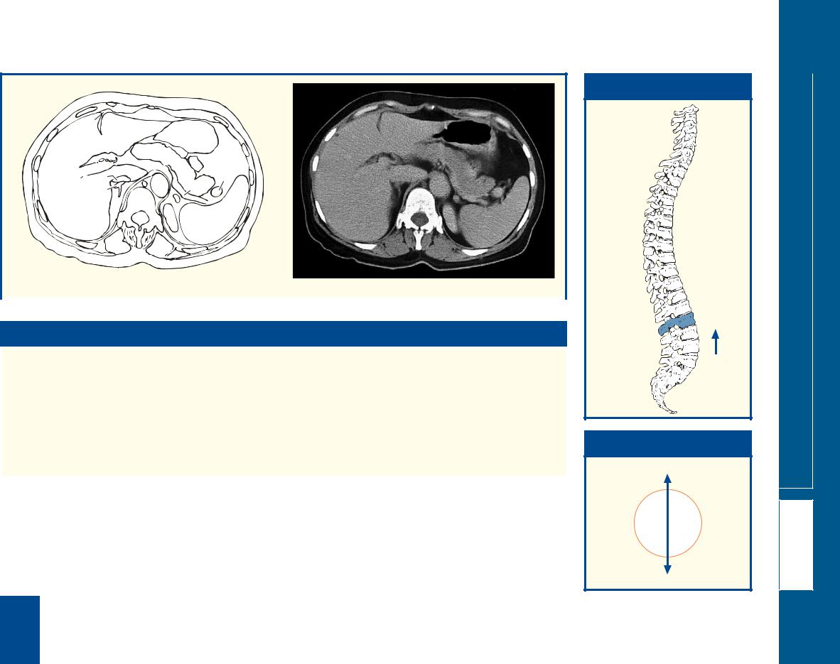
Axial computed tomogram (CT)
■ Notes
This section passes through the body of the tenth thoracic |
the cranial margin of this fissure, the lesser omentum |
vertebra (21) and anteriorly transects the xiphoid (4). |
reaches the diaphragm, where its two layers separate to |
The oesophagogastric junction (12) is seen in |
surround the lower end of the oesophagus. |
longitudinal section. This acts as a physiological sphincter in |
The ligamentum venosum is the thrombosed cord of the |
the prevention of reflux. The fundus of the stomach (11) |
ductus venosus, which, in fetal life, connects the left portal |
contains air in the erect position but in the supine position |
vein to the anterior aspect of the inferior vena cava. |
is normally full of fluid. It is opaque in the CT image |
The spleen (13) lies against the diaphragm (7) opposite |
because of the ingested radio-opaque iodinated material. |
ribs 9 (30), 10 and 11. This section demonstrates clearly |
The lesser omentum is the fold of peritoneum that |
how a stab wound of the left lower chest posteriorly might |
extends to the liver from the lesser curvature of the |
traverse the pleural cavity, injure the lower lobe of the lung |
stomach and the commencement of the duodenum. |
(14), traverse the diaphragm and lacerate the spleen. |
Superiorly it attaches to the porta hepatis and to the |
Similarly, a stab wound of the right chest at this level might |
bottom of the fissure for the ligamentum venosum (37). At |
injure the liver (32). |
|
|
■ Section level
T10
View
■ Orientation
Anterior
Right 
 Left
Left
Posterior
Male – 1 section Axial
ABDOMEN
135

136
|
1 |
2 |
3 |
|
|
|
4 |
|
|
||
|
|
|
|
|
|
39 |
40 |
|
|
5 |
6 |
|
|
|
7 |
||
|
|
38 |
|
|
8 |
|
37 |
|
|
|
|
|
|
|
9 |
|
|
|
|
|
|
|
|
|
36 |
|
|
|
|
32 |
|
35 |
|
10 |
|
|
|
|
|
||
|
|
|
|
|
|
|
|
34 |
|
|
|
31 |
|
33 |
|
11 |
|
|
|
|
|
||
|
|
|
|
|
|
|
|
24 |
23 |
|
12 |
30 |
|
|
|
||
|
20 |
|
22 |
||
|
|
|
13 |
||
|
|
18 |
19 |
|
|
|
25 |
|
|
||
|
|
|
21 |
5 |
14 |
|
|
|
|
|
|
|
5 |
26 |
17 |
16 |
|
29 |
27 |
|
|
|
15 |
|
28 |
1 |
Seventh costal cartilage |
11 |
Splenic pedicle |
20 |
Thoracic duct |
31 |
Right lobe of liver |
39 |
Sixth costal cartilage and |
2 |
Xiphoid |
12 |
Spleen |
21 |
Intercostal vein |
32 |
Seventh rib |
|
rib |
3 |
Rectus abdominis |
13 |
External oblique |
22 |
Left suprarenal gland |
33 |
Inferior vena cava |
40 |
Falciform ligament |
4 |
Superior epigastric artery |
14 |
Latissimus dorsi |
23 |
Aorta |
34 |
Caudate lobe of liver |
|
|
|
and vein |
15 |
Erector spinae |
24 |
Right crus of diaphragm |
35 |
Lesser omentum in fissure |
41 |
Portal vein |
5 |
Diaphragm |
16 |
Lower lobe of left lung |
25 |
Right suprarenal gland |
|
for ligamentum venosum |
42 |
Pancreas |
6 |
Pericardial fat |
17 |
Spinal cord within dural |
26 |
Head of eleventh rib |
36 |
Hepatic vein |
43 |
Left colic (splenic) flexure |
7 |
External oblique |
|
sheath |
27 |
Lower lobe of right lung |
37 |
Left lobe of liver medial |
44 |
Splenic vein |
8 |
Greater omentum |
18 |
Body of eleventh thoracic |
28 |
Tenth rib |
|
segment |
45 |
Left crus of diaphragm |
9 |
Body of stomach |
|
vertebra |
29 |
Ninth rib |
38 |
Left lobe of liver lateral |
46 |
Median arcuate ligament |
10 Left gastric artery branches |
19 |
Intercostal artery |
30 |
Eighth rib |
|
segment |
|
|
|
ABDOMEN
Male – 2 section Axial

|
|
|
|
|
|
|
|
|
|
|
38 |
|
■ Orientation |
|
|||
|
|
|
|
|
|
|
|
|
|
|
42 |
|
|
|
|
|
|
|
|
|
|
|
|
|
|
|
|
|
10 |
|
|
|
|
|
|
|
|
|
|
|
|
|
|
|
|
|
|
|
|
|
|
||
|
|
|
40 |
|
|
43 |
|
|
|
41 |
9 |
|
Anterior |
|
|||
|
|
|
|
|
|
|
|
|
|
|
|
|
|
|
|
||
|
|
37 |
38 |
|
|
|
|
34 |
46 |
|
|
|
|
|
|
||
|
|
9 |
|
|
|
|
|
|
|
|
|
|
|||||
|
|
|
|
|
|
|
|
|
|
|
|
|
|
|
|||
|
|
|
|
42 |
|
|
|
|
|
|
33 |
42 |
|
|
|
|
|
|
|
|
41 |
|
|
|
|
|
|
23 |
|
|
|
|
|
|
|
|
|
|
44 |
|
|
43 |
|
|
|
|
Right |
|
|
|
Left |
||
|
31 |
|
34 33 |
|
|
|
|
|
24 |
44 |
|
|
|
||||
|
|
23 |
|
|
|
|
|
|
|
|
|
||||||
|
|
|
|
|
|
|
|
|
|
|
|
|
|
|
|
||
|
|
|
25 |
|
|
|
|
|
|
|
|
|
|
|
|
|
|
|
|
|
24 |
42 |
|
|
|
|
|
|
|
|
|
|
|
|
|
|
|
|
|
|
31 |
25 |
|
22 |
|
|
|
|
|
||||
|
|
|
|
22 |
|
12 |
|
|
|
|
|
|
|
||||
|
|
|
|
|
|
14 |
|
|
|
|
45 |
|
|
|
|
|
|
|
|
|
|
|
|
|
|
|
|
24 |
|
Posterior |
|
||||
|
|
|
|
|
|
|
|
|
|
|
|
|
|||||
|
|
|
|
|
|
|
|
|
|
|
|
|
|
||||
|
|
|
|
|
|
|
|
|
|
|
|
|
|
|
|||
A |
|
|
|
15 |
|
|
|
|
|
|
|
|
|
|
|
|
|
|
|
|
|
|
|
B |
|
|
|
|
|
|
|
|
|
||
|
|
|
|
|
|
|
|
|
|
|
|
|
|
|
|
||
|
|
|
|
|
|
|
|
|
|
|
|
|
|
|
|
|
|
|
|
|
|
|
|
|
|
|
|
|
|
|
|
|
|
|
|
A |
|
B |
|
|
|
Axial computed tomograms (CTs)
■ Notes
This section passes through the body of the eleventh thoracic vertebra (18) and the xiphoid (2).
This is the most caudal section that transects intrathoracic viscera – note the pericardial fat (6) anteriorly and the lower lobe of the left lung (16).
The suprarenal glands (22, 25) have a constant relationship to the diaphragmatic crura (24, 45). Note on the CT images that the separate limbs of the suprarenal glands are demarcated.
The right crus of the diaphragm (24) on the CT image is often bulky. The crura change shape during respiration; normally they are bulkier on inspiration.
On the CT image, the pancreas (42) is just visible as it enters the plane of this section. It is seen better in more caudal sections. As the pancreas occupies only part of the section, its outlines are not demarcated sharply. This is another example of the ‘partial volume’ effect.
■ Section level
T11
View
Male – 2 section Axial
ABDOMEN
137

138
41
40
1
42
|
|
|
43 |
|
44 |
|
|
|
34 |
33 |
32 |
|
|
31 |
28 |
36 |
35 |
30 |
|
29 |
|
||
|
|
||
|
|
|
|
|
|
20 21 |
|
|
|
18 |
|
|
|
22 |
|
39 |
|
|
15 |
|
17 |
|
|
|
|
|
|
|
8 |
|
|
|
38 |
37 |
13 |
|
|
|
|
3 2
4
4
5
26 27
6
9
25
24
23 |
7 |
19
16
8
10
14
11
12
1 |
Linea alba |
11 |
Serratus posterior inferior |
18 |
Thoracic duct |
29 |
Inferior vena cava |
40 |
Ninth rib |
2 |
Rectus abdominis |
12 |
Erector spinae |
19 |
Left crus of diaphragm |
30 |
Caudate lobe of liver |
41 |
Eighth rib |
3 |
Superior epigastric artery |
13 |
Spine of eleventh thoracic |
20 |
Right crus of diaphragm |
31 |
Portal vein |
42 |
Seventh costal cartilage |
|
and vein |
|
vertebra |
21 |
Aorta |
32 |
Hepatic artery |
43 |
Left lobe of liver (lateral |
4 |
Greater omentum |
14 |
Conus medullaris |
22 |
Right suprarenal gland |
33 |
Common bile duct |
|
segment) |
5 |
Body of stomach |
|
surrounded by cauda |
23 |
Left suprarenal gland |
34 |
Radicle of portal vein |
44 |
Left lobe of liver (medial |
6 |
Left colic (splenic) flexure |
|
equina within dural sheath |
24 |
Tail of pancreas |
35 |
Hepatic artery branch |
|
segment) |
7 |
Spleen |
15 |
Body of twelfth thoracic |
25 |
Splenic vein |
36 |
Right lobe of liver |
|
|
8 |
Diaphragm |
|
vertebra |
26 |
Splenic artery |
37 |
Twelfth rib |
45 |
Gall bladder |
9 |
External oblique |
16 |
Left kidney |
27 |
Body of pancreas |
38 |
Eleventh rib |
46 |
Ligamentum teres |
10 Latissimus dorsi |
17 |
Right kidney |
28 |
Left gastric artery and vein |
39 |
Tenth rib |
47 |
Jejunum |
|
|
|
|
|
|
|
|
|
|
|
ABDOMEN
Male – 3 section Axial

|
|
2 |
|
|
|
|
46 |
5 |
|
|
|
|
44 |
43 |
|
|
|
|
45 |
|
|
|
|
|
|
32 |
27 |
47 |
|
|
|
31 |
|
||
|
|
47 |
|
||
36 |
|
29 |
|
6 |
|
22 |
|
|
|||
21 |
24 |
|
|
||
|
|
|
|
|
|
|
17 |
19 |
|
23 |
|
|
20 |
16 |
|
7 |
|
10
12
Axial computed tomogram (CT)
■ Notes
This section passes through the body of the twelfth thoracic |
on the anterior surface and by the fissures for the ligamentum |
vertebra (15). It demonstrates well the relationships of the |
teres and ligamentum venosum on its visceral surface. This is |
structures at the porta hepatis – the common bile duct (33) |
simply a gross anatomical descriptive term, with no morphologi- |
anterior and to the right, the hepatic artery (32) anterior and |
cal significance. Two subsidiary additional lobes are marked out |
to the left, and the portal vein (31) posterior to these |
on the visceral aspect of the liver – the quadrate lobe anteriorly, |
structures. The inferior vena cava (29) lies immediately behind |
between the gall bladder fossa and the fissure for the ligamen- |
the portal vein; between the two is the epiploic foramen, or |
tum teres, and the caudate lobe posteriorly, between the groove |
the aditus to the lesser sac (the foramen of Winslow). The |
for the inferior vena cava and the fissure for the ligamentum |
division between the cortex (peripheral) and medulla (central) |
venosum. The transverse fissure for the porta hepatis separates |
of the kidneys (16, 17) is shown well; in the plane of this |
the quadrate and caudate lobes. The distribution of the right |
division run the small arcuate vessels, which can just be |
and left branches of the hepatic artery and of the hepatic duct |
identified in this section. Post-mortem changes account for the |
shows that the morphological division of the liver is into a right |
discrepancy in the differentiation between cortex and medulla |
and left lobe demarcated by a plane that passes through the |
in the left kidney. |
fossa of the gall bladder and the fossa of the inferior vena cava |
|
(the median plane of the liver). Morphologically, the quadrate |
Note on the lobes of the liver |
lobe and the left half of the caudate lobe are part of the mor- |
The gross anatomical division of the liver is into right and left |
phological left lobe of the liver. Further subdivision into hepatic |
lobes, demarcated by the attachment of the falciform ligament |
segements is made by the Couinaud system (segments I–VIII). |
|
|
■ Section level
T12
View
■ Orientation
Anterior
Right 
 Left
Left
Posterior
Male – 3 section Axial
ABDOMEN
139

140
|
|
1 |
|
44 |
|
|
41 |
|
43 |
40 |
4 |
|
45 |
|
|
|
39 |
28 |
|
42 |
|
|
|
|
38 |
|
|
26 |
|
|
|
|
||
|
|
27 |
|
|
37 |
|
|
23 |
|
|
30 |
|
|
|
|
|
|
22 |
|
36 |
|
|
|
|
|
|
|
|
|
32 |
31 |
21 |
20 |
19 |
|
|
|
|
|
|
|
18 |
|
17 |
33 |
|
|
|
|
35 |
|
|
|
16 |
34 |
|
|
|
|
2
3
5
6
29 |
7 |
|
|
25 |
7 |
|
|
24 |
|
8
15 |
9 |
13 |
14 |
|
10 |
12 |
11 |
|
1 |
Linea alba |
14 |
Left kidney |
19 |
Left crus of diaphragm |
31 |
Right renal artery |
42 |
Ninth rib |
2 |
Rectus abdominis |
15 |
Left renal vein (intrarenal |
20 |
Thoracic duct |
32 |
Right renal vein |
43 |
Eighth costal cartilage |
3 |
Transversus abdominis |
|
portion); see also 23 |
21 |
Right crus of diaphragm |
33 |
Right kidney |
44 |
Ninth costal cartilage |
4 |
Stomach, body/antrum |
16 |
Conus medullaris |
22 |
Aorta |
34 |
Twelfth rib |
45 |
Left lobe of liver (medial |
5 |
Transverse colon |
|
surrounded by cauda |
23 |
Left renal vein |
35 |
Eleventh rib |
|
segment) |
6 |
External oblique |
|
equina within dural sheath |
24 |
Superior mesenteric artery |
36 |
Tenth rib |
|
|
7 |
Jejunum |
17 |
Part of intervertebral disc |
25 |
Splenic vein |
37 |
Right lobe of liver |
46 |
Right colic (hepatic) |
8 |
Lower pole of spleen |
|
between the twelfth |
26 |
Portal vein |
38 |
Gall bladder |
|
flexure |
9 |
Descending colon |
|
thoracic and first lumbar |
|
(commencement) |
39 |
First part of duodenum |
|
|
10 Latissimus dorsi |
|
vertebrae, with part of |
27 |
Common bile duct |
|
(cap) |
|
|
|
11 Serratus posterior inferior |
|
body of twelfth thoracic |
28 |
Head of pancreas |
40 |
Left lobe of liver (lateral |
|
|
|
12 Erector spinae |
|
vertebra |
29 |
Neck of pancreas |
|
segment) |
|
|
|
13 Quadratus lumborum |
18 |
Psoas major |
30 |
Inferior vena cava |
41 |
Falciform ligament |
|
|
|
ABDOMEN
Male – 4 section Axial

|
|
2 |
1 |
|
|
|
|
|
|
|
|
|
|
3 |
|
4 |
|
|
45 |
40 |
|
|
|
|
|
|
|
||
38 |
41 |
|
|
5 |
|
|
|
|
|||
|
|
|
|
|
|
|
|
39 |
29 |
|
|
|
|
46 |
|
7 |
|
|
|
|
|
||
37 |
|
26 |
|
25 |
|
|
30 |
22 |
9 |
||
|
|
21 |
19 |
||
|
|
|
|
|
|
|
|
33 |
|
|
8 |
|
|
|
|
14 |
|
10 |
|
|
|
|
|
|
|
|
|
12 |
|
Axial computed tomogram (CT)
■ Notes
This section transects the intervertebral disc between the |
superior to the vein. These features are demonstrated well |
twelfth thoracic and the first lumbar vertebrae (17). The |
on the CT image in Axial section 5. In exposure of the |
spinal cord tapers into the conus medullaris (16), which |
abdominal aorta (22), the surgeon can divide the left renal |
terminates, in this subject, at the level of the body of the |
vein (23) in order to obtain additional access. The left |
first lumbar vertebra. The site of termination is variable, the |
kidney is not infarcted if this is done because the left renal |
range being from the disc between the twelfth thoracic |
vein receives the terminations of the left gonadal and left |
and first lumbar vertebrae to the lower border of the |
suprarenal veins, so that venous drainage of the left kidney |
second lumbar vertebra. |
can take place via collaterals from these vessels. |
The plane of this section passes through the left renal |
Note the circular folds of mucous membrane that project |
vein (23) and demonstrates well the close relationship of |
into the lumen of the small intestine transversely to its long |
this vein to the superior mesenteric artery (24), which |
axis (7). These are termed the plicae circulares. Radiologists |
passes forward from its aortic origin (22) immediately |
and clinicians refer to these as valvulae conniventes. |
|
|
■ Section level
T12–L1
View
■ Orientation
Anterior
Right 
 Left
Left
Posterior
Male – 4 section Axial
ABDOMEN
141
142
41
38
37
36
35
|
|
1 |
|
2 |
|
|
|
|
|
|
|
|
42 |
|
|
3 |
|
40 |
43 |
|
|
6 |
|
|
|
|
|
7 |
|
|
4 |
5 |
|
4 |
|
|
|
|
|
||
|
|
|
|
7 |
|
39 |
|
|
28 |
|
|
|
|
29 |
27 |
4 |
8 |
|
|
|
7 |
|
9 |
|
|
|
|
|
|
|
31 |
30 |
|
7 |
|
|
24 |
|
26 |
|
|
|
25 |
23 |
|
||
|
22 |
|
|
||
|
|
21 |
|
10 |
|
|
|
20 |
|
16 |
|
|
|
19 |
|
|
|
|
32 |
17 |
|
|
|
|
|
|
|
||
|
|
|
15 |
|
|
|
|
|
|
|
|
33 |
|
|
|
14 |
|
|
|
18 |
|
11 |
|
|
|
|
|
||
|
|
|
|
12 |
|
|
|
|
|
|
|
34 |
|
|
|
13 |
|
1 |
Linea alba |
13 |
Erector spinae |
20 |
Right sympathetic chain |
32 |
Commencement of right |
42 |
Falciform ligament |
2 |
Rectus abdominis |
14 |
Quadratus lumborum |
21 |
Right crus of diaphragm |
|
ureter |
43 |
Left lobe of liver (lateral |
3 |
Transversus abdominis |
15 |
Left kidney |
22 |
Aorta |
33 |
Right kidney |
|
segment) |
4 |
Greater omentum |
16 |
Left ureter |
23 |
Para-aortic lymph node |
34 |
Twelfth rib |
|
|
5 |
Antrum of stomach |
17 |
Psoas major |
24 |
Cisterna chyli |
35 |
Renal fascia |
44 |
Left renal vein |
6 |
Transverse colon |
18 |
Cauda equina within dural |
25 |
Inferior vena cava |
36 |
Eleventh rib |
45 |
Renal cyst |
7 |
Jejunum |
|
sheath |
26 |
Inferior mesenteric vein |
37 |
Right lobe of liver |
46 |
Uncinate process pancreas |
8 |
Internal oblique |
19 |
Body of first lumbar |
27 |
Superior mesenteric artery |
38 |
Tenth rib |
|
|
|
|
||||||||
9 |
External oblique |
|
vertebra, with portion of |
28 |
Superior mesenteric vein |
39 |
Gall bladder |
|
|
10 Descending colon |
|
intervertebral disc between |
29 |
Head of pancreas |
40 |
Left lobe of liver (medial |
|
|
|
11 Latissimus dorsi |
|
the first and second lumbar |
30 |
Common bile duct |
|
segment) |
|
|
|
12 Serratus posterior inferior |
|
vertebrae |
31 |
Duodenum |
41 |
Ninth costal cartilage |
|
|
|
ABDOMEN
Male – 5 section Axial

|
|
|
|
2 |
|
|
6 |
|
|
6 |
|
|
|
|
|
||
|
|
28 |
26 |
||
37 |
29 46 |
27 |
7 |
||
31 |
25 |
22 |
10 |
||
|
|||||
|
|
|
|||
|
44 |
21 |
|
||
|
|
|
|
||
|
|
|
|
15 |
|
A |
13 |
|
|
|
|
A
Axial computed tomogram (CT)
|
|
|
|
|
|
■ Orientation |
|
||
6 |
|
6 |
|
|
|
|
|
|
|
|
|
|
|
|
|
|
|||
|
|
|
|
Anterior |
|
||||
|
|
|
|
|
|
||||
|
|
29 |
28 |
|
|
|
|
||
|
|
27 |
|
|
|
|
|
||
|
|
|
|
|
|
|
|
|
|
31 |
|
|
7 |
|
|
|
|
|
|
|
|
|
10 |
|
|
|
|
||
25 |
|
|
|
|
|
|
|
||
44 |
22 |
|
|
|
|
|
|
||
37 |
|
|
Right |
|
|
Left |
|||
|
21 |
|
|
|
|
||||
|
|
|
|
|
|||||
33 |
|
45 |
15 |
|
|
|
|
||
|
|
|
|
|
|
||||
|
|
|
|
13 |
|
|
|
|
|
B |
|
|
|
|
|
|
Posterior |
|
|
|
|
|
|
|
|
|
|
||
|
|
|
|
|
|
|
|
|
|
B
Axial magnetic resonance image (MRI)
■ Notes
This section passes through the body of the first lumbar |
anterior to the kidney and its vessels and merges with the |
vertebra (19), with a small portion of the intervertebral disc |
connective tissue anterior to the aorta and inferior vena |
between the first and second lumbar vertebrae. |
cava. The posterior layer extends medially in front of the |
The kidneys (15, 33) are embedded in a mass of fatty |
fascia covering quadratus lumborum (14) and psoas major |
connective tissue termed the perirenal (perinephric) fat, |
(17) and to the vertebrae and intervertebral discs. The |
which is thickest at their medial and lateral borders. The |
perirenal fat and renal fascia (35) are surrounded by further |
fibro-areolar tissue surrounding the kidney and perirenal fat |
retroperitoneal (pararenal) fatty connective tissue. The |
condenses to form a sheath termed the renal fascia (35). At |
amount varies with the relative obesity of the subject. |
the lateral border of the kidney, the two layers of the renal |
In this section, a tiny portion of the lateral segment of the |
fascia are fused. The anterior layer is carried medially |
left lobe of the liver can be seen (43). |
|
|
■ Section level
L1–2
View
Male – 5 section Axial
ABDOMEN
143

144
32
31
30
|
|
|
1 |
|
2 |
|
|
|
|
41 |
|
|
|
|
|
|
|
|
|
|
|
3 |
|
|
|
|
|
|
|
|
|
|
|
|
|
|
|
|
|
4 |
|
|
|
|
|
|
|
|
|
|
5 |
|
|
|
|
|
39 |
|
5 |
|
3 |
6 |
|
|
|
|
|
|
|
||
|
|
|
|
|
5 |
|
7 |
|
|
|
|
|
|
|
|
||
40 |
|
|
38 |
37 |
|
|
5 |
8 |
|
|
35 |
|
|
|
|
||
|
|
36 |
|
|
|
|
||
|
|
|
|
|
|
|
||
|
|
|
5 |
|
5 |
|
|
|
|
|
|
|
|
5 |
|
||
|
34 |
|
|
|
|
|
|
|
|
|
|
|
|
|
|
|
|
33 |
25 |
|
24 |
22 |
|
19 |
|
|
|
23 |
21 |
|
9 |
|
|||
|
29 |
|
18 |
20 |
|
|
||
28 |
|
15 |
|
|
|
|||
|
|
|
17 |
|
|
|||
27 26 |
|
|
|
|
|
|||
|
|
|
16 |
|
|
|||
|
|
|
|
|
|
|
||
14 |
11 |
|
13 |
||
|
||
|
10 |
|
|
12 |
1 |
Linea alba |
13 |
Quadratus lumborum |
|
(arrowed) |
32 |
Right lobe of liver |
40 |
Gall bladder |
2 |
Rectus abdominis |
14 |
Cauda equina within dural |
21 |
Left testicular vein |
33 |
Right colic (hepatic) flexure |
41 |
Falciform ligament |
3 |
Greater omentum |
|
sheath |
22 |
Para-aortic lymph node |
34 |
Duodenum – second part |
|
|
4 |
Transverse colon |
15 |
Body of second lumbar |
23 |
Left lumbar vein |
|
(with ampulla marked with |
42 |
Ascending colon |
5 |
Jejunum |
|
vertebra |
24 |
Aorta |
|
a white bristle) |
43 |
Right crus of diaphragm |
6 |
External oblique |
16 |
Psoas major |
25 |
Inferior vena cava |
35 |
Head of pancreas |
44 |
Left renal vein |
7 |
Internal oblique |
17 |
Left kidney |
26 |
Right lumbar vein |
36 |
Uncinate process of |
45 |
Left renal artery |
8 |
Transversus abdominis |
18 |
Left ureter |
27 |
Right ureter |
|
pancreas |
|
|
9 |
Descending colon |
19 |
Left colic artery – ascending |
28 |
Right kidney |
37 |
Superior mesenteric artery |
|
|
10 Latissimus dorsi |
|
branch |
29 |
Right testicular vein |
38 |
Superior mesenteric vein |
|
|
|
11 Renal fascia |
20 |
Inferior mesenteric vein, |
30 |
Twelfth rib |
39 |
Mesentery with mesenteric |
|
|
|
12 Erector spinae |
|
with origin of left colic vein |
31 |
Eleventh rib |
|
vessels |
|
|
|
ABDOMEN
Male – 6 section Axial

|
33 |
4 |
|
4 |
|
|
|
|
|
|
|
|
|
38 |
37 |
|
|
|
|
|
|
|
|
|
35 |
36 |
44 |
20 |
5 |
|
34 |
|
|
||
32 |
25 |
24 |
|
9 |
|
|
42 |
43 |
|
|
|
|
|
|
|
|
|
|
28 |
|
|
16 |
17 |
|
|
|
|
|
|
|
|
|
|
12 |
|
A
A
Axial computed tomogram (CT)
|
1 |
|
■ Orientation |
|
|
4 |
|
|
|
|
|
|
|
|
|
|
|
|
|
|
Anterior |
|
|
38 |
37 |
|
|
|
|
|
|
|
|
|
|
35 |
44 |
5 |
|
|
|
34 |
|
|
|
|
|
|
|
|
|
|
|
25 |
24 |
9 |
Right |
|
Left |
|
|
||||
32 |
45 |
|
|||
|
|
|
|
||
|
|
|
|
|
|
2843
 17
17
10
12
Posterior
B
B
Axial magnetic resonance image (MRI)
■ Notes
This section passes through the body of the second lumbar vertebra (15). The plane of section passes through a prominent left lumbar vein (23) as it passes posterior to the aorta (24) to drain into the inferior vena cava (25). Occasionally, it may constitute the principal venous return from the left kidney, when it is termed a retro-aortic renal vein.
The right testicular vein (29) drains directly into the inferior vena cava, whereas the left testicular vein (21) (together with the left suprarenal vein) drains into the left renal vein.
This section passes through the second part of the duodenum (34). The orifice of the ampulla of Vater on its papilla is marked with a white bristle.
On both the section and the CT image, the uncinate process of the pancreas (36) is seen clearly. This lies posterior to the superior mesenteric artery (37) and vein (38) and is related closely to the entry point of the left renal vein (44) into the inferior vena cava (25).
■ Section level
L2
View
Male – 6 section Axial
ABDOMEN
145

146
3
4
32 31
3
|
31 |
|
31 |
25 |
24 |
|
||
|
23 |
22 |
|
30 |
|
29
1 |
|
2 |
|
|
|
|
|
|
|
3 |
|
4 |
|
|
|
|
|
5 |
|
|
5 |
|
|
|
27 |
28 |
|
26 |
|
|
5 |
|
|
|
|
17 |
|
|
|
18 |
21 |
20 |
5 |
|
|
|
19
16 |
|
|
15 |
|
14 |
13 |
12 |
|
|
|
11 |
6
7
5
8
53
9
10
1 |
Linea alba |
10 |
Latissimus dorsi |
|
the second and third |
23 |
Right ureter |
30 |
Right kidney lower pole |
2 |
Rectus abdominis |
11 |
Erector spinae |
|
lumbar vertebrae |
24 |
Inferior vena cava |
31 |
Ascending colon and right |
3 |
Greater omentum |
12 |
Quadratus lumborum |
17 |
Aorta |
25 |
Right testicular vein |
|
colic (hepatic) flexure |
4 |
Transverse colon |
13 |
Cauda equina within dural |
18 |
Para-aortic lymph node |
26 |
Duodenum, third part |
32 |
Right lobe liver |
5 |
Jejunum |
|
sheath |
19 |
Left ureter |
27 |
Superior mesenteric artery |
|
|
6 |
External oblique |
14 |
Root of second lumbar |
20 |
Inferior mesenteric vein |
|
and vein |
33 |
Ascending colon |
7 |
Internal oblique |
|
nerve |
21 |
Left testicular artery and |
28 |
Mesentery with mesenteric |
34 |
Left kidney |
8 |
Transversus abdominis |
15 |
Psoas major |
|
vein |
|
vessels |
35 |
Ileum |
9 |
Descending colon |
16 |
Intervertebral disc between |
22 |
Right sympathetic chain |
29 |
Renal fascia |
|
|
ABDOMEN
Male – 7 section Axial

2
8 |
4 |
|
|
|
|
|
|
5 |
|
||
|
|
27 |
|
|
|
35 |
|
|
|
|
|
33 |
17 |
26 |
|
5 |
|
24 |
20 |
|
|||
|
|
|
|
|
|
30 |
|
15 |
19 |
9 |
|
|
34 |
||||
|
|
|
|
|
|
|
|
|
|
12 |
|
|
|
|
11 |
|
10 |
A |
|
|
|
||
|
|
|
|
|
|
|
|
|
|
|
|
A
Axial computed tomogram (CT)
|
|
|
|
1 |
|
2 |
|
|
|
|
|
|
|
|
|
|
|
|
|
6 |
8 |
|
|
|
|
5 |
|
|
|
|
|
|
27 |
|
|
|
|
||
7 |
|
35 |
|
|
|
|
|
|
|
33 |
|
|
|
|
|
|
|
||
|
|
26 |
26 |
28 |
|
|
5 |
||
|
|
|
|
|
|||||
|
|
|
24 |
17 |
|
|
|
||
|
|
|
|
|
|
|
|
||
|
|
|
|
|
5 |
|
|
||
|
|
|
|
|
|
|
|
||
|
30 |
|
|
|
15 |
19 |
34 |
9 |
|
|
|
|
|
|
|
||||
|
|
|
|
|
|
|
12 |
|
|
11 |
10 |
|
B
B
Axial computed tomogram (CT)
■ Orientation |
■ Section level |
|
|
|
|
Anterior |
|
|
Right 
 Left
Left
Posterior
L2–3
View
■ Notes
This section passes through the intervertebral disc |
produces obstruction of the third part of the |
the internal oblique; the posterior sheath is made up |
between the second and third lumbar vertebrae |
duodenum (duodenal ileus). |
of the aponeurosis of the transversus abdominis |
(16). It transects the most caudal part of the right |
Seen clearly in this section are the three layers of |
reinforced by the posterior portion of the internal |
lobe of the liver (32). The caudal extent of this lobe |
muscles that constitute the lateral part of the |
oblique. Below a line roughly halfway between the |
is variable and may project downwards in some |
anterior abdominal wall – the external oblique (6), |
umbilicus and the pubis, the posterior sheath is |
subjects for a considerable distance as a broad |
internal oblique (7) and transversus abdominis (8). |
deficient and all three aponeuroses pass in front of |
tongue-like process (Riedel’s lobe). |
Medially, their aponeuroses form the sheath that |
the rectus to form the anterior sheath. These |
Note the third part of the duodenum (26) lying in |
surrounds the rectus abdominis (2). The anterior |
muscles are demonstrated well on the CT image in |
the inverted V between the aorta (17) and the |
sheath comprises the aponeurosis of the external |
Axial section 8. |
superior mesenteric vessels (27). Occasionally, this |
oblique together with the split anterior portion of |
|
|
|
|
Male – 7 section Axial
ABDOMEN
147

148
3
31
31
31
3 |
4 |
|
27
29
28
1 |
|
|
2 |
|
|
|
|
|
|
|
|
|
|
31 |
|
|
|
3 |
|
|
|
|
4 |
|
|
||
|
|
|
4 |
|
||
|
|
|
|
|
|
|
|
|
|
|
|
5 |
|
|
30 |
|
|
|
5 |
|
|
|
|
|
|
5 |
|
25 |
|
|
|
|
3 |
8 |
|
|
24 |
|
|
||
|
18 |
23 |
|
5 |
7 |
|
|
|
|
5 |
|
||
|
|
|
|
22 |
6 |
|
|
|
|
|
|
||
26 |
|
19 |
20 21 |
|
9 |
|
17 |
|
|
||||
15
16
14
13
10
12
11
1 |
Linea alba |
10 |
Quadratus lumborum |
17 |
Body of third lumbar vertebra |
25 |
Duodenum, third part |
2 |
Rectus abdominis |
11 |
Erector spinae |
18 |
Aorta |
26 |
Right sympathetic chain |
3 |
Greater omentum |
12 |
Cauda equina within dural sheath |
19 |
Left sympathetic chain |
27 |
Inferior vena cava |
4 |
Ileum |
13 |
Dorsal root ganglion of third |
20 |
Left ureter |
28 |
Right ureter |
5 |
Jejunum |
|
lumbar nerve |
21 |
Left testicular artery and vein |
29 |
Ascending colon |
6 |
Transversus abdominis |
14 |
Ventral ramus of second lumbar |
22 |
Left colic artery and inferior |
30 |
Mesentery with mesenteric vessels |
7 |
Internal oblique |
|
nerve |
|
mesenteric vein |
31 |
Transverse colon |
8 |
External oblique |
15 |
Psoas major |
23 |
Para-aortic lymph node |
|
|
9 |
Descending colon |
16 |
Third lumbar artery |
24 |
Inferior mesenteric artery |
|
|
ABDOMEN
Male – 8 section Axial

1
|
|
|
2 |
|
|
|
4 |
|
5 |
|
|
|
|
|
|
29 |
4 |
|
5 |
|
|
|
||
|
|
25 |
|
|
|
|
|
|
|
7 |
6 |
27 |
18 24 |
5 |
|
15 |
|||
8 |
|
|
9 |
|
|
|
|
||
|
|
|
12 |
|
10
11
|
Axial computed tomogram (CT) |
|
|
■ Notes |
|
|
|
This section passes through the body of the third lumbar |
The linea alba (1) is wide above the umbilicus and |
vertebra (17). This is just distal to the origin of the inferior |
becomes quite narrow below this level (see page 164). This |
mesenteric artery (24) from the anterior aspect of the aorta |
line marks the almost avascular blending of the rectus |
(18) posterior to the third part of the duodenum (25). This |
sheaths on either side and gives the surgeon rapid access |
section is now caudal to the liver and the kidneys. |
to the abdominal cavity. The incision can, if necessary, be |
The ventral ramus of the second lumbar nerve (14) is |
extended from the xiphoid to the pubic symphysis. The |
seen in this section as it passes downwards and laterally |
falciform ligament (see page 152) lies to the right-hand |
into the psoas major (15). The first three lumbar nerves and |
side of the incision. |
the greater part of the fourth lumbar nerve form the |
Note the marked disparity between the patulous |
lumbar plexus within the posterior part of the psoas major |
ascending colon (29) and the thick-walled, narrow |
in front of the transverse processes of the lumbar vertebra. |
descending colon (9). |
|
|
■ Section level
L3
View
■ Orientation
Anterior
Right 
 Left
Left
Posterior
Male – 8 section Axial
ABDOMEN
149

150
1
|
|
|
|
|
2 |
|
|
|
37 |
|
3 |
|
|
|
|
|
|
|
36 |
|
|
|
21 |
|
35 |
|
28 |
|
|
|
|
|
|
|
|
|
31 |
30 |
|
28 |
22 |
|
33 32 |
29 |
|||
34 |
|
17 |
|
23 |
|
16 |
|
13 |
26 |
27 |
|
|
|
|
|
4 |
|
|
|
|
14 |
|
|
|
15 |
11 |
|
|
|
|
12 |
|
|
||
|
18 |
10 |
|
|
|
|
|
|
24 |
||
|
|
|
|
|
|
|
|
|
9 |
|
25 |
19 |
20 |
|
|
|
|
|
|
|
7 |
8 |
5 |
|
|
|
|
|
6 |
1 |
Linea alba |
8 Dorsal root ganglion of |
14 |
Left renal artery |
24 |
Perirenal fat within renal |
32 |
Lymph node in porta |
|
2 |
Eighth costal cartilage |
|
first lumbar nerve |
15 |
Right renal artery |
|
fascia |
|
hepatis |
3 |
Ninth rib/costal cartilage |
9 Part of body of first lumbar |
16 |
Inferior vena cava |
25 |
Spleen |
33 |
Hepatic artery |
|
|
junction |
|
vertebra |
17 |
Left renal vein |
26 |
Left suprarenal gland |
34 |
Right lobe of liver |
4 |
Tenth rib |
10 |
Part of intervertebral disc |
18 |
Right renal vein |
27 |
Splenic vein |
35 |
Common bile duct |
5 |
Eleventh rib |
|
between the first and |
19 |
Kidney |
28 |
Splenic artery |
36 |
Quadrate lobe of medial |
6 |
Twelfth rib |
|
second lumbar vertebrae |
20 |
Right ureter |
29 |
Superior mesenteric artery |
|
segment of left lobe of |
7 |
Cauda equina and |
11 |
Right crus of diaphragm |
21 |
Body of stomach |
30 |
Termination of splenic vein |
|
liver |
|
termination of spinal cord |
12 |
Left crus of diaphragm |
22 |
Greater omentum |
31 |
Commencement of portal |
37 |
Left lobe of liver, lateral |
|
within dural sheath |
13 |
Aorta |
23 |
Tail of pancreas |
|
vein |
|
segment |
ABDOMEN
Female – 1 section Axial

37 |
21 |
36
13
34 |
11 |
|
|
|
25 |
|
Axial computed tomogram (CT) |
|
|
■ Notes |
|
|
|
Axial sections 1 and 2 through the female abdomen should |
lateral. Anomalies of the hepatic artery are common. In 12 |
be compared with the male abdominal sections. There are |
per cent of cases, the right hepatic artery derives from the |
wide individual variations in both the sexes, but a |
superior mesenteric artery. The left hepatic artery or an |
comparison of the ‘typical’ male and female abdomens |
accessory hepatic artery may originate from the left gastric, |
reveals a greater accumulation of subcutaneous fat in the |
splenic or superior mesenteric artery. Occasionally, one or |
female in contrast to a higher proportion of intraperitoneal |
other of these vessels derives directly from the aorta. |
fat in the male subject. |
Note the caudal tip of the left suprarenal gland (26), |
This section passes through the intervertebral disc |
which may extend down to the left renal vein. |
between the first and second lumbar vertebrae. This |
This section demonstrates the fascial layers that enclose |
section shows well the quadrate lobe of the liver (36). |
the kidney (19). The kidney itself is enclosed in its renal |
Although the common bile duct (35) is usually the most |
capsule, which is readily stripped from the healthy organ. |
anterolateral structure in the free (right) edge of the lesser |
Surrounding this is the perirenal fat, contained within the |
omentum, variations are common. In this elderly female, |
renal fascia (24). A closed rupture of the kidney is usually |
the hepatic artery (33) is tortuous and thus is unusually |
contained and tamponaded by this fascial sheath. |
|
|
■ Section level
L1–2
View
■ Orientation
Anterior
Right 
 Left
Left
Posterior
Female – 1 section Axial
ABDOMEN
151
152
|
|
|
|
1 |
|
|
|
|
21 |
|
|
|
|
|
|
20 |
|
22 |
|
|
|
|
|
|
|
|
|
|
|
|
|
|
23 |
|
|
17 |
|
|
28 |
|
|
|
|
19 |
|
25 |
27 |
24 |
|
|
|
|
|
|
|
|
16 |
|
18 |
26 |
|
|
|
|
|
|
|
||
|
|
|
|
|
2 |
|
15 |
|
13 |
|
12 |
|
|
|
|
|
29 |
|||
|
14 |
|
|
|||
|
|
|
11 |
|
|
|
|
|
|
|
|
|
|
|
10 |
|
|
7 |
|
|
|
|
|
|
|
|
|
|
9 |
|
|
10 |
9 |
30 |
|
|
|
|
|||
|
|
|
8 |
|
|
|
|
|
|
|
|
3 |
|
|
|
|
|
6 |
|
|
|
|
|
|
|
5 |
|
|
|
|
|
|
|
4 |
1 |
Linea alba |
10 |
Ureter |
19 |
Common bile duct |
28 |
Body of pancreas |
2 |
Tenth rib |
11 |
Cisterna chyli |
20 |
Ligamentum teres |
29 |
Transverse colon |
3 |
Eleventh rib |
12 |
Aorta |
21 |
Falciform ligament |
30 |
Descending colon |
4 |
Twelfth rib |
13 |
Inferior vena cava |
22 |
Left lobe of liver (lateral segment) |
|
|
5 |
Quadratus lumborum |
14 |
Caudate lobe of liver |
23 |
Body of stomach |
31 |
Spleen |
6 |
Cauda equina within dural sheath |
15 |
Right lobe of liver |
24 |
Greater omentum |
32 |
Right suprarenal gland |
7 |
Body of second lumbar vertebra |
16 |
Neck of gall bladder |
25 |
Splenic vein |
|
|
|
|
||||||
8 |
Psoas major |
17 |
Left lobe of liver (medial segment) |
26 |
Superior mesenteric artery |
|
|
9 |
Kidney |
18 |
Lymph node in porta hepatis |
27 |
Splenic artery |
|
|
ABDOMEN
Female – 2 section Axial
21 |
|
22 |
|
17 |
|
23 |
|
|
|
||
|
|
28 |
|
|
13 |
14 |
27 |
|
12 |
||
|
32 |
25 |
|
15 |
|
||
|
|
|
|
|
|
9 |
31 |
|
Axial computed tomogram (CT) |
|
|
■ Notes |
|
|
|
This section lies just caudal to the left colic (splenic) flexure, |
segment (17). The visceral aspect of this, between the |
which joins the transverse colon (29) to the descending |
falciform ligament and the gall-bladder bed (16), forms the |
colon (30). |
anatomical quadrate lobe. |
The tip of the papillary process of the caudate lobe of |
Although the left extremity of the transverse colon (29) |
the liver (14) can be seen as a separate structure in the gap |
and the upper extremity of the descending colon (30) are |
medial to the right lobe of the liver. The ligamentum teres |
seen at this level, which is immediately inferior to the |
(20) is the fibrotic remnant of the obliterated left umbilical |
splenic flexure, this is above the level of the hepatic flexure |
vein. The falciform ligament divides the morphological left |
of the right colon, which is displaced downwards by the |
lobe of the liver into a lateral segment (22) and a medial |
right lobe of the liver. |
|
|
■ Section level
L2
View
■ Orientation
Anterior
Right 
 Left
Left
Posterior
Female – 2 section Axial
ABDOMEN
153

ABDOMEN Selected images – CT colonogram
154

Selected images – CT colonogram |
|
|
ABDOMEN |
|
|
|
|
|
|
4 |
6 |
|
7 |
5 |
|
3 |
|
8 |
|
2 |
|
1 |
1 |
Tube in distal |
■ Orientation |
|
|
|
rectum |
|
|
|
|
|
|
|
|
2 |
Sigmoid colon |
|
Anterior |
|
3 |
Descending colon |
|
|
|
4 |
Splenic flexure |
|
|
|
5 |
Transverse colon |
|
|
|
6 |
Hepatic flexure |
Right |
|
Left |
7 |
Ascending colon |
|
||
|
|
|
||
8Caecum
Posterior
■ Notes
This CT colonogram was obtained in the following |
was identical. This three-dimensional dataset can be |
way. First, the large bowel was cleaned by the oral |
analysed in a variety of ways – many people find |
administration of a standard purgative. The bowel |
software-generated virtual colonoscopy images |
was then distended by air via a small tube inserted by |
helpful, where colour-rendered images allow a ‘fly- |
rectum. The wall of the bowel was enhanced by the |
through’ approach that simulates what the |
use of a standard iodinated contrast agent |
endoscopist sees at standard colonoscopy. Others find |
administered intravenously. A spiral CT dataset was |
standard multiplanar two-dimensional reconstructions |
obtained on a multidetector CT system. Next, the |
helpful. For all such viewing, a roadmap of the whole |
individual thin slices were loaded together to form a |
colon is a valuable tool for orientation – hence this |
three-dimensional volume, with each voxel isometric |
reconstructed image, which looks uncannily like the |
so that the x, y and z resolution of the resulting pixels |
double-contrast barium enema of old. |
|
|
155
156
A
Coronal computed tomogram (CT)
|
4 |
|
|
|
|
|
|
1 |
2 |
3 |
5 |
|
|
|
|||
|
|
|
|
||
|
|
|
8 |
|
|
|
|
|
|
18 |
|
7 |
33 |
19 |
30 |
|
26 |
22 |
|
||||
|
|
|
|
|
|
|
25 |
31 15 |
|
|
|
|
|
|
14 |
|
|
23
|
22 |
|
28 |
|
28 |
A |
42 |
|
B
Coronal computed tomogram (CT)
4
|
9 |
|
|
|
5 |
|
|
|
|
|
|
|
|
13 16 |
|
|
|
7 |
10 |
|
32 |
26 |
|
|
14 |
|
|
|
|
|
|
21 |
22 |
|
|
20 |
|
12 |
|
|
27 |
|
|
|
|
||
|
|
|
|
|
|
31 |
|
|
|
|
|
20 |
10 |
15 |
|
|
|
|
|
22 |
|
||
|
|
|
|
|
|
24 |
|
|
|
|
|
23 |
44 |
|
|
|
50 |
|
|
|
|
||
|
|
|
|
|
|
11 |
|
52 |
|
51 |
|
|
|
|
|||
|
|
|
|
53 |
|
|
28 |
|
|
46 |
|
|
|
|
|
|
|
B |
42 |
|
48 |
|
|
|
|
47 |
|
||
|
|
|
|
|
|
C
Coronal computed tomogram (CT)
4
|
|
|
5 |
|
|
6 |
|
|
|
18 |
33 |
7 |
|
36 |
|
34 |
|
|
|
|
|
|
|
|
40 |
|
|
|
41 |
|
38 |
|
|
|
|
|
|
|
39 |
|
|
|
37 |
|
45 |
52 |
|
|
|
|
54
|
28 |
|
43 |
|
43 |
C |
29 |
|
ABDOMEN
CTs abdominal Coronal – images Selected

1 |
Right atrium |
14 |
Superior mesenteric |
27 |
Colon – descending |
41 |
Renal pelvis (distended |
2 |
Right ventricle |
|
artery |
|
part |
|
on right) |
3 |
Left ventricle |
15 |
Abdominal aorta |
28 |
Sigmoid colon |
42 |
Bladder (urinary) |
4 |
Diaphragm (right side) |
16 |
Splenic vein |
29 |
Rectum |
43 |
Seminal vesicle |
5 |
Diaphragm (left side) |
17 |
Superior mesenteric vein |
30 |
Body of pancreas |
44 |
Body of third lumbar |
6 |
Ascending aorta |
18 |
Fundus of stomach |
31 |
Head of pancreas |
|
vertebra |
7 |
Right lobe of liver |
19 |
Duodenum – cap (also |
32 |
Tail of pancreas |
45 |
Thecal sac containing |
8 |
Left lobe of liver |
|
known as D1) |
33 |
Gall bladder |
|
cauda equina |
9 |
Inferior vena cava – |
20 |
Duodenum – second |
34 |
Left and right crus of |
46 |
Ilium (and iliac crest) |
|
suprahepatic |
|
part (also known as D2) |
|
diaphragm |
47 |
Head of femur |
10 Inferior vena cava – |
21 |
Duodenum – fourth |
35 |
Right suprarenal |
48 |
Acetabulum |
|
|
infrahepatic |
|
part (also known as D4) |
|
(adrenal) gland |
49 |
Transversus abdominis |
11 Confluence of common |
|
joining: |
36 |
Left suprarenal |
50 |
Internal oblique |
|
|
iliac veins |
22 |
Jejunum |
|
(adrenal) gland |
51 |
External oblique |
12 Left renal vein |
23 |
Ileum |
37 |
Renal (Gerota) fascia |
52 |
Psoas major |
|
13 Coeliac trunk – hepatic, |
24 |
Colon – ascending part |
38 |
Perirenal space |
53 |
Iliacus |
|
|
left gastric and splenic |
25 |
Colon – hepatic flexure |
39 |
Pararenal space |
54 |
Gluteus maximus |
|
arteries |
26 |
Colon – splenic flexure |
40 |
Kidney |
|
|
|
|
|
|
|
|
|
|
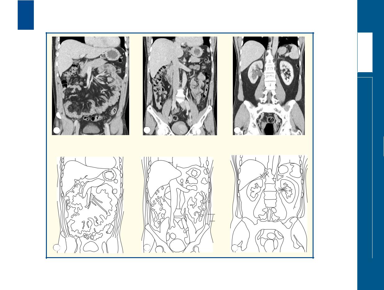
■ Notes
A spiral CT dataset of the abdomen was obtained on a |
standard investigation for a wide range of abdominal |
multidetector CT system. The individual thin slices were |
conditions, the radiologist has to scroll through hundreds |
loaded together to form a three-dimensional volume, with |
of axial images on a monitor. Some lesions are depicted |
each voxel isometric, so that the x, y and z resolution of |
better on coronal rather than axial images (e.g. asymmetry |
the resulting pixels is identical. This three-dimensional |
of the pelvicalyceal systems in the two kidneys in this case). |
dataset can be analysed in a variety of ways – here in |
To non-radiologists, such coronal views are a more intuitive |
coronal multiplanar two-dimensional reformats. |
method of looking at the abdomen than the source axial |
Now that CT of the abdomen has become such a |
images. |
|
|
■ Section level |
A B C |
View |
■ Orientation
Superior
Right 
 Left
Left
Inferior
CTs abdominal Coronal – images Selected
ABDOMEN
157
|
158 |
|
|
|
|
|
|
|
|
|
|
|
|
|
|
|
|
|
|
|
|
|
|
|
|
|
|
|
|
|
|
|
|
|
|
|
|
|
|
|
|
|
|
|
|
|
|
|
|
|
|
|
|
|
|
|
|
6 |
1 |
|
|
|
||
|
|
|
|
|
|
|
|
|
|
|||
|
|
|
|
|
|
5 |
|
|
|
|
||
|
|
|
|
|
|
|
|
|
L4 |
|
2 |
|
|
|
|
|
|
|
8 |
|
|
|
|||
|
|
|
|
|
|
|
|
|
|
|||
|
|
|
|
|
|
9 |
7 |
|
|
|
||
|
|
|
|
|
|
|
|
|
|
|
|
|
|
|
|
|
|
|
|
|
|
16 |
|
3 |
|
|
|
|
|
|
|
10 |
|
|
||||
|
|
|
|
|
|
|
|
|
11 |
4 |
|
|
|
|
|
|
|
|
|
|
|
|
|
|
|
|
|
A |
|
|
|
A |
|
|
|
|
||
|
|
|
|
|
|
|
|
6 |
1 |
|
|
|
|
|
|
|
|
|
|
|
|
|
|||
|
|
|
|
|
|
|
|
|
|
|||
|
|
|
|
|
|
|
|
|
|
|
||
|
|
|
|
|
|
|
5 |
|
|
|
|
|
|
|
|
|
|
|
|
|
|
L4/5 |
2 |
|
|
|
|
|
|
|
|
|
|
L4 |
|
|
||
|
|
|
|
|
|
|
|
|
|
|
||
|
|
|
|
|
|
|
15 |
|
|
|
|
|
|
|
|
|
|
|
|
|
|
7 |
14 |
|
|
|
|
|
|
|
|
|
|
|
|
|
|
|
|
|
|
|
|
|
|
|
|
16 |
12 |
13 |
|
|
|
|
|
|
|
|
|
|
10 |
3 |
|
|
|
|
|
|
|
|
|
|
|
|
|
||
|
|
|
|
|
|
|
|
|
11 |
|
4 |
|
|
|
B |
|
|
|
B |
|
|
|
|||
|
|
|
|
|
|
|
|
|
||||
|
|
|
|
|
|
|
|
|
|
|
|
|
|
|
|
|
|
|
|
|
|
|
|
|
|
|
|
|
|
|
|
|
6 |
1 |
|
|
|
|
|
|
|
|
|
|
|
|
|
||||
|
|
|
|
|
5 |
|
|
|
|
|||
|
|
|
|
|
|
|
|
|
L4/5 |
|
2 |
|
|
|
|
|
|
|
|
|
|
|
|
|
|
|
|
|
|
|
8 |
|
|
9 |
|
|||
|
|
|
|
|
|
|
|
|
7 |
|
|
|
|
|
|
|
|
|
|
13 |
|
|
|
||
|
|
|
|
|
14 |
10 |
|
|
||||
|
|
|
|
|
12 |
3 |
|
|||||
|
|
|
|
|
|
|
|
|
11 |
|
4 |
|
|
|
C |
|
|
|
C |
|
|
|
|
||
|
|
|
|
|
|
|
|
|
|
|
|
|
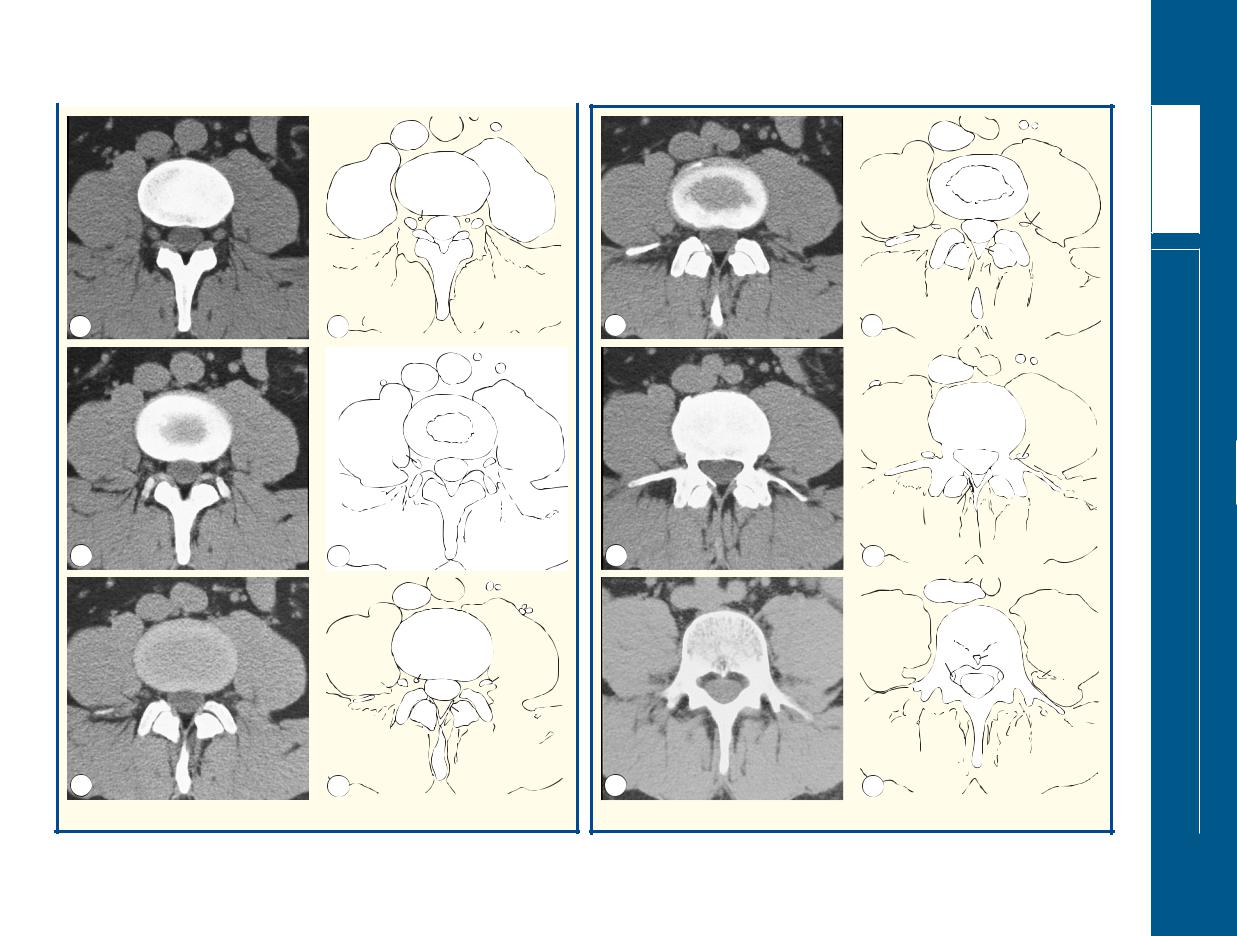
Axial computed tomograms (CTs)
|
|
|
6 |
1 |
|
|
|
|
|
|
|
|
|
|
|
|
|
5 |
|
|
|
|
|
|
|
|
|
L4/5 |
2 |
|
|
|
|
|
|
|
|
||
|
|
|
L5 |
|
|
9 |
|
|
|
|
|
|
|
|
|
|
|
|
13 |
7 |
|
|
|
|
|
|
14 |
10 |
|
|
|
|
|
|
12 |
3 |
|
||
|
|
|
|
|
|
4 |
|
|
|
|
|
11 |
|
|
|
D |
|
D |
|
|
|
|
|
|
|
|
|
|
|
|
|
|
|
|
|
|
|
|
|
|
|
|
6 |
1 |
2 |
|
|
|
|
|
|
|
|
||
|
|
|
|
|
|
|
|
|
|
5 |
|
|
|
|
|
|
|
|
|
L5 |
|
3 |
|
|
|
|
|
|
|
|
|
|
|
|
9 8 |
|
|
15 |
|
|
|
12 |
7 |
|
|
|
|
|
|
10 |
|
|
|
||
|
|
|
14 |
|
|
|
|
|
|
|
11 |
|
|
|
|
|
|
|
|
|
|
|
|
|
|
|
13 |
|
|
|
|
|
|
|
|
|
|
4 |
|
E |
|
E |
|
|
|
|
|
|
|
|
|
|
|
|
|
|
|
|
|
|
|
|
|
|
|
|
16 |
|
2 |
|
|
|
|
|
|
|
|
|
|
|
|
|
|
L5 |
|
3 |
|
|
|
|
|
|
|
|
|
|
|
|
17 |
|
18 |
15 |
|
|
|
|
9 |
|
14 |
|
|
|
|
|
7 |
|
|
||
|
|
|
|
|
|
|
|
|
|
|
19 |
|
|
|
|
|
|
|
|
20 |
|
|
|
|
|
|
|
|
|
4 |
|
F |
|
F |
|
|
|
|
|
|
|
|
|
|
|
|
|
Axial computed tomograms (CTs)
ABDOMEN
spine Lumbar – CTs Axial – images Selected

Images A–B |
13 Superior facet L5 |
9 Ventral ramus L4 |
6 Inferior vena cava |
|||
1 |
Aorta |
14 Capsule L4/L5 facet joint |
10 Flaval ligament |
7 Dural sheath |
||
15 L4 nerve |
11 Spinous process L4 |
8 Pouch for L5 root |
||||
2 |
Psoas major |
|||||
16 Epidural fat |
12 Inferior facet L4 |
9 Pedicle L5 |
||||
3 |
Quadratus lumborum |
|||||
|
13 Superior facet L5 |
10 |
Superior facet L5 |
|||
4 |
Erector spinae |
|
||||
Images C–D |
14 Epidural fat |
11 |
Inferior facet L4 |
|||
5 |
Psoas minor |
|||||
|
12 |
Transverse process of L5 |
||||
|
|
|||||
6 |
Inferior vena cava |
|
|
|||
1 Aortic bifurcation |
Images E–F |
13 |
Flaval ligament |
|||
7 |
Dural sheath |
|||||
14 |
Epidural fat |
|||||
2 Psoas major |
|
|||||
8 |
Epidural vein |
|
||||
3 Quadratus lumborum |
1 Right common iliac |
15 |
Ventral ramus L4 |
|||
9 |
Dorsal root ganglion L4 |
|||||
4 Erector spinae |
artery |
16 |
Confluence of common |
|||
|
in foramen between L4 |
|||||
|
5 Psoas minor |
2 Left common iliac artery |
|
iliac veins |
||
|
and L5 |
|
||||
|
6 Inferior vena cava |
3 Psoas major |
17 |
L5 nerve root sheath |
||
10 Lamina L4 |
||||||
7 Dural sheath |
4 Erector spinae |
18 |
Basi-vertebral vein |
|||
11 Spinous process L4 |
||||||
8 Epidural vein |
5 Psoas minor |
19 |
Lamina L5 |
|||
12 Inferior facet L4 |
||||||
|
|
20 |
Spinous process L5 |
|||
|
|
|
|
|||
|
|
|
|
|
|
|
■ Notes
This series of six computed tomograms (A–F) demonstrates the key anatomical features of a segment of the lumbar spine. Although all the features can also be demonstrated by magnetic resonance imaging (MRI), which is now the preferred test, computed tomography (CT) is perhaps easier to understand: bone appears white, soft tissues appear grey and fat appears black.
Images A–B
Image a traverses the slightly sclerotic endplate of L4. The dorsal root ganglion (9) lies in the foramen, immediately caudal to the L4 pedicle. Note how the dorsal root ganglion is demarcated clearly by normal epidural fat.
Images C–D
Image C traverses the L4/L5 disc. Note that the posterior aspect of the disc is concave with respect to the dural sheath (7). A normal disc at this anatomical level has either a concave or flat interface with the sheath. A convex disc here is indicative of an annular bulge. Note how the L4 ventral ramus (9) is now heading towards the psoas muscle
in which the lumbar plexus is formed. The dorsal ramus is too small to be resolved by CT; it would pass just lateral to the superior facet of L5.
Image D shows a portion of the L5 endplate surrounding the inferior aspect of the L4/L5 disc.
Images E–F
Image E transects the sclerotic endplate of L5. The flaval ligaments (13) running from the L4 to L5 laminae are shown well. Surgeons often operate through small openings in the flaval ligaments without the full laminectomy that used to be the standard approach for spinal surgery.
Image F passes through the body of the L5 vertebra – the normal bony architecture can be appreciated. The veins running through the body converge on the basi-vertebral vein (18), which has a small bony hood guarding its passage so that venous blood passes to the epidural veins (see 8 in image C). The right L5 root sheath (17) hugs the medial aspect of the pedicle (9).
■ Section level
A
B
C D E F
View
■ Orientation
Anterior
Right 
 Left
Left
Posterior
spine Lumbar – CTs Axial – images Selected
ABDOMEN
159

160
|
|
10 |
|
|
12 |
11 |
|
|
13 |
|
15 |
|
16 |
|
|
1 |
|
|
|
|
|
|
|
19 |
|
|
|
10 |
|
|
17 |
6 |
|
|
|
8 |
23 |
20 |
|
|
5 |
7 |
|
|
|
|
|
|
21
25
A |
22 |
A |
14 9
11
12
110
15 |
16 |
18
2 |
17 |
20 |
23 |
||
4 |
|
|
3 |
|
|
|
21 |
24 |
|
|
|
B |
22 |
B |
Coronal magnetic resonance image (MRI)
Coronal magnetic resonance image (MRI)
ABDOMEN
spine Lumbar – MRIs Coronal – images Selected

1 |
L1 vertebral body |
8 |
Thecal sac |
15 |
Right kidney |
21 |
Iliacus |
2 |
L4 vertebral body |
9 |
Aorta |
16 |
Left kidney |
22 |
Ilium |
3 |
L5 vertebral body |
10 |
Retroperitoneal fat |
17 |
Psoas |
23 |
Iliac crest |
4 |
L4/L5 intervertebral disc |
11 |
Crus of diaphragm |
18 |
Lumbar vein |
24 |
Gluteal muscles |
5 |
Pedicle |
12 |
Liver |
19 |
Descending colon |
25 |
S1 vertebral body |
6 |
Nerve root sheath L4 |
13 |
Spleen |
20 |
Anterior abdominal-wall |
|
|
7 |
Dorsal root ganglion L4 |
14 |
Right adrenal gland |
|
musculature |
|
|
|
|
|
|
|
|
|
|
■ Notes
Two coronal T1-weighted images elegantly show the |
well; this is particularly useful in patients with lumbosacral |
relationship of the lumbar spine to the psoas muscles and |
anomalies (around 25 per cent of people). |
kidneys (within retroperitoneal fat). Note that the kidneys |
Also apparent are the segmental lumbar veins, which |
lie in an oblique orientation, aligned to the lateral margins |
drain blood from the epidural veins. These run anteriorly |
of the psoas muscles; thus, the upper poles lie in a more |
within a narrow, but important, fat plane alongside each |
medial sagittal plane than the lower poles. Because of the |
vertebral body. |
lumbar lordosis, the upper poles lie in a more posterior |
The psoas muscles (17) are particularly prominent in this |
coronal plane than the lower poles. |
individual. The superior attachment to the lateral aspects of |
Because of the lumbar lordosis, the thecal sac and |
the disc at the thoracolumbar junction is seen well. One |
emerging nerve root sheaths can be seen in the L4 region, |
can appreciate how a disc-space infection (often |
while vertebral bodies are demonstrated more superiorly |
tuberculous) at this level could track inferiorly in and |
and inferiorly. Note the way in which each nerve root |
around the psoas muscle down to the iliacus and |
sheath hugs the medial and inferior aspects of its |
eventually present as a cold abscess in the inguinal region. |
associated pedicle (L4 root inferomedial to the L4 pedicle). |
The close relation of the adrenal to the right crus of the |
The expansion for the dorsal root ganglion can just be |
diaphragm (labelled 11 on the left) is apparent. The adrenal |
appreciated. The fairly constant relationship of the L4/L5 |
gland originates at this site, while the kidneys ‘ascended’ |
disc space with the level of the superior iliac crest is shown |
by differential growth during fetal life. |
|
|
■ Section level
AB
View
■ Orientation
Superior
Right 
 Left
Left
Inferior
spine Lumbar – MRIs Coronal – images Selected
ABDOMEN
161
162 |
|
|
|
|
|
|
|
|
|
|
|
|
|
|
|
|
|
|
4 |
|
|
Sagittal T1- |
|
|
|
|
|||
|
|
|
|
|
weighted |
|
|
|
|
|
magnetic |
|
5 |
|
|
resonance |
|
|
10 |
|
|
image (MRI) |
|
|
1 |
|
|
|
|
|
|
|
9 |
|
|
13 |
6 |
8 |
|
|
|
|
|
|
|||
|
|
|
|
||
|
7 |
|
|
|
|
|
2 |
|
|
|
|
A |
3 |
|
A |
|
|
|
|
|
|
|
|
|
|
|
|
|
|
4 |
Sagittal T2- |
|
weighted |
|
magnetic |
20 |
5 |
resonance |
|
||
|
image (MRI) |
|
|
|
|
1 |
|
|
|
8 |
|
|
6 |
|
|
7 |
|
|
2 |
|
B |
3 |
B |
|
Sagittal T2weighted magnetic resonance image (MRI)
10 1
7
|
17 |
|
18 |
|
|
|
|
|
2 |
|
|
C |
|
3 |
C |
|
|
|
|
Sagittal T1- |
|
|
weighted |
|
|
magnetic |
|
|
resonance |
14 |
|
image (MRI) |
|
|
|
12 |
1 |
15 |
|
|
11
16
19
2
3
D D
ABDOMEN
spine Lumbar – MRIs Sagittal – images Selected

1 |
L1 vertebral body |
7 |
Cerebrospinal |
11 |
Inferior vena cava |
vertebra |
18 Spinous process L4 |
|
2 |
L5 vertebral body |
|
fluid within thecal |
12 |
Right renal artery |
16 L3 nerve root |
19 |
Pars |
3 |
S1 vertebral body |
|
sac |
13 |
Retro-aortic left |
sheath/dorsal root |
|
interarticularis L5 |
4 |
Spinal cord |
8 |
Epidural fat |
|
renal vein |
ganglion |
20 |
Basi-vertebral vein |
5 |
Conus medullaris |
9 |
Spinous process L1 |
14 |
Crus of diaphragm |
17 Nerve roots within |
|
|
6 |
Cauda equina |
10 Aorta |
15 |
Pedicle of L1 |
cerebrospinal fluid |
|
|
|
|
|
|
|
|
|
|
|
|
■ Notes
Images A–B
These midline sagittal magnetic resonance images are of key importance in evaluating the lumbar spine (one of the commonest anatomical sites examined by MRI).
The anteroposterior diameter of the spinal canal can be assessed readily. This normally measures around 15 mm from the posterior aspect of the vertebral body to the anterior aspect of the laminar arch; values under 11.5 mm indicate a degree of spinal stenosis. The height of the disc spaces can be evaluated, as can the degree of hydration within. The normal disc space yields high signal intensity on T2 weighting (image B); a degenerate disc returns low signal intensity and becomes narrower. In these images, the L5/S1 is slightly degenerate, as judged by the slight reduction of signal. There is a slight increase in fat content in the superior portion of S1 vertebral body, suggesting a longstanding disc abnormality.
These sagittal images also demonstrate clearly the slight expansion of the distal cord at the T12/L1 level (the conus medullaris). The collection of nerve roots that forms the cauda equina (‘horse’s tail’) is seen well posteriorly within the canal when the patient lies supine (as during MRI). Because normal roots move freely within the cerebrospinal fluid, lumbar puncture is generally a very safe procedure at any level caudal to the conus medullaris.
Image C
A T2-weighted magnetic resonance image about 10 mm to the left of the median sagittal plane shown in the images A and B. Here, the segmental roots can be seen traversing the cerebrospinal fluid towards their respective nerve root sheaths and exit foramina. The aorta can just be seen anterior to the vertebral bodies.
Image D
A T1-weighted sagittal magnetic resonance image even more lateral than in image C. This is to the right of the midline, however, as the right renal artery can be seen passing anterior to the diaphragmatic crus and posterior to the inferior vena cava. This plane shows the exit foramina at several segmental levels. The classical shape has been said to resemble that of the human ear. The pedicles of two adjacent vertebral bodies form the superior and inferior boundaries of the foramen. The anterior margin is formed by the vertebral body superiorly and the posterolateral portion of the intervertebral disk inferiorly. Posteriorly lie the pars interarticularis, the flaval ligament and the facet joint. Narrowing of the disc space and degenerative changes in the facet joints will reduce the capacity of the foramen, and the flaval ligament gets thicker as the disc space narrows; all of these changes can contribute to nerve-root compression.
The nerve root sheaths, dorsal root ganglion and segmental nerve lie in the superior portion of the foramen. There are commonly two epidural veins in each foramen – a superior vein between the nerve root and the body/pedicle, and a second vein that usually lies much more caudally within the foramen. Remember that in the lumbar (and thoracic and sacral) spine, the segmental nerve root escapes caudal to its numbered vertebral body. For example, the L5 nerve root escapes caudal to the L5 pedicle through the L5/S1 foramen. Although the L5 root can be affected by a lateral L5/S1 disc herniation or facet joint degeneration, much more commonly it will be affected by a more central herniation at the L4/L5 level.
■ Section level
AB
D

View
C
■ Orientation
Superior
Anterior 
 Posterior
Inferior
spine Lumbar – MRIs Sagittal – images Selected
ABDOMEN
163