

Sehu - Ophthalmic Pathology-2005
.pdf110 C H A P T E R 5
Pathological classification of orbital lymphoproliferative disease
Currently a pathologist may require fresh unfixed tissue in order to apply modern techniques such as immunophenotyping, messenger RNA by in situ hybridisation, or DNA abnormalities (Southern blot, PCR, fluorescence in situ hybridisation (FISH), and gene sequencing). This is required for better classification of lymphoid tumour subgroups in relation to prognosis following treatment.
Lymphoproliferative disease is usually well controlled by radiotherapy if localised and by chemotherapy if systemic.
Lacrimal gland
Normal lacrimal gland
In order to appreciate the morphology of primary tumours of the lacrimal gland, histology of the normal gland is illustrated (Figure 5.48). Neoplasms can arise from acini (adenocarcinoma) or ducts (pleomorphic adenomas/adenocarcinomas). The term “pleomorphic” is applied because tumours which arise from ducts can contain cells which have arisen in epithelial cells and mesenchymal cells (myoepithelium – Figure 5.49). The normal presence of lymphoid tissue around the acini explains the relatively high incidence of primary lymphoid neoplasms in the lacrimal gland.
Inflammatory diseases of the lacrimal gland
Enlargement of the lacrimal gland may be due to various inflammatory processes and these are rare. Biopsies are occasionally performed on acute inflammations that do not resolve. Important inflammatory conditions are described below.
Dacryoadenitis
There have been many reports of infection by bacteria and viruses which can lead to unilateral or bilateral swellings of the gland.
Sarcoidosis
Non-caseating granulomas within the lacrimal gland have been described in systemic sarcoidosis – see Figure 5.9.
Mikulicz’s syndrome
Consists of bilateral swellings of both salivary and lacrimal glands secondary to sarcoidosis, lymphoid neoplasia, and leukaemia.
Sjögren’s syndrome (keratoconjunctivitis sicca with dry mouth)
A systemic autoimmune disease affecting mainly elderly women in whom there is idiopathic chronic inflammatory destruction of salivary and lacrimal gland tissue leading to
deficiency in saliva and tear production (see Chapter 3 for changes in the conjunctival epithelium). Labial gland biopsies are preferred over the lacrimal gland (Figure 5.50) due to convenience of access. Serological investigations include ANA, rheumatoid factor, SS-A, and SS-B as part of the work-up.
Tumours of the lacrimal gland
Most lacrimal gland tumours present by mass effect within the orbit. The diagnosis and management of lacrimal tumours are heavily dependent on high resolution orbital imaging. The remainder of this section will be confined to epithelial tumours of the lacrimal gland. Lymphoid malignancies, inflammatory and metastatic diseases in the lacrimal gland are identical to those occurring in the orbit. Lacrimal gland tumours are summarised in Table 5.4.
Benign
Pleomorphic adenoma
Other name: benign “mixed” tumour of the lacrimal gland – this term was used to account for the variable histology as described below.
A pleomorphic adenoma presents as a painless exophthalmos that has been slowly progressive over 12 months. CT scans show a well circumscribed tumour in the lacrimal fossa and, if sufficiently large, remoulding of the adjacent orbital bone without infiltration. The incidence is between the second and fifth decades (peak in the fourth) with a sex ratio of 1.5 : 1 (male : female).
Although classified as benign, the surgical treatment for pleomorphic adenoma relies on a complete excision (including the pseudocapsule) by means of a lateral orbitotomy. If the surgical procedure leaves residual tumour within the orbit there is a high risk of recurrence with the possibility of malignant transformation.
Macroscopic examination reveals a single solid pale-grey mass with a bosselated surface. The cut surface shows solid tumour with mucoid cystic spaces and areas of haemorrhage (Figure 5.51).
Table 5.4 Summary of lacrimal gland tumours.
Tissue origin |
Conditions |
|
|
Epithelial (20–25%) Benign mixed tumour: pleomorphic adenoma (50% of epithelial tumours)
Adenoid cystic adenoma Adenocarcinoma
Lymphoid Benign reactionary lymphoid hyperplasia Atypical lymphoid hyperplasia Malignant lymphoma
Metastatic

O R B I T A N D O P T I C N E R V E 111
Orbit - Lacrimal gland
Normal
ducts
lymphatics
acini
hilum
Figure 5.48
Orbit - Lacrimal gland
Sjögren’s syndrome
lymphocytic infiltration
advanced periacinar fibrosis
periacinar fibrosis
decreased density of acini
Figure 5.50
Figure 5.48 The primary unit within a lacrimal gland is a lobule (shown in outline), which contains acini, ductules, lymphatics, and nerves. The hilum is the central core which connects the lobules. The larger lacrimal ducts which drain into the fornix are present in the hilum.
Figure 5.49 Acini are circular structures formed by cuboidal cells with a central lumen – these are the secretory units for tears and the cells also transport immunoglobulins produced by the adjacent plasma cells. The tear fluid is passed into ductules and ducts which are lined by cuboidal epithelial cells and a surrounding contractile cell layer (myoepithelium, inset) resembling a sweat gland duct. Contraction of the ducts is responsible for reflex tearing. Benign and
arteriole
duct
acini
|
venule |
|
lymphatic |
|
|
tubular acini leading |
|
|
into a ductule |
|
|
|
|
|
|
cuboidal epithelium |
|
plasma cells and lymphocytes |
myoepithelium |
|
Orbit - Lacrimal gland / Normal |
||
|
Figure 5.49
Orbit - Lacrimal gland
Pleomorphic adenoma
pseudocapsule |
mucoid cyst |
nodular cut surface
tumour extending
to excision line
nodular surface
Figure 5.51
malignant tumours may be derived from both types of cells in the ducts and, therefore, they contain two or more cell types, i.e. epithelial and mesenchymal cells.
Figure 5.50 At a late stage of Sjögren’s syndrome, there is extensive periacinar fibrosis and a marked reduction in the number of acini (compared with
Figures 5.48, 5.49). A lymphocytic infiltration is present within the fibrous tissue.
Figure 5.51 In this excision of a pleomorphic adenoma, the tumour was not adequately cleared. The surface of the tumour is in part covered by a pseudocapsule, but elsewhere tumour tissue extends to the excision line. The surgeon and patient were fortunate that the tumour did not recur.

112 C H A P T E R 5
Microscopic examination shows the tumour to consist of two different components:
1Epithelial: the tumour cells adopt a glandular pattern in which cords and ducts resemble the ducts in a normal lacrimal gland (Figures 5.52, 5.53). The lumen of some of the ducts may be filled with an eosinophilic proteinaceous secretion.
2Stromal: this tissue is formed by myoepithelial cells which break away from ducts and proliferate to form connective tissue (Figures 5.52, 5.53). The components
are primarily fibrous with myxoid areas but the tumour may also contain fat and cartilage (Figure 5.54).
It is important to note that the pseudocapsule in a pleomorphic adenoma is merely a fibrous condensation which contains infiltrating tumour. It is the responsibility of the pathologist to take multiple samples of the pseudocapsule to ascertain the possibility of incomplete clearance, which implies that there is residual tumour in the orbit.
Malignant
Malignant tumours of the lacrimal gland differ clinically from the benign by a relatively faster onset of growth (less than 12 months) and an often painful proptosis. Orbital imaging often shows destruction of adjacent bone with reactionary ossification.
Malignant pleomorphic adenoma
This tumour usually arises after several incomplete excisions and the most malignant component is a squamous carcinoma or an adenocarcinoma.
Adenoid cystic carcinoma
Adenoid cystic carcinoma is the most common malignant variant of lacrimal gland carcinoma with a peak incidence in the fourth decade.
The macroscopic appearance of this tumour is that of a pale-grey mass similar to a pleomorphic adenoma, although the capsule may not appear to be formed or be intact.
Microscopic examination shows solid cords of hyperchromatic cuboidal cells with a high mitotic rate, proliferating around cystic spaces containing myxoid material – hence the “Swiss cheese” appearance (Figure 5.55). This classical cribriform pattern is only one of several different patterns described, but these are in the province of the pathologist.
This tumour has a propensity for perineural invasion which explains the common symptom of pain and leads to considerable difficulties in assessing clearance margins. Treatment is by total excision or exenteration with adjunctive radiotherapy, but the prognosis is poor.
Adenocarcinoma
The cellular constituents of an adenocarcinoma are the same as those seen in an adenoid cystic carcinoma but the pattern is that of solid masses of tumour tissue (Figure 5.55 inset).
Metastases
A careful history and a thorough general examination are mandatory to ascertain the possibility of metastatic disease when an orbital tumour is identified. Metastases usually present with rapid proptosis. Note that a metastatic scirrhous carcinoma of the breast (Figure 5.56) or stomach may result in an enophthalmos due to orbital fibrous tissue contraction.
Other primary sources for metastatic disease include: lung, prostate, gastrointestinal tract, and kidney, but occasionally no source is found.

O R B I T A N D O P T I C N E R V E 113
epithelial tumour cells
stromal tumour cells
Orbit - Lacrimal gland
Pleomorphic adenoma
Figure 5.52
Orbit - Lacrimal gland Pleomorphic adenoma
primitive cartilage cells (binucleated chondrocyte)
proliferating epithelial cells
vacuolated fat cells (lipocytes)
Orbit - Lacrimal gland |
myoepithelial cells |
|
Pleomorphic adenoma |
migrating into and |
|
|
forming stroma |
|
hyperplastic |
myoepithelial cells |
|
epithelial cells |
||
|
proteinaceous secretion by epithelial cells
Figure 5.53
Orbit - Lacrimal gland
Adenoid cystic carcinoma
adenoid cystic carcinoma
pseudocapsule
extraocular muscle |
solid |
|
adenocarcinoma |
||
|
Figure 5.54
Orbit - Tumours / Metastasis
Primary breast carcinoma
indian file pattern of tumour infiltration
fibrous tissue
EMA immunolabelling PAP
Figure 5.56
Figure 5.55
Figure 5.52 A low power illustration of the wide variation in cell morphology in a pleomorphic adenoma. The tumour is formed by the proliferation of epithelial cells and stromal cells (connective tissue including fibrocytes, chondrocytes, and lipocytes). The stromal cells are derived from myoepithelial cells in the ducts.
Figure 5.53 A higher magnification from the specimen shown in Figure 5.52 illustrates the proliferation of myoepithelial cells to form the stromal component in a pleomorphic adenoma. The epithelial component is hyperplastic and is also proliferating as nests of cells. Note the resemblance to normal ducts of the lacrimal gland (see Figure 5.49).
Figure 5.54 Pleomorphic adenomas display wide variations in cellular morphology. In the left example, epithelial cells predominate, while in the right example, the myoepithelial cells have formed mesenchymal tissue (resembling cartilage and fat).
Figure 5.55 Malignant tumours derived from the epithelial cells of acini and ducts may form solid masses (adenocarcinoma) or may form cystic and ductlike structures (adenoid cystic carcinoma). The inset shows a solid adenocarcinoma adjacent to the cystic variant.
Figure 5.56 In a metastatic scirrhous carcinoma of the breast, enophthalmos is due to contraction of the fibrous tissue proliferation which accompanies tumour cell infiltration. In this example, the cancer cells adopt a single “Indian-file” pattern within the fibrous tissue. By immunohistochemistry (inset), the tumour is identified as being of epithelial origin by the epithelial membrane antigen (EMA) marker.

114 C H A P T E R 5
Optic nerve pathology
Normal anatomy
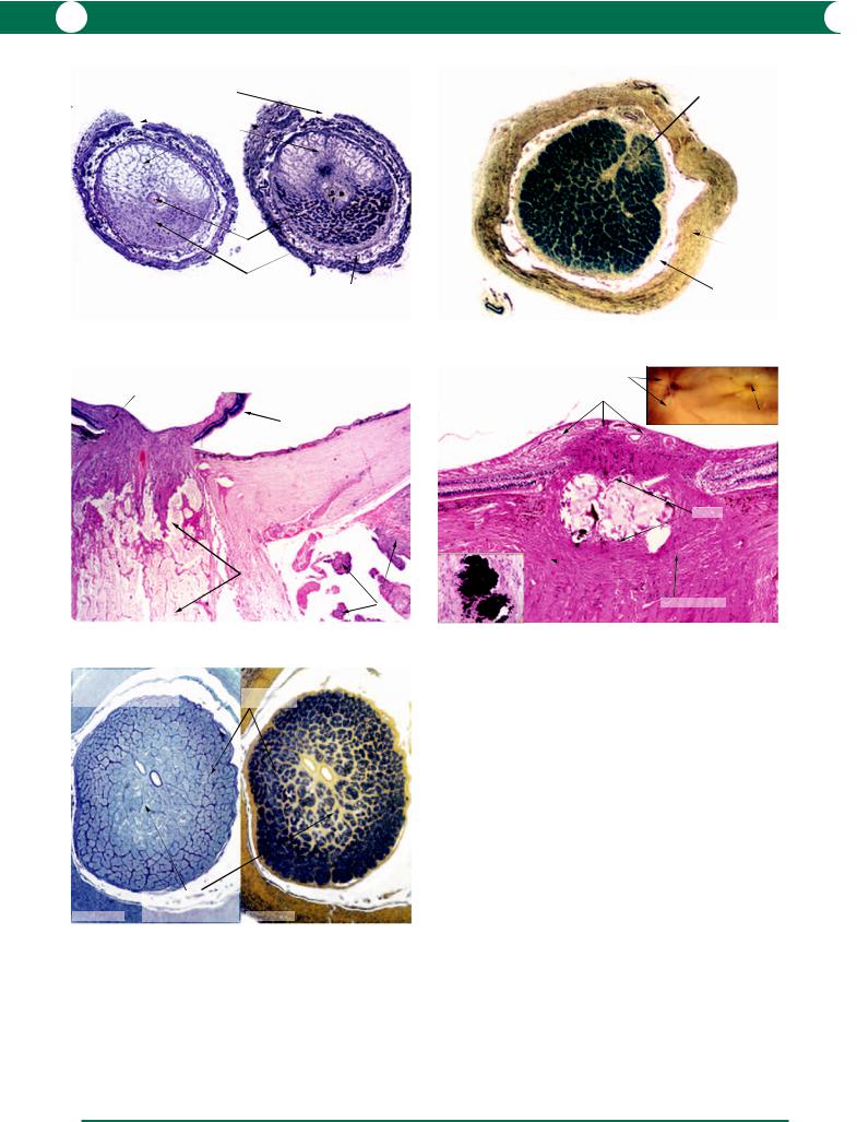
The normal optic nerve contains 1.2 million axons with supporting glial cells and blood vessels. The surrounding layers are formed by the pia mater, the arachnoid mater, and the dura mater (Figure 5.57). The pathologist relies on the H&E stain for the identification of glial cell replacement of atrophic myelinated axons (Figure 5.58). Special stains for the identification of axons and myelin are also employed (Figures 5.59, 5.60).
Optic disc swelling/optic atrophy
The numerous causes of unilateral and bilateral optic disc swellings and atrophy are comprehensively documented in clinical textbooks. In this prioritised account of the relevant
pathology (clinicopathological incidence), optic disc swelling in pathological specimens is mostly observed in the acute phase of a disease process. Disc swelling will be followed by loss of myelinated axons in the optic nerve if treatment (for example for acute glaucoma) is unsuccessful – it is at this stage that the majority of pathological specimens are encountered. Often there is total loss of myelinated axons with replacement gliosis, but in some conditions the geographical pattern of axonal loss is diagnostic:
•Glaucoma: swelling occurs in acute glaucoma (see Figure 7.53). Atrophy in chronic forms is characterised by posterior cupping of the lamina cribrosa and loss of prelaminar neural tissue (see Figures 7.54–7.57).
•Trauma: mechanical, for example swelling in hypotonia following penetrating injury (Figure 5.61). Optic atrophy follows any process which leads to extensive retinal atrophy, for example post-traumatic pseudoretinitis pigmentosa (see Chapter 9).
pial septae
myelinated axons within nerve bundles
pia mater
pial vessel penetrating nerve
|
artefactual |
arachnoid mater |
space |
Optic Nerve - Normal |
dura mater |
|
|
Meninges / Transverse section |
|
Figure 5.57 |
|
Optic Nerve - Normal & atrophic |
Bodian stain |
Longitudinal section |
for axons |
|
|
|
pial septae |
|
axons cut longitudinally |
Normal
Transverse section
thickened pial septae
normal pial septae
Optic Nerve - Normal & atrophic |
gliotic nerve |
|
Meninges / Longitudinal section |
||
bundles |
pia mater |
thickened pial septae |
thickened |
|
|
pia mater |
|
distended |
|
arachnoid |
|
mater |
arachnoid mater |
|
|
dura mater |
Normal Atrophic
Figure 5.58
Figure 5.57 This transverse section of an optic nerve is from a young patient who died from pneumococcal meningitis – polymorphonuclear leucocytes are present in the arachnoid layer. Note that the thin pial septae surround the nerve bundles and contain capillaries (derived from the network within the pia mater).
Figure 5.58 In a longitudinal section through an optic nerve, the myelinated axons are parallel to the pia mater (left). Advanced optic atrophy (right) is characterised by gliotic replacement of the nerve bundles and fibrous thickening of the pial septae. The atrophic nerve shrinks away from the dura so that the arachnoid space becomes enlarged.
Figure 5.59 A stain which demonstrates axons, in this example the Bodian stain, can be useful in demonstrating the normal density (upper and lower left) and axonal loss in optic atrophy (lower right).
normal axon density |
axons in reduced numbers |
|
Normal Atrophy |
||
|
Figure 5.59

O R B I T A N D O P T I C N E R V E 115
•Ischaemic neuropathy: any vascular disease, degenerative or inflammatory, can lead to an insufficient blood supply to the optic nerve, for example giant cell arteritis, anterior ischaemic optic neuropathy, ophthalmic artery occlusion, diabetes, and radiation vasculopathy. A total infarction of the optic nerve in GCA has been illustrated earlier in Figure 5.16. Two branches from the ophthalmic artery form the posterior ciliary arteries. Occlusion of one of these branches leads to hemi-infarction of the optic nerve. The clinical manifestation is that of an altitudinal hemianopia which is mirrored in a pathological specimen (Figure 5.62).
•Inflammatory/demyelinating disease: optic neuritis, multiple sclerosis, sarcoidosis, syphilis, and tuberculosis.
Chronic inflammation within the optic nerve results in focal or generalised demyelination and atrophy (Figure 5.63). In some cases, as in multiple sclerosis, there is survival of axons.
•Compressive lesion: orbital or intracranial masses (see Tables 5.2, 5.3) may directly compress the optic nerve or chiasm. Mechanical pressure can cause atrophy by direct compression, or by interference with the blood supply of the nerve (Figure 5.64). Optic nerve/disc drusen are included in this section because the retrolaminar masses interfere with axoplasmic flow and cause disc swelling. On clinical examination, drusen appear as localised refractile masses which autofluoresce within the optic disc. The pathogenesis is thought to be the result of abnormalities in axoplasmic flow leading to calcification. The histological appearance is that of an
irregular basophilic mass within the nerve (Figure 5.65). This condition is associated with an abnormal central retinal vasculature and a predisposition to central retinal vein occlusion. Drusen are also found in association with angioid streaks and pseudoxanthoma elasticum.
•Toxic/nutritional: alcohol and many drugs (for example ethambutol, chloramphenicol), toxins (for example cyanide), combined with nutritional deficiencies (vitamins B1, B2, B12, and folate) produce a characteristic pattern of atrophy of the papillomacular bundle within the optic nerve (Figure 5.66). Visual field investigations detect a characteristic bilateral centrocaecal scotoma. The common pathway that is theorised is a disturbance of mitochondrial metabolism, which is thought to be more sensitive within the papillomacular bundle.
•Metabolic: for example Leber’s hereditary optic neuropathy. Identical clinical and pathological findings described for toxic amblyopia occur in young males in the second and third decades: females are unaffected. This condition is maternally inherited (via mitochondrial DNA) and is included as an example of an inherited disorder of mitochondrial enzyme systems. The point mutations in mitochondrial DNA have been identified. The abnormality presents initially as a unilateral acutely progressive loss of central vision. This is followed within weeks by involvement of the second eye. The major clinical feature is an apparent swelling of the nerve fibre layer surrounding the disc, with radiating telangiectatic vessels. Pathologically at a later stage, there is atrophy of the papillomacular bundle (Figure 5.67).
Optic Nerve - Normal |
Loyez stain for |
Transverse section |
myelin |
pial septae |
|
myelin cylinders cut transversely
Figure 5.60
Figure 5.60 A stain for myelin (Loyez stain) at low power reveals a uniform distribution of myelinated axons in the normal optic nerve (inset). At higher magnification, myelinated axons appear as black circles surrounding the nonstaining axons.
Optic Nerve - Swelling
Penetrating trauma / Hypotonia
thickening |
exudates |
of NFL |
|
|
displacement |
|
|
of |
|
|
photoreceptors |
|
termination of |
normal |
|
nerve |
||
Bruch’s membrane |
||
bundles |
||
|
||
|
normal |
|
lamina cribrosa |
CRA |
|
|
Figure 5.61
Figure 5.61 In a case of penetrating trauma, there may be severe hypotonia which interferes with the blood supply of the prelaminar part of the optic nerve and results in the interruption of axoplasmic flow. Swelling of the nerve fibre layer (NFL) is such that the photoreceptors are displaced away from the termination of Bruch’s membrane. The central retinal artery (CRA) is patent. Note the normal architecture of the retrolaminar part of the optic nerve. (Compare with Figure 7.53.)
116 C H A P T E R 5
Optic Nerve - Atrophy
Hemi-infarct |
pathologist’s notch to indicate superior dura |
|
total infarction of superior half
of nerve
central vessels
surviving myelinated axons
in inferior half of nerve widening of subarachnoid space
Bodian stain |
Loyez stain |
Figure 5.62
Optic Nerve - Infarction
Acute ischaemia from tumour compression mild optic
disc swelling artefactual retinal detachment
|
retolaminar |
|
|
infarction |
|
|
tumour |
|
|
mass |
|
Figure 5.64 |
|
|
Optic Nerve - Atrophy |
|
|
Toxic Amblyopia |
normal nerve |
|
Alcohol/Nutritional |
||
bundles |
||
|
|
atrophy of nerve fibres |
|
in papillomacular |
Bodian stain |
bundle located centrally Loyez stain |
Figure 5.66
Optic Nerve - Atrophy |
Loyez stain for myelin |
|
Focal demyelination |
plaque of demyelination |
|
Multiple sclerosis |
||
|
thickened dura
widened subarachnoid space due to nerve atrophy
Figure 5.63
Optic Nerve - Disc drusen |
white |
|
masses |
|
swollen NFL |
macula
drusen
lamina cribrosa
Figure 5.65
Figure 5.62 A patient died following cerebral infarction after he had complained of a unilateral altitudinal inferior field defect. The nerve was removed at autopsy and a transverse section revealed an infarction in the superior half (left: Bodian stain for axons; right: Loyez stain for myelin). Compare with the normal optic nerve in Figure 5.60 (note that the Loyez stain in this case does not have a yellow counterstain).
Figure 5.63 Focal areas of demyelination are characteristic of plaques in multiple sclerosis.
Figure 5.64 A patient developed optic disc swelling due to compression from a large malignant fibrous tumour of the orbit which was subsequently treated by an exenteration. Histology showed that the retro-ocular tumour had caused thrombosis of the ophthalmic artery and its posterior ciliary branches (not illustrated). This resulted in an infarction of the retrolaminar part of the optic nerve.
Figure 5.65 In an autopsy specimen, optic disc drusen appeared as pale nodules within the optic disc (upper inset). Because drusen are heavily calcified, the material is hard and even small drusen fragment on microtomy (lower inset). Decalcification exposes the underlying undisturbed matrix of a drusen in the prelaminar portion of the optic nerve. NFL nerve fibre layer.
Figure 5.66 An autopsy specimen from a patient with end-stage liver failure due to chronic alcoholism. Atrophy of the macular ganglion cells is followed by retrograde atrophy of the papillomacular bundle, which is central at this level of the optic nerve. At the periphery of the nerve, the myelinated axons are present within bundles of normal size. In the centre, the nerve bundles are atrophic (left: Bodian stain; right: Loyez stain).

O R B I T A N D O P T I C N E R V E 117
•Raised intracranial pressure with transfer of pressure to the subarachnoid space around the nerve produces bilateral optic disc swelling (papilloedema). Papilloedema may be either primary (for example benign idiopathic intracranial hypertension – BIIH) or secondary (for example space occupying lesion, inflammation, infection). The mechanism is assumed to be the combined effects of both mechanical pressure and ischaemia (due to interference with vasculature) impeding axoplasmic flow at the level of the lamina cribrosa. Pathological examination reveals widening of the subarachnoid space and atrophy of the optic nerve and disc (Figure 5.68).
•Primary optic nerve tumours: for example glioma/meningioma (see below).
Primary optic nerve tumours
Glioma
The term glioma refers to a group of tumours derived from the supporting cells in neural tissue, for example astrocytomas grades I–IV, oligodendroglioma, and glioblastoma multiforme. In the optic nerve, the gliomas in children are equivalent to intracranial astrocytoma type I and are low grade.
Therefore in ophthalmic practice, gliomas of the optic nerve are usually low grade, unilateral, and are most common in children. Bilateral cases are almost pathognomonic for neurofibromatosis type 1.
The clinical presentation will depend on the location of this tumour. Most cases have decreased vision, altered colour perception, and a relative afferent pupillary defect. The optic disc may be normal, exhibit swelling or atrophy (see above), and with sufficient compression result in a central retinal vein occlusion. Intraorbital gliomas present with proptosis, whereas those in the chiasm may have bilateral visual field disturbances or symptoms due to secondary effects on the central nervous system.
High resolution orbital imaging (CT or MRI) demonstrates a fusiform enlargement of the nerve with axial proptosis along the line of the optic nerve, and this is important in diagnosis and treatment planning.
A meningioma should be considered in the differential diagnosis.
Surgery appears to be the only effective treatment as the tumour is unresponsive to radiotherapy and chemotherapy, and this creates much controversy, especially with regards to the risks and morbidity of surgery for those tumours located in the optic chiasm or the intracranial part of the optic nerve. It is for this reason that most cases of
resection encountered by an ophthalmic pathologist would be in the intraorbital location.
Previously, the specimens took the form of a globe and nerve containing a glioma (Figure 5.69). Currently attempts are made to excise the tumour, leaving behind the globe if possible. Microscopically, gliomas consist of neoplastic astrocytes with oval or spindle shaped nuclei and branching cytoplasmic processes (Figures 5.70, 5.71). Neoplastic proliferation of astrocytes destroys myelinated axons within the nerve bundles.
Gliomas in adults are rarer, but are more aggressive in behaviour and their histopathological characteristics are those of a poorly differentiated astrocytoma.
Meningioma
These slowly growing neoplastic proliferations of meningothelial cells occur as primary tumours within the meninges of the optic nerve. Alternatively a meningioma in the skull may spread into the orbit. Unlike optic nerve gliomas, meningiomas more commonly present in middle-aged adults, usually women.
Symptoms will depend on the location of the tumour – an orbital location would result in a posterior mass effect with proptosis and corneal exposure. Location within the optic canal may result in visual loss, altered colour perception, or an afferent pupillary defect, and the initial optic disc swelling is followed by atrophy.
Diagnosis is very much dependent on high resolution CT/MRI.
Management of orbital meningiomas is usually conservative with serial imaging and visual fields to assess growth and tumour extension. Active treatment depends on the severity of optic nerve compression and the location of the tumour. Although the tumour is partially responsive to radiotherapy and hormonal therapy, surgery is still the treatment of choice and can vary from local excision to exenteration combined with craniotomy for intracranial extension.
The tumour usually surrounds the optic nerve, but there may also be an eccentric mass which distorts the orbital tissues (Figure 5.72). Histologically, the commonest form of orbital meningioma is the meningothelial type
(Figure 5.73). In this tumour, fibrous septae surround small spindle cells which do not have prominent cytoplasm – these cells resemble those in the normal arachnoid mater. Psammomatous meningiomas contain numerous pink-staining “psammoma” bodies (Figure 5.73). Within the cranial cavity, meningiomas have a variety of appearances – this classification is in the province of the neuropathologist.

118 C H A P T E R 5
Optic Nerve - Atrophy |
|
artefactual stain |
temporal |
Leber’s hereditary optic neuropathy |
demyelination of |
side |
|
nasal side |
residual disc |
papillomacular bundle |
|
swelling |
|
|
|
|
|
|
|
surviving myelinated axons on nasal side
widened subarachnoid space |
Loyez stain |
Figure 5.67
optic nerve thickened by tumour infiltration
optic disc swelling
arachnoid proliferation
Optic Nerve - Tumours Glioma
Figure 5.69
Optic Nerve - Tumours
Glioma
Immunohistochemistry
S-100 PAP |
Neurofilaments PAP |
Figure 5.71
atrophic gliotic optic disc
dilated subarachnoid space
atrophy
Optic Nerve - Atrophy of nerve bundles Benign idiopathic
intracranial hypertension
Figure 5.68
Optic Nerve - Tumours
Glioma
long cytoplasmic
processes nuclei with irregular chromatin distribution
glioma
arachnoid
crush proliferation artefact
Figure 5.70
Figure 5.67 In the retrolaminar part of the optic nerve, the papillomacular bundle is on the temporal side. In this example of Leber’s hereditary optic neuropathy, there is extensive loss of myelinated axons in the papillomacular bundle with some survival on the nasal side. The arachnoid space is widened because the optic nerve is atrophic (Loyez stain).
Figure 5.68 A patient suffering from benign idiopathic intracranial hypertension died from meningitis after an infection of a ventriculo-peritoneal shunt. The case serves to illustrate the pathology of the effects of longstanding raised intracranial pressure. The subarachnoid space is widened, and the optic nerve is atrophic.
At this stage, the optic disc is also atrophic and gliotic. The peripapillary retina is drawn inwards (in contrast with the initial outward displacement of photoreceptors seen in optic disc swelling).
Figure 5.69 An optic nerve glioma is ovoid or fusiform in shape, in this case, measuring 20 mm in diameter and 35 mm in length. The cut surface reveals indistinct enlargement of the optic nerve surrounded by arachnoidal proliferation. A superficial biopsy containing only arachnoidal proliferation can be deceptive for the pathologist who might consider the diagnosis of a meningioma. In this specimen, the tumour extends to the excision line which implies the risk of spread into the optic foramen and chiasm. The inset shows optic disc swelling.
Figure 5.70 In a glioma of the optic nerve, myelinated axons are replaced by proliferating astrocytes which are recognised by their long cytoplasmic processes. Mitotic figures are only rarely found. The inset shows a low power view of a glioma which was clamped behind the globe: note the diffuse infiltration within the optic nerve and the reactionary arachnoidal proliferation.
Figure 5.71 Immunohistochemistry confirms the presence of neoplastic astrocytes which react positively with S-100 antibody (left). S-100 labels any cells of neural crest origin and a more specific marker is the antibody against intermediate contractile filaments (neurofilaments) which are present in astrocytes (right). Note the branching cytoplasmic processes.

O R B I T A N D O P T I C N E R V E 119
Optic Nerve - Tumours
Meningioma
lens
posterior wall of
compressed globe
Figure 5.72
extraocular muscle
compressed and displaced optic nerve
solid white lobular tumour
Figure 5.72 Meningiomas within the orbit can grow to a large size and cause severe distortions of the optic nerve and globe requiring exenteration. The solid pale grey lobules in close proximation to the nerve hint at the diagnosis of a meningioma.
Optic Nerve - Tumours
Meningioma
artefactual break
calcified psammoma bodies
fibrovascular septae
proliferating
nest of meningothelial cells
Meningothelial (syncytial) Psammomatous
Figure 5.73
Figure 5.73 The commonest histological subtypes of intraorbital meningiomas are meningothelial or syncytial (left) and psammomatous (right). The meningothelial tumour is formed by neoplastic arachnoidal cells which do not have extended cytoplasmic processes. The tumour cells are surrounded by fibrovascular septae. A tumour which contains several spherical pink bodies is classified as a psammomatous meningioma – similar pink bodies may be seen in the normal arachnoid mater.