

Sehu - Ophthalmic Pathology-2005
.pdf190 C H A P T E R 9
Penetrating injury
A penetrating injury involves entry into a structure without traversing the entire substance. Hence, a penetrating injury of the cornea does not involve the entire thickness, whereas in a penetrating injury of the globe either the cornea or the sclera is perforated (Figure 9.12). In the remainder of this section, penetrating or perforating trauma is discussed in relation to the globe.
Perforating injury
Perforation differs from penetration, in that there are both entry and exit wounds in the globe (see Figure 9.2).
Disorganisation of intraocular contents
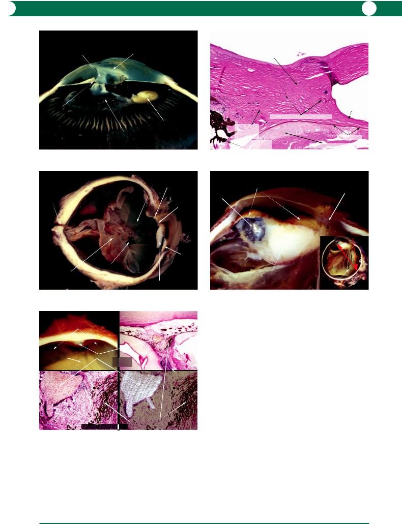
These include tears in the uveal tract, laceration of the lens capsule, retinal wounds, vitreous detachment, and intraocular haemorrhage (Figure 9.16).
Lens induced uveitis
Trauma to the lens is one of the commoner causes of autoimmunity to lens matter (see Chapter 8).
Sympathetic ophthalmitis
Unilateral trauma involving a corneoscleral wound with uveal prolapse is associated with bilateral granulomatous choroiditis (see Chapter 8).
Secondary complications
Epithelial ingrowth
Failure to close a penetrating wound of the cornea allows epithelial migration into the interspace (Figure 9.13). When epithelium grows into a perforating wound of the cornea, the cells migrate across the chamber angle and secondary open angle glaucoma is the result (see Chapter 7).
Fibrous ingrowth
The cells in the corneoscleral envelope react to traumatic laceration by proliferation to form dense scar tissue within the globe (Figures 9.14, 9.15).
Intraocular foreign body (IOFB)
By definition, an IOFB can only occur following penetrating injury of the globe, but the foreign body may not be located in the line of trajectory due to a ricochet (Figure 9.17).
Organic
The most commonly encountered organic IOFB is wood. Penetration of the globe by sharp splinters introduces microorganisms into the eye such as fungi and bacteria leading to intractable endophthalmitis. This scenario is regarded as a surgical emergency. In the absence of secondary pyogenic infection, a wood particle becomes surrounded by a giant cell granulomatous reaction (Figure 9.18).
Trauma - Penetrating & perforating
Corneal wounds
|
epithelial debris |
pannus |
surface epithelial |
|
loss |
penetrating wound of cornea
perforating wound of cornea
(full thickness) filled with granulating tissue
Figure 9.12
Figure 9.12 An assault with a broken glass bottle can cause multiple wounds to the eye – in this case examples of both penetrating and perforating wounds to the cornea are illustrated. The edges of the penetrating wound are not in apposition and the space is filled by fibrovascular tissue and inflammatory cells derived from the adjacent pannus. Note that a perforating wound of the cornea is also a penetrating wound of the globe.
Trauma - Penetrating wound of cornea |
epithelial ingrowth in |
Epithelial ingrowth |
unhealed partial thickness |
|
penetrating wound of cornea |
absent endothelium & Descemet’s membrane (lamellar keratoplasty)
Figure 9.13
Figure 9.13 In general, a penetrating wound heals by fibrosis (see Figure 4.94), but occasionally, there is a failure of apposition and epithelium grows into
the defect. This is a lamellar keratoplasty specimen which does not include the posterior corneal layers.
WO U N D H E A L I N G A N D T R A U M A 191
Trauma - Penetrating globe Fibrous ingrowth
fibrousfibrousingrowthingrowth growinggrowingoverover vitreousvitreouswickwick
fforwardr rd ttentingti off pupilil
corneal scar
nuclear lens remnants
capsular bag remnant
Figure 9.14
Trauma - Perforating wound of globe |
opaque |
|
vitreous |
|
entry wound |
exit wound |
|
|
absent iris leaf |
inferior iris leaf
artefactual hole
detached retina
lens remnants
Figure 9.16
Trauma - IOFB
Wood
subretinal exudate
retina fragment of wood
subretinal exudate
retina
proliferating RPE
multinucleate giant |
cells |
Polarised light |
Figure 9.18
Trauma - Penetrating globe |
PAS stain |
Fibrous ingrowth |
|
source of fibrous |
|
tissue ingrowth |
|
artefactual fold
|
fibrous |
|
|
tissue growing |
|
|
onto lens |
|
|
surface |
|
|
defect in Descemet’s membrane |
|
fibrous |
|
|
tissue |
|
|
replacing iris |
lens capsule |
|
remnant of |
||
degenerate lens cortex |
||
ciliary processes |
Figure 9.15
Trauma - Intraocular foreign body (IOFB) Shotgun pellet
intact iris
entry wound
shotgun pellet
line of trajectory
intumescent lens matter
vitreous strands
Figure 9.17
Figure 9.14 A patient developed secondary angle closure glaucoma some years after a penetrating injury resulting in a corneal wound, a lens perforation, and a disrupted anterior vitreous face. A vitreous wick has provided a scaffold for a fibrous ingrowth.
Figure 9.15 A broad peripheral corneal wound can be identified in a PAS stained section which illustrates the edges of Descemet’s membrane. A fibrous ingrowth lines the surface of a degenerate lens. The iris was lost during trauma and the defect is now replaced with fibrous tissue.
Figure 9.16 A man was stabbed in the eye with a fine chisel. The entry and exit wounds are in line but the site of retinal perforation is obscured by a retinal detachment. The inferior iris leaf is present, but the superior leaf is absent. This section is taken through the pupil and optic nerve (PO block) of the globe, but in making the second cut an artefactual hole was made in the retina.
Figure 9.17 The momentum of a small metallic particle is markedly reduced by a ricochet within the globe. This shotgun pellet came to rest behind the iris and damaged the lens, which over the next few days became swollen and opaque. The proposed trajectory route is shown in the inset.
Figure 9.18 A young boy was hit in his only seeing eye by a homemade arrow. The arrow was extracted but a small fragment of wood was left behind which induced a giant cell reaction and fibrosis in the choroid. The retinal pigment epithelium (RPE) became metaplastic and was transformed into a fibrous tissue scar which tacked down the retina. Once the retinal defect had been sealed, the retinal detachment converted from a rhegmatogenous to an exudative type (upper left). The tiny wood fragment is surrounded by inflammatory and fibrous tissue (upper right and lower left). The cellulose in the wood is birefringent in polarised light (bottom right).

192 C H A P T E R 9
Table 9.2 Common inorganic intraocular foreign bodies and reactions.
Material |
Reaction |
|
|
Glass |
Minimal reaction. Usually from civil trauma |
Plastic |
Minimal reaction. Intraocular lenses would be most common |
Iron |
Ferrous ions (Fe2 ) are retinotoxic, leading to siderosis bulbi. Histological effects are similar to those |
|
described for haemosiderosis* |
Copper |
Copper produces a massive acute sterile inflammatory reaction: acute chalcosis (Figure 9.19). Chronic |
Brass (copper/tin) |
chalcosis is due to slow leakage of Cu2 ions with the formation of opacities in the lens (sunflower |
|
cataracts). Peripheral corneal stromal copper staining (Kayser–Fleischer ring) may also be found in |
|
Wilson’s disease, which is a systemic disturbance in copper metabolism |
Lead |
Usually from a retained airgun pellet injury associated with gross disorganisation of the globe |
|
(Figure 9.20). Diffusion of lead salts is minimal. Massive intraocular fibrosis is a non-specific response |
|
to a perforating injury of the globe (Figure 9.21) |
|
|
* The term siderosis also describes deposition of iron salts from an intraocular foreign body and should not be confused with haemosiderosis (Figure 9.9) which is secondary to retained blood (see above).
Trauma - IOFB
Brass
site of IOFB
extracted brass foreign body
uveal  effusion
Figure 9.19
subretinal exudate
optic nerve
exit wound
Trauma - IOFB
Perforating wound of globe Air gun pellet
Figure 9.21
subretinal exudate
exudate in anterior chamber
opaque vitreous
intact iris and ciliary body
entry wound
lens fragment
Trauma - IOFB
Penetrating wound of globe Air gun pellet
subretinal haemorrhage
detached retina
|
|
irido- |
air gun pellet |
outline of |
cyclectomy |
air gun pellet |
|
|
embedded in |
|
|
|
|
|
optic nerve |
|
|
|
entry wound |
|
Figure 9.20
Figure 9.19 Brass may contain different concentrations of copper in the copper/tin alloy. In certain concentrations of copper, the response to a brass foreign body leads to a sterile purulent exudate. In this example, pus surrounds the site from which the foreign body was removed (located adjacent to the globe). The vitreous is completely opaque due to inflammatory cell infiltration and the secondary effects are exudative retinal detachment and an exudate in the anterior chamber.
Figure 9.20 An airgun pellet injury is extremely destructive. In this macroscopic and microscopic example, the pellet rotated on entry into the globe and was embedded within the optic nerve. The iris and ciliary body are destroyed at the point of entry and intraocular haemorrhage detached the retina. The lens presumably was lost through the entry wound after decompression at the
time of injury.
Figure 9.21 If an airgun is fired at close range, there is sufficient momentum in the pellet to produce a perforating wound of the globe. In this example, the damage to the scleral wall is extensive and surgical closure is incomplete. As a result, massive fibrovascular tissue proliferation leads to distortion and shrinkage of the affected segment of the eye. A fragment of lens matter is displaced and there is histological evidence of an early lens induced inflammation.

WO U N D H E A L I N G A N D T R A U M A 193
Inorganic
The most commonly encountered inorganic IOFBs are metallic, plastic, or glass (Table 9.2).
Surgical pathology
Cataract surgery
Many examples of surgical pathology have been illustrated in other chapters. In the following section the pathological complications of cataract surgery will be discussed.
Historically, cataract surgery developed from the displacement of the lens into the vitreous (couching) and was later followed by surgical lensectomy in toto (intracapsular) which involved the complete removal of lens matter and capsule. These two procedures had high visual morbidity outcomes due to the required aphakic spectacle correction which distorted and reduced the visual field. The development of
plastic intraocular lenses (IOLs) with insertion into the anterior chamber (Figure 9.22) and later into the capsular bag have achieved superior visual outcomes. Currently lens matter is most commonly removed by extracapsular cataract extraction (ECCE) with the intention of leaving an intact lens capsule to house a posterior chamber intraocular lens (PCIOL, Figure 9.23). Newer techniques have allowed smaller surgical wounds by means of phacoemulsification of lens matter and insertion of foldable lenses.
Complications
Although complication rates are low, the following are the most important of those recorded.
Endophthalmitis This complication can occur following any form of intraocular surgery. After cataract surgery, the widely quoted incidence of endophthalmitis is approximately 1 in 1000 (Figure 9.24).
Trauma - Lens surgery
haptic
ACIOL
optic
ACIOL
cobblestone degeneration of retina
indentation ACIOL from ACIOL
haptic
artefactual tear in iris
iris
Figure 9.22
Trauma - Lens surgery
IOL / Endophthalmitis
corneal abscess
hypopyon
corneolimbal rupture
uveal effusion
 haptic
vitreous abscess
edge of IOL
pars plicata
PCIOL |
iris |
|
|
|
atrophy |
residual cortical
Trauma - Lens surgery lens matter PCIOL / Retained cortical lens matter
Figure 9.23
Figure 9.22 In an autopsy globe the first cut disturbed an anterior chamber intraocular lens (ACIOL) which led to an artefactual tear in the iris (left). The design of ACIOLs has changed with time and one of the more recent developments is to use thin extensions (haptics) which slot into the anterior chamber angle to fix the lens (right inset). Processing for paraffin histology dissolves the plastic to leave an empty space (right).
Figure 9.23 For technical reasons, it may not always be possible to completely remove lens matter during an extracapsular cataract procedure. In this autopsy case, there was rupture of the posterior capsule with vitreous prolapse into the anterior chamber. Further cortical clean up was difficult and the intraocular lens was inserted into the sulcus.
Figure 9.24 Within a few days after intraocular lens (IOL) implantation, a fulminating endophthalmitis occurred and Streptococcus pneumoniae was identified. Despite administration of broad spectrum intraocular antibiotics, the infection was not contained and an enucleation was carried out. The ocular compartments were filled with pus and the intraocular lens was displaced during the initial cut but was still identifiable.
Figure 9.24

194 C H A P T E R 9
Retained lens matter Evidence of inadequate lens matter clearance is easily identified macroscopically (Figure 9.23). The term “doughnut cataract” describes the residual lens matter at the cortical rim. Histology reveals degenerate lens matter which may be totally or partially enclosed by a residual capsule (Figure 9.25). If the equatorial lens epithelium is not removed, fibrous metaplasia occurs and opaque membranes grow across the posterior capsule (posterior capsular opacity (PCO); Figure 9.26). Photodisruption by a YAG laser is used to treat this condition.
Cystoid macular oedema/edema (CMO/CME) The visual results following cataract surgery are sometimes disappointing due to the development of oedema and cyst formation in the macula (CMO): the aetiology is uncertain. Three theories have been proposed: (1) vitreomacular traction; (2) diffusion of inflammatory mediators (prostaglandins) from the anterior segment; and (3) damage to the integrity of the blood–retinal barrier. CMO is identified by fluorescein angiography which reveals a petaloid pattern of fluid accumulation around the fovea. In the majority of cases, fluid is resorbed and only a small percentage (1%) of cases progress to chronic visual impairment. Studies of acute pathology are rare, but occasionally specimens are received and these show cyst formation in the outer plexiform layer with expansion to involve all the layers (Figure 9.27).
Chemical
The chemicals most commonly involved in eye injuries are acids and alkalis. A useful review article is Wagoner MD (1997) Chemical injuries of the eye: current concepts in pathophysiology and therapy. Surv Ophthalmol 41:275–313.
Acid
Examples: sulphuric (H2SO4), hydrofluoric (HF), acetic (CH3COOH), and hydrochloric (HCl) acids.
Acids damage the ocular surface by denaturation of proteins in the epithelium leading to cell death. Protein precipitation creates a barrier which usually limits further diffusion into the eye. It is for this reason that the resultant damage from an acid burn is superficial scarring. A pathological specimen would most commonly be encountered in the form of a host corneal disc following a penetrating keratoplasty for visual rehabilitation. The pathology at the end stage is non-specific with epithelial instability, stromal fibrosis, and vascularisation. Very concentrated acids, however, may diffuse into the eye with effects similar to alkalis (see below).
Alkali
Examples: ammonia (NH3), sodium hydroxide (NaOH, Lye), potassium hydroxide (KOH), magnesium hydroxide (Mg[OH]2), and lime (Ca[OH]2).
In comparison with acids, alkalis create more damage to the ocular tissues by saponification of fatty acids within the cell membranes by the hydroxyl (OH–) ion, resulting in cell wall disruption. Alkali diffusion into the eye is progressive and may damage the cells in the cornea, trabecular meshwork, iris, lens, ciliary body, retina, and optic nerve.
In addition to the primary insult, the secondary effects of alkali burns are destructive to the eye for the following reasons:
1Destruction of conjunctival cells leads to fibrosis in the fornices and lid distortion (symblepharon).
2A loss of corneal limbal stem cells reduces the capacity for re-epithelialisation of the corneal surface with migration of surviving conjunctival cells onto the corneal surface.
3The damage is often made more extensive due to an exaggerated inflammatory and healing response. Stromal melting and thinning from extensive liberation of metalloproteinases from both keratocytes and neutrophils can occur.
4Fibrosis in the anterior segment tissues is followed by complicated sequelae which include glaucoma and cataract formation.
Severe cases of alkali burns are more likely to reach the pathologist at the end stage of the disease (Figure 9.28).
Physical
Thermal
The damage is mainly superficial to the cornea and episclera. Tissue destruction and subsequent reactions from accidental thermal burns are similar to chemical injuries and result in tissue lysis and scarring.
Thermotherapy using heated applicators or needles has also been employed to form scars in order to promote adhesions between the retina and choroid in retinal detachment treatment. In addition, ciliary body thermoablation has been applied in end-stage glaucoma. Currently lasers such as argon and diode (see below) have replaced direct heat application techniques.
Radiation
The electromagnetic spectrum is categorised according to wavelength, which has varying effects on the ocular tissues by nature of its penetrative ability and amount of energy delivered. Radiation from any source will suppress cell division by its effect on nuclear DNA with chromosome fracture. In addition, the release of free radicals such as superoxides also results in cell damage. Therefore, the ocular tissues which are constantly proliferating (for example corneal epithelium and lens epithelium) are at risk while cells with a low turnover (for example retinal neurones, RPE, corneal endothelium) are radioresistant.

WO U N D H E A L I N G A N D T R A U M A 195
corneal wound
iris open wick angle
sulcus
retained cortical lens matter
ruptured capsule |
sphincter pupillae |
|
|
|
fibrous tissue |
Trauma - Lens surgery |
|
PCIOL / Retained cortical lens matter |
PAS stain |
Figure 9.25 |
|
Trauma - Lens surgery |
cystoid spaces in inner macula |
Cystoid macular oedema (CMO) |
|
large cystoid space in the fovea
cyst and exudate formation in OPL
large cyst in macula
cyst formation in the OPL
Figure 9.27
Figure 9.25 This is an example of failure to remove all the cortical lens matter during cataract surgery. The lens capsule does not completely surround the lens matter, which suggests a capsular rupture during the procedure. Similarly, the presence of iris tissue adjacent to the corneal wound implies an incarceration of the iris at a deeper level of the cornea.
Figure 9.26 This eye was removed at post mortem in 1982 with a presumed history of an extracapsular cataract extraction. The posterior chamber intraocular lens (PCIOL) is surrounded by a capsular bag in which there are dense fibrous strands. The membranes are the result of proliferation of lens epithelial cells undergoing fibrous metaplasia. Contraction of such fibrous tissue may displace the intraocular lens and create traction lines on the posterior capsule. In current practice, such posterior capsular opacities would be disrupted by means of a YAG laser.
PCIOL
fibrous tissue within |
linear lines of |
lens capsule |
|
Trauma - Lens surgery |
traction of |
PCIOL / Posterior capsular opacity |
posterior capsule |
Figure 9.26
Trauma - Chemical burn |
neovascularisation |
||
Ammonia/alkali |
|||
|
|
||
End stage |
|
|
|
epithelial oedema |
|
|
|
and separation |
|
|
|
|
artefactual split of Bowman’s layer |
PAS stain |
|
|
|
||
artefactual splitting of lamellae |
|
||
|
band keratopathy |
||
|
acellular stroma |
||
|
thickened |
|
|
|
Descemet’s |
attenuated |
|
|
membrane |
||
|
endothelium |
||
|
|
||
Figure 9.28
Figure 9.27 In advanced cystoid macular oedema, small cysts are present in the outer plexiform layer (OPL) initially and these expand to form larger central cysts (upper and lower). In this and the other cases on file, it has been impossible to demonstrate vitreomacular traction, which is one of the proposed mechanisms for this condition. Similarly, there is an absence of inflammatory cell infiltration, which has also been implicated.
Figure 9.28 Enucleation following an alkali burn takes place usually many years after the injury when secondary complications supervene. In this example, an ammonia burn occurred during childhood and enucleation was carried out in adult life for pain and secondary glaucoma. The cornea is vascularised and scarred (inset). In the histological example (lower), there is calcification of Bowman’s layer (band keratopathy), an acellular stroma, and an attenuated endothelium behind a thickened Descemet’s membrane. These end-stage findings are non-specific.

196 C H A P T E R 9
Microwave
This has the longest wavelength and is cataractogenic.
Infrared
Unprotected workers in industries depending on the intense heat in processing are at risk: glass blowers, foundry workers, and iron smelters. Splits in the anterior lens capsule (true exfoliation) and degenerative changes in the lens cortex are identified clinically and pathologically.
Ultraviolet
This is absorbed in the corneal epithelium, and excessive exposure (for example from arc welding, reflected light from snow/water/sand) can lead to epithelial necrosis and separation. These cases heal spontaneously and thus would not be presented to a pathologist.
Visible light
If sufficiently intense, visible light can damage the retina. An example of this type of injury would be “sun staring”, which destroys the photoreceptors of the macula. A therapeutic form of this type of energy is the xenon-arc photocoagulator which was used for retinal ablation in diabetic retinopathy; this has now been replaced by lasers.
Laser
Laser stands for light amplification by stimulated emission of radiation. A detailed description of lasers and their applications are outside the scope of this text.
The three principle effects of lasers are dependent upon the amount of energy and wavelength:
1 Photocoagulation causes heat destruction of target tissues. Examples include the use of the argon laser (green 457, 488, 514 and 610 nm) for panretinal photocoagulation (PRP; see Chapter 10), trabeculoplasty, and treatment of choroidal neovascular membranes. Other lasers commonly used are the krypton laser and diode laser (the latter works in the infrared range and is used for ciliary body ablation and transpupillary thermotherapy).
2Photodisruption: an example is the YAG laser (infrared, 1064 nm) which produces a high intensity energy which mechanically disrupts tissues. Most commonly this is applied to disrupt membranes which form within the capsule after an intraocular lens implant (YAG capsulotomy).
3Photoablation: the high ultraviolet energy from an excimer laser (ultraviolet, 193 nm) directly vaporises tissues by disrupting the molecular bonds. The excimer laser is used in photorefractive surgery.
Ionising
Therapeutic ionising radiation in ophthalmology is used in the treatment of primary and secondary tumours of the eye and orbit.
•Beta irradiation has low penetration and is used for surface tumours of the globe, for example dysplasia, pterygium, and melanocytic tumours of the conjunctiva.
•Proton beams can be collimated and therefore accurately focused on intraocular tumours. The most appropriate use is teletherapy for uveal melanomas.
•X-rays have higher penetration and are used in the treatment of retinoblastomas, lymphomas, and metastatic tumours.
•Gamma irradiation is used in plaque brachytherapy such as ruthenium-109 in treatment essentially for melanomas and for other intraocular tumours where appropriate. Collimated gamma rays (gamma knife) are also used for external radiation in teletherapy for melanomas.
Effects of radiation on normal ocular tissues
Dose-related effect
The severity of damage is proportional to the total dose delivered or the size of fractionated dose in the case of teletherapy, and increases sharply once a threshold is reached (see below). In general, retinal vasculopathy is least likely to develop in eyes receiving doses less than 25 Gy in fractions of 2 Gy or less.
Although many parts of the eye are affected by irradiation, much of the damage is secondary to the primary effects on the vasculature and is, therefore, not pathognomonic. However, accidental irradiation in sufficiently high doses (20 Gy in one dose) may result in acute necrosis of the corneal keratocytes, endothelial cells, and scleral fibrocytes, resulting in necrotising keratitis or scleritis (Figures 9.29, 9.30).
Long term sequelae
The conditions described below are long term sequelae from therapeutic radiotherapy for other conditions.
Eyelid and conjunctiva Skin changes are characterised by atrophy of adnexal glands, endarteritis obliterans, and telangiectasia with collagen necrosis. Following radiation, a mild non-specific conjunctivitis is a common early reaction.
Lacrimal gland Dry eye can follow destruction of lacrimal gland tissue with secondary keratoconjunctivitis sicca. Inadequate lubrication may lead to bacterial infection, corneal ulceration, and perforation. (40 Gy)
Lens As low a dose as 5 Gy can cause a posterior subcapsular cataract (Figure 9.31).
Retinal vasculature Vascular disease secondary to radiation occurs months or years after exposure. This is due to radioresistance in the vascular endothelium which is said to replicate every 3 years. Failure of the retinal vascular endothelium to regenerate means that there is a progressive breakdown of the blood–retinal barrier with leakage of lipid-rich plasma constituents. Pathologically, the retina contains areas of lipid-rich proteinaceous exudates (Figures 9.32–9.34). The most important sequel to this ischaemic retinopathy is secondary neovascular glaucoma. Diabetes may exacerbate the severity of the condition, and is an important differential diagnosis in radiation retinopathy. (50 Gy)

WO U N D H E A L I N G A N D T R A U M A 197
Trauma - Radiation |
iridocorneal loss of epithelium |
Acute necrosis |
contact |
axial corneal
defect haemorrhagic vitreous
lymphoma
of conjunctiva
|
iridocorneal contact |
Figure 9.29 |
|
Trauma - Radiation |
corneal ulceration |
Keratitis and cataract |
|
cortical spokes
nuclear |
posterior |
cataract |
subcapsular |
|
cataract |
Figure 9.31
Trauma - Radiation |
retinal oedema |
Radiation retinopathy |
|
atrophy of inner and outer nuclear layers
foamy macrophages
cholesterol clefts
Figure 9.33
Figure 9.29 In treatment of a conjunctival lymphoma, a dose of 20 Gy was delivered unintentionally in one session. This was followed within a few days by an acute corneal perforation with prolapse of the lens and vitreous; the globe was subsequently enucleated. Note the absence of inflammatory infiltration (inset).
Figure 9.30 Histology from the cornea shown in Figure 9.29 reveals pyknosis of the keratocytes and advanced atrophy and degeneration in the corneal epithelium.
Figure 9.31 A radiation cataract and corneal ulceration occurred after deep X-ray therapy for nasopharyngeal carcinoma.
Figure 9.32 An inferior choroidal melanoma was treated with ruthenium (106Ru) plaque brachytherapy. After 3 years, the eye was enucleated due to continuing
degenerate epithelium Bowman’s layer
pyknotic nuclei of keratocytes
Trauma - Radiation
Acute necrosis
Figure 9.30
Trauma - Radiation
Radiation retinopathy
clear lens
extensive exudates in retina
open angles
|
preretinal haemorrhage |
location of |
choroidal |
ruthenium |
|
plaque |
melanoma |
Figure 9.32
Trauma - Radiation
Radiation retinopathy
Frozen section/Oil red O stain
neutral fat in macrophages |
birefringent |
|
cholesterol |
artefactual separation |
crystals |
|
of choroid and RPE |
non-polarised |
polarised |
Figure 9.34 |
|
growth of the tumour. The macroscopic appearance illustrates the extensive lipoidal exudates secondary to radiation vasculopathy.
Figure 9.33 In a paraffin section taken from the specimen shown in Figure 9.32, the lipids were dissolved during routine processing. The presence of an extensive lipid exudate was identified by macrophages and cholesterol clefts in the tissue. The layers of the outer retina have been disrupted by the exudate.
Figure 9.34 Frozen retinal tissue (case shown in Figure 9.32) was embedded in gelatin and sectioned in order to preserve the lipids in the exudate. Using the Oil red O stain, neutral lipids are red in colour and the cholesterol crystals are birefringent in polarised light.

198 C H A P T E R 9
Optic neuropathy Tumouricidal radiotherapy to the orbit results in endarteritis of the ophthalmic artery and its branches with damage to the microvasculature of the optic nerve. The optic atrophy is therefore based on ischaemia rather than radiation induced necrosis of the myelinated nerve fibres. (60 Gy)
Non-accidental injury of infants (NAI)
Other terms: battered baby syndrome (BBS) and shaken baby syndrome (SBS).
Children may be injured by direct force leading to broken bones and organ damage or may suffer chemical or thermal injuries, all of which may be accidental or malicious. A more specific form of abuse occurs when an infant is violently shaken but does not have any further evidence of trauma.
The brain and eye are usually well protected from sudden impacts but are particularly vulnerable to sudden acceleration and deceleration forces giving rise to subdural and retinal haemorrhages, and there is a good correlation between these findings. Repeated translational or torsional forces cause direct trauma to the brain as well as intracranial haemorrhage from the fragile vessels that traverse the subdural and subarachnoid spaces. The main mechanism of brain injury, however, is said to be secondary to hypoxia, caused by stretch (neuraxis) injury at the craniocervical junction with resultant apnoea and hypoxia.
The mechanisms for retinal haemorrhage are uncertain. One theory implicates the oscillation of the lens and vitreous causing tractional tearing to the retinal vessels. Another possibility is that the back pressure from raised intracranial pressure causes retinal haemorrhage by compressing the central retinal vein in the meninges. High morbidity and mortality (up to 33%) are correlated with the presence of retinal haemorrhages.
It is important to note that there are no ocular findings that are pathognomonic of NAI but an ophthalmological examination is often requested in suspect cases in which an infant is admitted in a comatose state or is moribund. Additional evidence of injury to the skeleton or viscera supports the diagnosis of non-accidental injury.
Currently there is much contention regarding the reliability of eye and brain findings alone as evidence in the diagnosis of SBS, as there are cases of accidental death in which retinal haemorrhages occurred following seemingly minor injuries. The reader should be well aware of the medicolegal consequences which can arise in cases of suspected NAI and should refer to the latest publications reflecting the current opinion.
Pathology
It is common for eyes to be enucleated at post mortem for investigation of unexplained neonatal or childhood deaths for exclusion of NAI. On preliminary macroscopic examination, subdural and subarachnoid haemorrhages may be identified in the optic nerves (Figure 9.35). Bleeding in the retina and vitreous may be evident (Figure 9.36). Retinal folds occur in eyes when fixation is delayed or may be due to retinal oedema. Microscopically, haemorrhages may be present in the vitreous or at any level of the retina (Figure 9.37). The Prussian blue stain demonstrates the presence of iron in areas of previous haemorrhage.
Differential diagnosis
Blood dyscrasias and septicaemia should be excluded.
Terson’s syndrome
SBS should be distinguished from Terson’s syndrome, which is the presence of intraocular haemorrhage secondary to intracranial haemorrhage from many other causes and usually occurs in adults. The popular theory for Terson’s syndrome is that excess CSF pressure within the subarachnoid space of the nerve sheath compresses the retrobulbar portion of the optic nerve, and indirectly the retinochoroidal anastomoses and the central retinal vein – the impeded venous drainage results in stasis and haemorrhage.
The shrunken eye (atrophia/phthisis
bulbi)
The term “phthisis” is defined as a progressive wasting disease and “phthisical eye” is often written as a clinical diagnosis in the pathology request form to describe a shrunken eye. Trauma is the usual association but many other conditions (for example ocular infection and chronic non-specific inflammation) may be encountered. Hypotonia is the common pathogenic mechanism. The eye is usually enucleated for cosmesis, for relief of pain, or if there is suspicion of an underlying tumour. It should be noted that melanomas are occasionally encountered in phthisical eyes.
There are two separate pathological terms based on the degree of internal disorganisation which are used to describe the shrunken globe:
1 Atrophia bulbi: where there is preservation of the choroidal and retinal anatomy.
2Phthisis bulbi: where there is severe internal disorganisation, for example following penetrating trauma of the globe. The remainder of this section describes the phthisical eye.

WO U N D H E A L I N G A N D T R A U M A 199
Pathology
There is usually a long interval between the initial insult and enucleation. The history is often obscure and histopathological examination is often unrewarding in elucidating the primary mechanisms.
Reactive proliferation of three cell types predominates in the phthisical eye:
1 Fibroblasts: these cells can form contractile broad sheets within the eye. The source of these cells may be from the corneoscleral envelope, the iris, or the choroidal stroma following penetrating trauma.
2Retinal glial cells: these cells are activated Müller cells and perivascular astrocytes. Any process which destroys neural cells in the retina may be repaired by these proliferating spindle-shaped glial cells.
3Epithelium: these cells are derived from the ciliary body and retinal pigmented epithelium and have the capacity for reactionary proliferation and fibrous metaplasia.
Calcification or ossification can occur within fibrous pro-
liferation in both atrophia and phthisis bulbi. Such specimens would require decalcifying treatment with a weak acid prior to routine histological processing.
Trauma - NAI
Shaken baby syndrome
cut surface of optic nerve
posterior part of globe
blood in meninges
Figure 9.35
Trauma - NAI |
bleeding in NFL |
Shaken baby syndrome |
|
bleeding in INL
Trauma - NAI
Shaken baby syndrome
swollen optic disc
retinal haemorrhage extending from
disc to periphery
Figure 9.36
Figure 9.35 The routine practice of transecting the optic nerve reveals extensive bleeding into the meninges in the shaken baby syndrome. The optic nerve is normal.
Figure 9.36 The pathologist is most likely to encounter the shaken baby syndrome post mortem when there is often extensive haemorrhage within the retina. In the clinical situation, the haemorrhage may vary in severity. Usually fixation is delayed and the autolysed retina has a corrugated, thickened appearance which would not occur in vivo.
Figure 9.37 In the shaken baby syndrome, the distribution of bleeding is throughout the retina but is most extensive in the nerve fibre layer (NFL) and nuclear layers (INL inner nuclear layer, ONL outer nuclear layer).
bleeding in ONL  subretinal space
Figure 9.37