

Sehu - Ophthalmic Pathology-2005
.pdf160 C H A P T E R 8
Macrophages
These cells circulate in the bloodstream and are attracted to locations where phagocytic activity is required. These cells are larger than lymphocytes, plasma cells, and PMNLs (Figure 8.7). Most commonly macrophages are identified by intracytoplasmic phagocytosed debris or microorganisms. Many examples will be found in the section on secondary open angle glaucoma in Chapter 7.
Fibroblasts
These are spindle shaped and their function is to repair damaged tissue by synthesising collagen. The nuclei are relatively large and irregular in shape and the nuclear chromatin is irregularly distributed (Figure 8.8). The cytoplasm of the cell is elongated and it is often difficult to trace the limits since the entire cell often passes out of the plane of section.
In certain conditions the macrophages mature to form epithelial and multinucleate giant cells. The commonest examples of a granulomatous reaction are tuberculosis, sarcodosis, and sympathetic ophthalmitis.
3Non-granulomatous reactions: characterised by lymphocytic and plasma cell infiltrates with only minimal macrophagic involvement. This is often a feature of autoimmune diseases such as collections of lymphocytes (lymphorrhages) in extraocular muscle in thyroid eye disease and diffusely in the uveal stroma in many forms of uveitis.
Endophthalmitis/panophthalmitis
Any inflammatory process that involves some or all the tissues within the eye is referred to as an endophthalmitis. If the scleral and episcleral tissues are also involved, the term panophthalmitis is employed.
Classification of inflammatory reactions
While the identification of different types of inflammatory cells forms a basis for interpretation of patterns in inflammatory disease (for example acute versus chronic), it must be appreciated that the infiltrates almost always contain cells of two or more types.
A convenient broad separation of inflammatory processes is as follows:
1 Pyogenic reactions: essentially a polymorphonuclear infiltration in response to pyogenic bacteria such as staphylococci/streptococci and fungi.
2Granulomatous reactions: a chronic inflammatory condition in which the hallmark is the presence of foci of macrophages. These foci are surrounded by chronic inflammatory cells, mainly lymphocytes and plasma cells (and occasionally eosinophils in certain disorders).
Pyogenic reactions
Endophthalmitis may be classified into the following categories:
1Exogenous – for example postoperative and traumatic.
2Endogenous – for example blood borne.
Most commonly bacterial infections complicate (1) while
fungal infections are more common than bacterial in (2).
Clinical presentation
The history is most important, especially that of recent intraocular surgery (for example cataract or trabeculectomy) or penetrating eye trauma. Ocular discomfort is accompanied with loss of vision.
Endogenous infections are likely to occur in immunocompromised patients, intravenous drug abusers, and patients with systemic infections leading to septic emboli (for example subacute bacterial endocarditis).
Inflammatory disease - Fibroblasts
lymphocyte
macrophages |
macrophages |
with ingested |
fibroblasts |
PMNLs |
|
ingested red cell
PMNLs
collagenous matrix
Inflammatory disease - Macrophages
Figure 8.7 |
Figure 8.8 |
Figure 8.7 Macrophages are able to remove necrotic PMNLs after a bacterial infection has been contained. Numerous PMNLs are present within the cytoplasm of enlarged macrophages. A macrophage that has yet to be involved in phagocytosis has an eccentric nucleus and pink cytoplasm. Note the difference in size between a macrophage and a PMNL.
Figure 8.8 Fibroblasts are best identified by an elongated nucleus which is much larger than that of a lymphocyte. The cytoplasm of spindle-shaped fibroblasts is indistinct and often extends beyond the plane of section. The cells secrete a collagenous matrix.
I N F L A M M AT I O N 161
Clinical evidence of inflammation:
1Anterior (usually exogenous):
•a unilateral painful red eye
•corneal clouding
•anterior chamber flare and cells that may form a hypopyon.
2Posterior (exogenous):
•vitritis: cloudy vitreous with or without opacities.
3Posterior (endogenous):
•Roth’s spot – a pale spot within an area of retinal haemorrhage (see below)
•vascular sheathing
•vitreous abscess.
Causative organisms are described in detail in the section on microbiology under “Stains for microscopy” in Chapter 1.
Late-stage (saccular) postoperative endophthalmitis
This condition merits consideration due to the popularity of extracapsular cataract surgery. The commonest causative organism is Proprionibacterium acnes, an anaerobic Gram positive rod. This disease is characterised by recurrent attacks of anterior uveitis even after several months after surgery. The organism infects the residual cellular tissue left within the capsular bag. The inflammation subsides with topical steroids, but reappears upon cessation with anterior chamber activity (cells and flare) and hypopyon. Treatment requires intravitreal antibiotics, posterior capsulectomy, and vitrectomy (see below).
Pathogenesis
Tissue destruction is the result of toxins released from bacteria or fungi and by lytic enzymes released by necrotic
PMNLs. The most serious acute effects are lysis of the retina and generalised vasculitis, which lead to infarction of tissues in the anterior segment. The repair processes which follow, such as fibrosis in the anterior chamber, interfere with aqueous circulation and lead to glaucoma. Traction bands in the vitreous detach the retina and distort the ciliary body so that hypotonia may result.
Possible modes of treatment
The importance of prompt recognition of acute endophthalmitis can not be overemphasised. The management requires an urgent vitreous ( / anterior chamber) tap for microbiological identification followed by intraocular administration of broad spectrum antibiotic/antifungal agents.
Macroscopic and microscopic
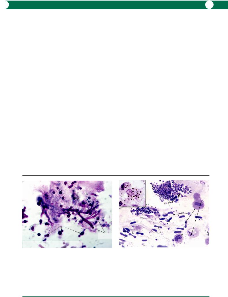
Vitreous biopsy anterior chamber tap A vitreous biopsy in the majority of exogenous cases will contain Gram positive cocci (Figure 8.2). In endogenous infections, fungal elements are more commonly identified (Figure 8.9).
Capsulectomy and vitrectomy In late stage chronic postoperative endophthalmitis (Proprionibacterium acnes) the histology shows phagocytosed organisms within macrophages – the pathogens are able to proliferate within the cells (Figure 8.10). With the accumulation of organisms, the macrophages increase in size to eventually rupture with the release of organisms and the inflammatory reaction is intensified. Microbiological diagnosis of P. acnes requires prolonged culture (at least 2 weeks) in anaerobic conditions. The bacteriologist should be given prior warning of the possibility of this diagnosis.
budding yeast |
|
|
non-septate |
|
hyphae |
Inflammatory disease - Fungal endophthalmitis |
|
Candida sp./Vitrectomy specimen |
PAS stain |
Figure 8.9
Figure 8.9 A vitreous biopsy was performed in a case of endogenous infection, in this case an intravenous drug abuser. The PAS stain is most commonly used to identify fungal elements. Candida sp. are characterised by the presence of budding yeasts and non-septate hyphae.
Gram stain |
toluidine blue stain |
Gram positive  intracytoplasmic
rods
dispersed P. acnes  bacteria
intracytoplasmic macrophage bacteria
 red cells
Inflammatory disease - Bacterial endophthalmitis
Late stage (saccular) - Proprionibacterium acnes
Figure 8.10
Figure 8.10 In this example of saccular endophthalmitis, Gram positive rods (Proprionibacterium acnes) are seen within the cytoplasm of macrophages (inset). The larger figure is taken from a plastic section stained with toluidine blue to show organisms within the cytoplasm of intact macrophages. The organisms are able to proliferate within macrophages, which increase in size to eventually rupture with release of large clumps of bacteria. This results in cyclical exacerbation of inflammation.

162 C H A P T E R 8
Evisceration Evisceration of ocular contents is preferred by some clinicians for acute uncontrollable infection. The motivation is to reduce the risk of the spread of infection along the optic nerve meninges. Such specimens contain fragments of intraocular tissues and pus. The histopathological abnormalities will be the same as that described in enucleation specimens.
Enucleation An enucleation will be performed for the late stages of infection.
1Postsurgical and trauma-related endophthalmitis (exogenous): purulent material within the vitreous cavity or extrusion through a wound makes the diagnosis of endophthalmitis obvious. The presence of a lens implant is an indication of previous cataract surgery (Figures 8.11, 8.12). If lens tissue is not identified, the possibility of nonsurgical trauma should be considered because the lens is often extruded through a scleral or corneal wound in sudden decompression. In a pathological specimen, purulent material is dense white or creamy-yellow in colour: the surrounding vitreous is less opaque (Figure 8.13).
The retina is often necrotic, and the uveal tract may be thickened due to hypotonic exudation (Figure 8.13) or to lymphocytic infiltration (Figure 8.14). Perforation of the cornea may be evident (Figure 8.11).
2 Blood-borne infection (endogenous): normal ocular tissues can be recognised in many cases, although there may be varying stages of necrosis. Initially a small septic embolus appears as a white nodule surrounded by a thin layer of haemorrhage (Roth’s spot – Figure 8.15). Later, the inflammatory process extends into the vitreous (Figure 8.16). An abscess in the vitreous releases inflammatory mediators which disrupt the integrity of the blood–- ocular barrier. Leakage of proteinaceous exudate causes retinal detachment. Retinal ischaemia stimulates the formation of preretinal fibrovascular membranes. Rupture of the anterior vitreous face and damage to the iris vessels is followed by exudation into the anterior compartment (Figures 8.17, 8.18).
Special investigations/stains
See Chapter 1 for specific stains for microorganisms.
Inflammatory disease - Bacterial endophthalmitis
Post surgical pseudophakia
|
corneal wound |
intraocular |
|
lens |
|
subchoroidal |
with prolapsing |
|
iris |
|
|
effusion |
|
|
|
|
exudative
retinal detachment with haemorrhagic infarct
abscess
choroidal
detachment subchoroidal effusion and
haemorrhage
Figure 8.11
Inflammatory disease - Bacterial endophthalmitis
|
dense |
detached retina |
abscess |
|
surrounding |
thickened choroid |
opaque vitreous |
Inflammatory disease - Bacterial endophthalmitis |
thickened choroid |
Post-surgical pseudophakia |
|
|
abscess |
|
space |
|
|
occupied |
|
subretinal |
by intraocular |
|
exudate |
lens |
|
necrotic |
hypopyon |
|
|
||
retina |
|
|
suprachoroidal |
fragment of lens |
|
haemorrhage |
||
|
Figure 8.12
Figure 8.11 This is an example of endophthalmitis (Streptococcus sp.) following complicated cataract surgery. A lens implant can be identified and there is a dehiscence at the superior limbus. The anterior chamber contains pus and the infection has spread into the cornea. The retina is detached by exudate and the collapsed vitreous is filled with purulent material. Secondary vasculitis has resulted in haemorrhagic infarction of the retina. The exudative detachment of the choroid is due to hypotonia.
Figure 8.12 In this case, cataract surgery was complicated by a dropped lens nucleus. An endophthalmitis (Staphylococcus sp.) developed around the lens fragment. The retina and anterior segment are infarcted. The presence of suprachoroidal exudation and haemorrhage confirms the subsequent hypotonia.
Figure 8.13 A vitreous abscess is sometimes localised and this dense white appearance differs from the secondary vitreous opacification. The uveal tract is the “lymph node” of the eye and the choroidal thickening is due to lymphocytic infiltration – see Figure 8.14.
Figure 8.13

I N F L A M M AT I O N 163
Inflammatory disease - Bacterial endophthalmitis
VITREOUS
necrotic retina
Figure 8.14
methenamine silver
Figure 8.16
preretinal membrane
subretinal exudate
SCLERA
non-granulomatous lymphocytic infiltrate
Inflammatory disease -
Endogenous fungal endophthalmitis
fungal hyphae |
fungal |
|
of Candida |
||
hyphae |
||
|
||
|
in clumps |
preretinal haemorrhages
closed angles
retinal folding
iris bombe
proteinaceous exudate
abcess
Inflammatory disease - Endogenous fungal endophthalmitis
Figure 8.18
Inflammatory disease - Endogenous fungal endophthalmitis
Roth’s spot
vascular sheathing
oedematous |
optic disc |
macula |
|
preretinal inflammatory focus
Figure 8.15
solid proteinaceous
subretinal exudate
detached retina
closed angles
|
iris bombe |
|
abscess |
proteinaceous |
|
exudate |
||
|
Inflammatory disease - Endogenous fungal endophthalmitis
Figure 8.17
Figure 8.14 In an infarcted retina due to an occlusive vasculitis, the tissue has disintegrated into basophilic fragments. The choroid contains a dense lymphocytic infiltrate.
Figure 8.15 The globe was removed at autopsy in this case of disseminated
Candida infection in a drug addict. The features of early septic embolism are small white areas surrounded by a ring of haemorrhage (Roth’s spots) and sheathing of the retinal vessels. The chorioretinal infection also spreads into the vitreous forming a preretinal inflammatory focus. In autopsy tissue, the retina swells particularly in the region of the macula.
Figure 8.16 This section is at the level of the preretinal nodule shown in Figure 8.15. The choroid and retina have been destroyed by the invading fungi and the inflammatory process. Fungi are not easily seen in an H&E section and the inset shows one of the special stains used to demonstrate fungal elements (methenamine-silver).
Figure 8.17 At the end stage of fungal infection, there is extensive disorganisation of the ocular tissues. Fungal abscesses are present in the anterior vitreous and there is exudation behind the iris with posterior synechiae formation. An iris bombé has resulted. The thickened white appearance of the retina is deceptive, the thickened tissue consists of folded retina behind a preretinal membrane which is the source of bleeding into the posterior vitreous (see Figure 8.18).
Figure 8.18 A section through the globe shown in Figure 8.17 illustrates the pathogenesis of disorganisation due to fungal infection. Damage to the endothelium of the blood vessels results in a massive leakage of proteinaceous fluid into all the ocular compartments. Subretinal exudation has contributed to the detachment but the traction by a preretinal fibrovascular membrane explains
the folding of the retina. Friable blood vessels within the preretinal membrane have ruptured and bleeding into the vitreous has occurred. This case illustrates two possible causes of retinal detachment – exudative and tractional.
164 C H A P T E R 8
Granulomatous reactions
In the introduction, the basic cellular constituents (lymphocytes, plasma cells, and macrophages) of a granulomatous reaction were simplified. It is important to appreciate that the various patterns described in the following section vary quite markedly but, in some conditions, the patterns can provide a specific diagnosis.
Infective
Tuberculosis
Infection in tuberculosis (along with sarcoidosis and syphilis) can present in any form of ocular granulomatous inflammatory disease, particularly anterior uveitis and chorioretinitis.
Ocular infections due to mycobacteria (Mycobacterium tuberculosis and Mycobacterium avium-intracellulare) are more common in individuals in whom the cell mediated immunity response is suppressed. The organisms are occasionally present in large numbers when examined by Ziehl–Neelsen stain (Figure 8.19).
In patients with an intact immune system, the histopathological pattern can vary between caseating and non-caseating granulomas. In both, Langhans’ multinucleate giant cells are characteristic (Figure 8.20). Caseation represents intensive necrosis in the infiltrating inflammatory cells (Figures 8.21, 8.22).
Leprosy
Infection by Mycobacterium leprae remains endemic in the tropics and subtropics. The ocular manifestations are variable. Involvement of nerves leads to paralytic lagophthalmos and is an indirect cause of exposure keratitis. Damage to autonomic nerve fibres leads to a mild iridocyclitis, and this is the most common manifestation of ocular leprosy. Anterior chamber cells and flare with sparse keratic precipitates are the usual features.
Infections vary depending on the immune status: tuberculoid leprosy (active cell mediated response) or lepromatous leprosy (poor response). In the tuberculoid form, the histopathological patterns are similar to those described in tuberculosis with non-caseating granulomas and scanty organisms. An infiltrate containing macrophages, lymphocytes, and overwhelming numbers of organisms is characteristic of lepromatous leprosy.
Syphilis
Like tuberculosis, syphilis has the potential to present in any form of ocular inflammatory disease. Pathological experience of this condition will be either in the form of a penetrating keratoplasty specimen or a globe, but is rare. Infection is by the spirochaete Treponema pallidum. It may be congenital or acquired. The latter is essentially a sexually transmitted disease but may also be acquired via blood transfusions and direct contact with a surface lesion. Although potentially treatable with antibiotics, the incidence of syphilitic infection is rising due to prophylactic and treatment complacency.
Inflammatory disease - Florid tuberculous choroiditis
lymphocyte
curved organisms forming “Chinese letters”
macrophages
Ziehl-Neelsen stain
Figure 8.19
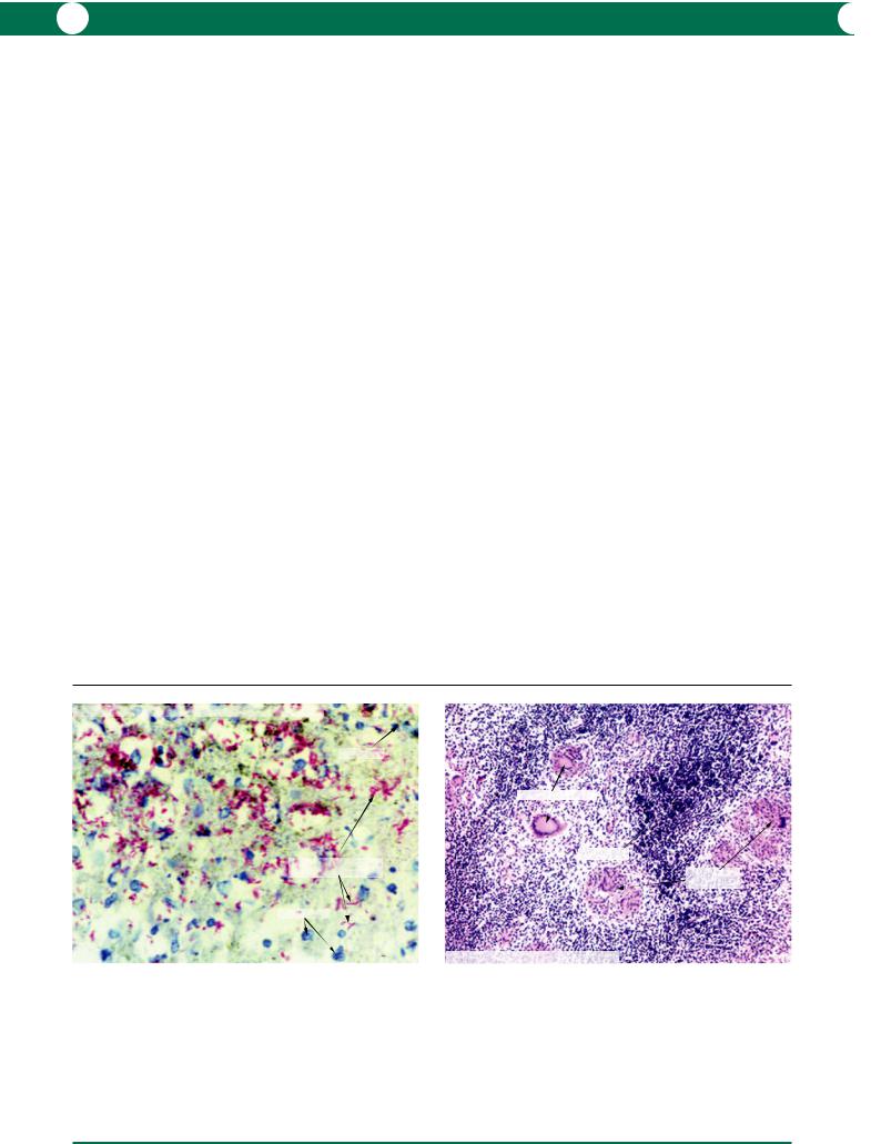
Figure 8.19 In an eye removed at the autopsy of an immunosuppressed patient, nodules were identified in the choroid. Histological examination revealed massive accumulations of slender tubercle bacilli. The organisms are curved and are red in colour with the Ziehl–Neelsen stain, and form patterns described previously but incorrectly as “Chinese letters”.
Langhans’ giant cells
lymphocytes
clusters of macrophages
Inflammatory disease - Tuberculosis
Figure 8.20
Figure 8.20 The histological pattern in tuberculosis varies according to the status of the cell-mediated immune response. In this example, there is no evidence of caseation but the clusters of macrophages contain Langhans’ giant cells. In this type of reaction, very few tubercle bacilli will be identified.
I N F L A M M AT I O N 165
Clinical manifestations of ocular syphilis will depend on the stage of disease:
1Primary: chancre of the eyelid or conjunctiva.
2Secondary: iridocyclitis, neuroretinitis, and chorioretinitis.
3Tertiary:
•Cornea: interstitial keratitis with the appearance of ghost vessels. The majority of cases are congenital (see Chapter 4).
•Uveal tract: the inflammation may be granulomatous or non-granulomatous.
•Visible dilated capillaries on the iris surface (roseola) are a characteristic feature. A chorioretinitis may be present which eventually resolves and takes the form of a pigmentary retinopathy.
•Retina: vasculitis with retinal exudation which may be bilateral, multifocal, or diffuse. Retinal oedema with cotton wool spots may also be present.
•Optic nerve: optic disc oedema with haemorrhage and exudate.
Non-infective
Sympathetic ophthalmitis
A rare bilateral autoimmune granulomatous choroiditis resulting from trauma to one eye. If untreated, the condition leads to bilateral blindness.
Clinical presentation
A history of a penetrating trauma to one eye (surgical and non-surgical) is essential.
By definition, this condition is bilateral, although it may be variable in severity and symmetry.
vitreous
haemorrhage massive inflammation in anterior chamber
showing caseation
caseation
detached |
lens |
|
retina |
||
|
Inflammatory disease - Tuberculosis
Figure 8.21
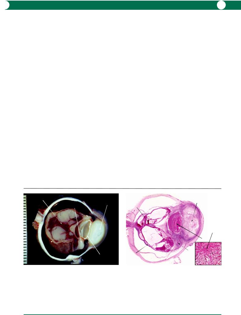
Figure 8.21 Some 15 years after treatment for spinal tuberculosis, a male patient developed an episcleral nodule and an anterior uveitis. This progressed to retinal detachment and end-stage disorganisation of the ocular tissues with caseation in the anterior chamber and anterior uvea due to tuberculosis.
The clinical signs vary from a localised uveitis to a panuveitis with features including:
1Anterior chamber: mutton-fat keratic precipitates.
2Choroid: diffuse thickening with white nodules (Dalen–Fuchs spots) representing focal elevated subretinal infiltrates. A vitritis, which obscures the view of the fundus, may be present.
Pathogenesis
The pathogenesis is poorly understood but it is generally considered to be the sensitisation of the systemic immune system to antigens present in the retina or uveal tissue. It is assumed that the proteins in chorioretinal tissue are sequestered and are exposed by trauma.
This condition is commonly associated with lens-induced uveitis (see below) by the nature of the trauma involved.
In animal models, it is possible to induce a uveitis by sensitising the immune system to retinal and uveal proteins in combination with Freund’s adjuvant. Unfortunately, the histological pattern is unlike human sympathetic ophthalmitis.
Possible modes of treatment
Prophylaxis Enucleation of the non-viable injured eye – there have been no published cases of sympathetic ophthalmitis when an injured eye was enucleated within 10 days of injury. As the incidence of sympathetic ophthalmitis is rare and is continuing to decline, it is common in many practices to conserve an injured eye that still maintains some vision.
Treatment With the recognition of sympathetic ophthalmitis, treatment would involve systemic corticorticosteroids and other immunosuppressive agents.
massive inflammation in anterior chamber
showing caseation
vitreous haemorrhage
caseation
lens
caseation
detached retina
Inflammatory disease - Tuberculosis
Figure 8.22
Figure 8.22 A histological section showing the globe in Figure 8.21. The inset shows amorphous eosinophilic material (caseation) and fragmentation of inflammatory cells. No more than four tubercle bacilli were identified in this case using the Ziehl–Neelsen stain.

166 C H A P T E R 8
Macroscopic
The only specimen likely to be submitted would be an enucleated eye. With the onset of this disease, in either the injured or non-injured (sympathising) eye, a prominent thickened choroid is evident macroscopically (Figures 8.23, 8.24).
At late stages, hypotonia due to ciliary body shutdown leads to uveal effusion and eventual phthisis.
Microscopic
The granulomatous reaction is confined to the choroid, but in an appropriate plane of section, extension into the scleral canals will be evident (Figure 8.25). The choriocapillaris is usually spared. Multinucleate giant cells are sparse and the accumulations of macrophages are less compact (Figure 8.26) than in sarcoidosis (see below). The macrophages may contain a fine dusting of melanin granules (Figure 8.27). Macrophagic infiltration beneath and within the retinal pigment epithelium (Figure 8.28) is a characteristic feature (Dalen–Fuchs nodule).
Immunohistochemistry/immunopathology
There are no specific markers but conventional immunohistochemistry has revealed T cells (CD3), B cells (CD20), and macrophages (CD68) within the infiltrate.
Sarcoidosis
A chronic multisystem disease of unknown aetiology characterised by the formation of non-caseating granulomas within the eye, conjunctiva, and orbit (see Chapters 3 and 5).
Clinical presentation
The first presentation of the systemic disease is usually within the third to sixth decades, and approximately 80% of cases have ocular involvement.
Any or all parts of the eye may be involved:
1Anterior uveitis: often bilateral, with “mutton-fat” keratic precipitates; formation of anterior and posterior synechiae; secondary open and closed angle glaucoma.
2Posterior uveitis: vitritis, chorioretinitis, neovascularisation, haemorrhage.
3Granulomas: note that granulomas can also occur in conjunctiva, eyelid, orbit, optic nerve, and lacrimal drainage systems.
4Vascular: peripheral retinal periphlebitis, exudates, “candle wax drippings”, neovascularisation, and haemorrhage, branch or central retinal vein occlusion.
5Optic nerve: swelling, granuloma, atrophy, and neovas-
cularisation. Field defects may be evident.
It is important to refer the patient for investigation of systemic involvement, especially the lung, liver, skin, and central and peripheral nervous systems.
Basic investigations include a chest X-ray for bilateral hilar lymphadenopathy, serology to detect a raised serum angiotensin converting enzyme (ACE), and a gallium scan. A confirmatory biopsy of a suspicious nodule is preferred, especially with the prospect of committing the patient to long term steroid treatment.
Pathogenesis
Unknown with considerable variation in disease onset and progression.
Genetics
•Gender: females males.
•Race: black to white, 10 : 1.
Possible modes of treatment
Treatment is mainly with corticosteroids and other immunosuppressive/antimetabolic therapy.
Figure 8.23 This globe was badly traumatised and enucleation was delayed for a month. Several months later, the patient developed choroiditis in the fellow non-traumatised eye and sympathetic ophthalmitis was diagnosed. In the enucleated globe, the retina was avulsed and the iris was incarcerated into the limbal wound. The thickened choroid is described as having a “marble-like” appearance. In this case, there is an associated lens-induced uveitis and the anterior uvea is thickened by inflammatory cell infiltration.
Figure 8.24 In this low power micrograph from the specimen in Figure 8.23, the choroid and anterior uvea contain a purple granulomatous infiltrate. Lens remnants were found within a fibrous ingrowth and this gave rise to the inflammatory infiltration in the anterior uvea. The avulsed retina forms a nodule surrounded by scar tissue from a fibrous ingrowth from the limbal wound.
Figure 8.25 A granulomatous reaction in sympathetic ophthalmitis is not confined to the choroid but spreads along the scleral canals through which the nerves and arteries penetrate.
Figure 8.26 In sympathetic ophthalmitis, the granulomatous reaction is not compact (compared with sarcoidosis, see below). The macrophages are loosely intermingled with lymphocytes. The multinucleated cells are useful diagnostic features.
Figure 8.27 Within multinucleate giant cells in sympathetic ophthalmitis (left), the nuclei are irregularly distributed unlike the Langhans’ giant cells in tuberculosis (see Figure 8.20). Macrophages are identified by their oval nuclei and light dusting of melanosomes in the cytoplasm (right).
Figure 8.28 Clusters of macrophages on the inner surface of Bruch’s membrane are the histological equivalent of the white spots (Dalen–Fuchs) as seen clinically in sympathetic ophthalmitis. Note the photoreceptor atrophy.

I N F L A M M AT I O N 167
subretinal exudate
thickened choroid
avulsed retina
optic nerve |
prolapsed |
|
uveal tissue |
|
through |
|
wound |
Inflammatory disease - Sympathetic ophthalmitis
Figure 8.23
SUBRETINAL SPACE lymphocytic infiltrate
granulomatous foci
extension along scleral canal (nerve and artery)
SCLERA
Inflammatory disease - Sympathetic ophthalmitis
Figure 8.25
retinal pigment epithelium
Bruch’s membrane |
|
multinucleated |
lymphocytes |
|
|
giant cells |
macrophages |
|
plasma cell
macrophages
Inflammatory disease - Sympathetic ophthalmitis
choroid thickened |
subretinal exudate |
|
by granulomatous |
||
|
||
inflammatory |
|
|
infiltrates |
avulsed retina |
|
|
fibrous ingrowth through wound
optic nerve |
|
|
massive |
|
inflammatory |
|
infiltration |
|
of iris and |
Inflammatory disease - Sympathetic ophthalmitis |
ciliary body |
Figure 8.24
retinal pigment epithelium
granuloma |
Bruch’s |
|
membrane |
lymphocytes
epithelioid macrophages
giant cells
Inflammatory disease - Sympathetic ophthalmitis
Figure 8.26
Dalen–Fuchs nodule - clusters of macrophages
retina
Bruch’s membrane |
retinal pigment |
|
epithelium |
Inflammatory disease - Sympathetic ophthalmitis
Figure 8.27 |
Figure 8.28 |

168 C H A P T E R 8
Macroscopic
The specimen presented would usually be in the form of a diagnostic/confirmatory biopsy, although enucleations may be performed for end-stage secondary complications. In an enucleated eye, perivascular granulomatous nodules (Figure 8.29) and perivascular sheathing are present. The anterior segment may show secondary changes of iritis with angle closure.
With the many new and established investigatory modalities, conjunctival “blind biopsies” are less commonly performed (see Chapter 3), and are usually restricted to areas where small suspicious granulomatous nodules are present.
Complications/secondary effects
1 Secondary glaucoma and cataract from persistent anterior uveitis – open and closed angle.
2 Central and branch retinal vein occlusion due to periphlebitis.
Microscopic
The hallmark pathological feature of sarcoidosis is a noncaseating granuloma with lymphocytes intermingled with epithelioid macrophages. Multinucleate cells are rarely seen.
The granulomas are usually small and can be identified in the retina around blood vessels, in the uveal tract, and in the vitreous (Figures 8.30–8.32). Sarcoid granulomas in the trabecular meshwork are rare (Figure 8.33).
Lens-induced uveitis
Lens-induced uveitis can be divided into the following categories:
1Phacolytic glaucoma.
2Phacoanaphylactic endophthalmitis.
The former is not a true uveitis and has been described
in Chapter 7. The remainder of this section refers to phacoanaphylaxis.
Phacoanaphylactic endophthalmitis is a severe autoimmune granulomatous reaction against lens matter exposed through a disrupted lens capsule by surgical and nonsurgical trauma. Occasionally, “spontaneous” cases are encountered.
Clinical presentation
The patient presents with a unilateral painful red eye. A history of surgical or non-surgical trauma (blunt or penetrating) is significant. The reaction may take several weeks to develop after the initial insult, but the onset may be within 24 hours if there was prior sensitisation to lens matter (for example cataract surgery in the other eye).
Severe granulomatous anterior uveitis appears as “muttonfat” keratic precipitates, synechiae, and hypopyon. There may be a secondary elevation of intraocular pressure. The posterior segment is relatively unaffected.
Infection and sympathetic endophthalmitis are important differential diagnoses.
Pathogenesis
The aetiology is not well understood. Exposure to certain components of (possibly degenerate) lens proteins sensitises and activates the humoral and cell mediated responses to the remaining exposed lens matter.
Possible modes of treatment
Corticosteroids may be used to decrease the immune response but ultimately surgical removal of residual lens matter is required.
Macroscopic
Opportunities for pathological studies are in the form of an irretrievably damaged globe (Figures 8.34, 8.35) or excised lens tissue.
Trabeculitis and anterior and posterior synechiae may lead to secondary open or closed angle glaucoma.
Inflammatory disease - Sarcoidosis
perivascular sarcoid granulomas
blood vessel
granuloma
swollen optic disc
|
photoreceptors |
|
Inflammatory disease - Sarcoidosis |
Figure 8.29 |
Figure 8.30 |
Figure 8.29 A patient suffering from sarcoidosis also experienced extreme pain |
Figure 8.30 In retinal sarcoidosis, the granulomatous reaction is compact and |
from scleritis. She requested bilateral enucleation. The retina contains small |
consists of macrophages and intermingled lymphocytes. Multinucleate giant |
white granulomatous foci located around blood vessels. The optic disc is |
cells are rare. |
swollen due to the accumulation of granulomatous foci. |
|
|
|

I N F L A M M AT I O N 169
Inflammatory disease - Sarcoidosis
previous location of artefactual crystalline body scratch
Bruch’s membrane |
sarcoid granuloma |
|
Figure 8.31
scleral sulcus filled with fibrovascular tissue
giant cell
sarcoid granulomas
Inflammatory disease - Sarcoidosis
Figure 8.33
condensed vitreous
inflammatory focus
plane of |
remnant |
section |
lens matter |
Inflammatory disease - Lens induced uveitis
Phacoanaphylactic endophthalmitis
Figure 8.35
granuloma in vitreous base
epithelioid macrophage giant cell
lymphocytes
pars plana pigmented
epithelium pars plana non-pigmented epithelium
Inflammatory disease - Sarcoidosis
Figure 8.32
Inflammatory disease - Lens induced uveitis |
inflammatory |
Phacoanaphylactic endophthalmitis |
exudate in |
|
anterior chamber |
lens cortex
thickened iris
lens nucleus
opaque vitreous
Figure 8.34
Figure 8.31 In this example of sarcoidosis, the granulomatous reaction involves the retinal pigment epithelium and the underlying choroid. Sometimes granulomas contain crystalline bodies – in this example the microtome knife has sheered off the crystalline body and has left a scratch mark in the retina.
Figure 8.32 Sarcoid granulomas can be found in the vitreous base adjacent to the pars plana. The macrophages with their pink cytoplasm are larger than the lymphocytes, which have heavily stained nuclei.
Figure 8.33 A multinucleate giant cell is present in one of the two small granulomas in the outer part of the trabecular meshwork.
Figure 8.34 An elderly female patient with a morgagnian cataract had accidental blunt trauma to her eye resulting in disruption of the lens and its contents. The brown sclerotic nucleus is surrounded by a yellow inflammatory infiltrate. The iris is thickened by inflammatory cell infiltration and the anterior chamber contains a dense inflammatory exudate.
Figure 8.35 An intracapsular cataract operation was complicated by lens zonule dehiscence, with dislocation of the lens remnants against the ciliary body. An intense inflammatory reaction ensued several days later, with intractable anterior uveitis and raised intraocular pressure. An endophthalmitis was suspected, but was resistant to treatment and the eye was subsequently enucleated. A section was taken in the plane of the black line and is shown in Figure 8.36.