

Erik_Kandel_-_V_poiskakh_pamyati_angl
.pdfThat neurotransmitter, we later discovered, is glutamate, also the major excitatory transmitter in the mammalian brain. By increasing the amount of glutamate a sensory cell sends to a motor cell, sensitization strengthens the synaptic potential elicited in the motor cell, thus making it easier for that neuron to fire an action potential and cause the gill to withdraw.
The synaptic potential between the sensory and motor neurons lasts only milliseconds, yet we had observed that a shock to Aplysia's tail enhances glutamate release and synaptic transmission for many minutes. How does this come about? As my colleagues and I focused on the question, we noticed something curious. The strengthening of the synaptic connection between the sensory and motor neuron is accompanied by a very slow synaptic potential in the sensory cell, one that lasts for minutes rather than the milliseconds typical of synaptic potentials in the motor neuron. We soon found that the shock to Aplysia's tail activates a second class of sensory neurons, one that receives information from the tail. These tail sensory neurons activate a group of interneurons that acts on the sensory neuron from the siphon. It is these interneurons that produce the remarkably slow synaptic potential. We
then asked ourselves: What neurotransmitter do the interneurons release? How does this second neurotransmitter lead to the release of more glutamate from the terminals of the sensory neuron, thus creating short-term memory storage?
We found that the interneurons activated by a shock to Aplysia's tail release a neurotransmitter called serotonin. Moreover, the interneurons form synapses not only on the cell body of the sensory neurons but also on the presynaptic terminals, and they not only produce a slow synaptic potential but also enhance the sensory cell's release of glutamate onto the motor cell. In fact, we could simulate the slow synaptic potential, the enhancement of synaptic strength, and the strengthening of the gill-withdrawal reflex simply by applying serotonin to the connections between the sensory and motor neurons.
We called these serotonin-releasing interneurons modulatory interneurons because they do not mediate behavior directly; rather, they modify the strength of the gill-withdrawal reflex by enhancing the strength of the connections between sensory and motor neurons.
These findings caused us to realize that there are two kinds of neural circuits important in behavior and learning: mediating circuits, which we had characterized earlier, and modulating circuits, which we were just beginning to characterize in detail (figure 16-1). Mediating circuits produce behavior directly and are therefore Kantian in nature. These are the genetically and developmentally determined neuronal components of the behavior, the neuronal architecture. The mediating circuit is made up of the sensory neurons that innervate the siphon, the interneurons, and the motor neurons that control the gill-withdrawal reflex. With learning, the mediating circuit becomes the student and acquires new knowledge. The modulating circuit is Lockean in nature; it serves as a teacher. It is not directly involved in producing a behavior but instead fine-tunes the behavior in response to learning by modulating—heterosynaptically—the strength of synaptic connections between the sensory and motor neurons. Activated by a shock to the tail, a completely different part of the body than the siphon, the modulating circuit teaches Aplysia to pay attention to a stimulus to the siphon that is important for its safety. Thus the circuit is, in essence,
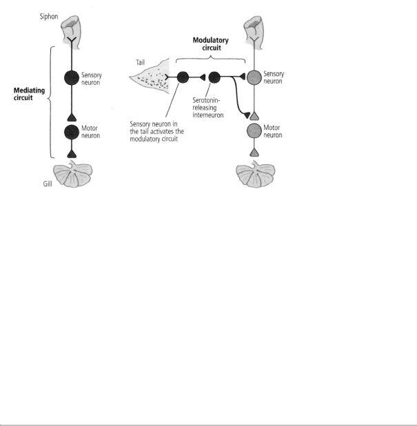
16-1 The two types of circuits in the brain. Mediating circuits produce behaviors. Modulatory circuits act on the mediating circuits, regulating the strength of their synaptic connections.
responsible for arousal or salience in Aplysia, just as analogous modulatory circuits are an essential component of memory in more complex animals, as we will see later.
That serotonin was a modulator for sensitization simply amazed me! Some of my first experiments with Dom Purpura in 1956 had focused on the action of serotonin. In fact, on Student Day at NYU Medical School in the spring of 1956, I had given a brief talk entitled "Electrophysiological Patterns of Serotonin and LSD Interaction on Afferent Cortical Pathways." Jimmy Schwartz had been kind enough to listen to a rehearsal of the talk and to help me improve it. I was now beginning to appreciate that life is circular. I had not worked on serotonin for almost twenty years, and here I was returning to it with renewed focus and enthusiasm.
ONCE WE KNEW THAT SEROTONIN ACTED AS A MODULATORY
transmitter to enhance the release of glutamate from the presynaptic terminals of the sensory neuron, the stage was set for a biochemical analysis of memory storage. Fortunately in Jimmy Schwartz I had an excellent guide and fellow traveler on this journey.
Before returning to NYU, Jimmy had worked at Rockefeller University on the bacterium Escherichia coli, the single-celled organism in which many fundamental principles of modern biochemistry and molecular biology were first worked out. In 1966 his interest had shifted to Aplysia, and he began his research by delineating the chemical transmitters used by a neuron in the abdominal ganglion. In 1971, we joined forces to study the molecular actions that accompany learning.
Jimmy was of inestimable help in this second major stage of my biological education. We were influenced by the work of Louis Flexner, who had shown a few years earlier that long-term memory in mice and rats requires the synthesis of new protein, whereas short-term memory does not. Proteins are the workhorses of the cell. They constitute its enzymes, ion channels, receptors, and transport machinery. Since, as we had found, long-term memory involves the growth of new connections, it is not surprising that the synthesis of new protein constituents is required for that growth.
Jimmy and I set out to test this idea in Aplysia and to do so at the level of the siphon sensory cell and its synapses on the motor neurons to the gill. If synaptic changes parallel changes in memory, then the short-term synaptic changes we had delineated should not require the synthesis of new protein. That is exactly what we found. What, then, mediates this short-term change?

Cajal had shown that the brain is an organ constructed of neurons wired to each other in specific pathways. I had seen this remarkable connection specificity in the simple neural circuits that mediate reflex behavior in Aplysia. But Jimmy pointed out that this specificity also extends to molecules—to the combinations of atoms that serve as the elementary units of cellular function. Biochemists had found that molecules can interact with one another within a cell and that these chemical reactions are organized in specific sequences known as biochemical signaling pathways. The pathways convey information in the form of molecules from the surface of the cell to the interior, much as one nerve cell conveys information to another. In addition, the pathways are "wireless." Molecules floating within the cell recognize and bind to specific molecular partners and regulate their activity.
My colleagues and I had not only fulfilled my early ambition of
trapping a learned response in the smallest possible population of neurons, we had trapped a component of a simple form of memory in a single sensory cell. But even a single Aplysia neuron contains thousands of different proteins and other molecules. Which of these molecules are responsible for short-term memory? As Jimmy and I began to discuss the possibilities, we focused in on the idea that the serotonin released in response to a shock to the tail might enhance glutamate release from the sensory neuron by launching a specific sequence of biochemical reactions in the sensory cell.
The sequence of biochemical reactions that Jimmy and I sought would have to serve two fundamental purposes. First, they would have to translate the brief action of serotonin into molecules whose signals would last for minutes within the sensory neuron. Second, those molecules would have to broadcast signals from the cell membrane, where serotonin acts, to the interior of the sensory cell, particularly to the specialized regions of the axon terminal involved in the release of glutamate. We elaborated on these thoughts in our 1971 article in the Journal of Neurophysiology and speculated on the possibility that a specific molecule known as cyclic AMP might be involved.
WHAT IS CYCLIC AMP? HOW DID WE HIT UPON IT AS A LIKELY
candidate? Cyclic AMP came to mind because this small molecule was known to serve as a master regulator of signaling within muscle and fat cells. Jimmy and I knew that nature is conservative— therefore, a mechanism used in the cells of one tissue is likely to be retained and used in the cells of another tissue. Earl Sutherland at Case Western Reserve University in Cleveland had already shown that the hormone epinephrine (adrenaline) produces a brief biochemical change at the surface membrane of fat and muscle cells that gives rise to a more enduring change inside the cells. That longer lasting change is brought about by an increase in the amount of cyclic AMP inside those cells.
Sutherland's revolutionary findings came to be described as the second-messenger signaling theory. The key to this biochemical signaling theory was his discovery of a new class of receptors at the cell surface of fat and muscle cells that responds to hormones. Earlier, Bernard
Katz had found the neurotransmitter-gated receptors known as ionotropic receptors; on binding a neurotransmitter, these receptors open or close the gate of an ion channel contained within the receptor, thus translating a chemical signal into an electrical signal. But the new class of receptors, called metabotropic receptors, has no ion channel within it to open or close. Instead, one region of these receptors protrudes from the outside surface of the cell membrane and recognizes signals from other cells, while another region protrudes from the inside of the cell membrane and engages an enzyme. When these receptors recognize and bind a chemical messenger on the outside of the cell, they activate an enzyme within the cell called adenylyl cyclase, which makes cyclic AMP.
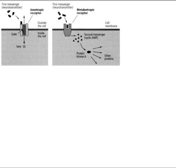
This process has the advantage of having greatly amplifying the cell's response. When one molecule of chemical messenger binds to a metabotropic receptor, that receptor stimulates adenylyl cyclase to make a thousand molecules of cyclic AMP. Cyclic AMP then binds to key proteins that trigger a whole family of molecular responses throughout the cell. Finally, adenylyl cyclase continues making cyclic AMP for minutes. The actions of metabotropic receptors therefore tend to be more powerful, more widespread, and more persistent than the actions of ionotropic receptors. Whereas ionotropic actions typically last milliseconds, metabotropic actions can last from seconds to minutes—one thousand to ten thousand times longer.
To distinguish between the two spatially distinct functions of metabotropic receptors, Sutherland called the chemical messenger that binds to the metabotropic receptor on the outside of the cell the first messenger, and the cyclic AMP activated inside the cell to broadcast the signal the second messenger. He argued that the second messenger conveys the signal at the cell surface from the first messenger into the interior of the cell and initiates the cell-wide response (figure 16-2). Second-messenger signaling suggested to us that metabotropic receptors and cyclic AMP might be the elusive agents connecting the slow synaptic potential in sensory neurons to the enhanced release of glutamate and consequently to the formation of short-term memory.
In 1968 Ed Krebs at the University of Washington provided the ini-
16-2 Sutherland's two classes of receptors. Ionotropic receptors (left) produce changes lasting milliseconds. Metabotropic receptors (e.g., serotonin receptors) act through second messengers (right). They produce changes that last seconds to minutes and are broadcast throughout the cell.
tial insight into how cyclic AMP produces its widespread effects. Cyclic AMP binds to and activates an enzyme that Krebs called cyclic AMP-dependent protein kinase, or protein kinase A (because it was the first protein kinase to be discovered). Kinases modify proteins by adding a phosphate molecule to them, a process known as phosphorylation. Phosphorylation activates some proteins and inactivates others. Krebs found that phosphorylation can be readily reversed and thus can serve as a simple molecular switch, turning the biochemical activity of a protein on or off.
Krebs next went about finding out how this molecular switch works. He discovered that protein kinase A is a complex molecule made up of four units—two regulatory and two catalytic. The catalytic units are designed to carry out phosphorylation, but the regulatory units normally "sit" on them and inhibit them. The regulatory units contain sites that bind cyclic AMP. When the concentration of cyclic AMP in a cell increases, the regulatory units bind the excess molecules. This action changes their shape and causes them to fall off the catalytic units, leaving the catalytic units free to phosphorylate target proteins.
These considerations raised a key issue in our minds: Was the mechanism discovered by Sutherland and Krebs specific to the action of hormones on fat and muscle cells, or could it also involve other
transmitters, including those present in the brain? If so, this would represent a previously unknown mechanism of synaptic transmission.
Here we were helped by the work of Paul Greengard, a gifted biochemist who was also trained in physiology and who had recently moved to Yale University from his former position as director of biochemistry at Geigy Pharmaceutical Research Laboratories. On the way to Yale, he stopped off for a year in Sutherland's department. Realizing the importance of a potentially novel signaling mechanism in the brain, Greengard began in 1970 to sort out metabotropic receptors in the brains of rats. Now, a wonderful coincidence emerged that was to link Arvid Carlsson, Paul Greengard, and me on a scientific journey that would lead the three of us to Stockholm in 2000 to share in the Nobel Prize in Physiology or Medicine for signal transformations (transduction) in the nervous system.
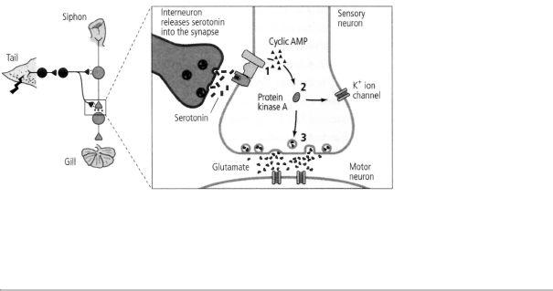
16-3 Biochemical steps in short-term memory. A shock to the tail of Aplysia activates an interneuron that releases the chemical messenger serotonin into the synapse. After crossing the synaptic cleft, serotonin binds to a receptor on the sensory neuron, leading to production of cyclic AMP (1). Cyclic AMP frees the catalytic unit of protein kinase A (2). The catalytic unit of protein kinase A enhances the release of the neurotransmitter glutamate (3).
In 1958 Arvid Carlsson, a great Swedish pharmacologist, discovered dopamine to be a transmitter in the nervous system. He then went on to show that when the concentration of dopamine is decreased in a rabbit, the animal develops symptoms that resemble Parkinson's disease. When Greengard started to explore metabotropic receptors in the brain, he started with a receptor for dopamine and found that it stimulates an enzyme that increases cyclic AMP and activates protein kinase A in the brain!
Based on these leads, Jimmy Schwartz and I discovered that cyclic AMP second-messenger signaling is also turned on by serotonin during sensitization. As we have seen, a shock to Aplysia's tail activates modulatory interneurons that release serotonin. Serotonin, in turn, increases the production of cyclic AMP in the presynaptic terminals of the sensory neurons for a few minutes (figure 16-3). Thus it all came together: the increase in cyclic AMP lasts about as long as the slow synaptic potential, the increase in synaptic strength between the sensory and motor neurons, and the animal's enhanced behavioral response to the shock applied to its tail.
THE FIRST DIRECT CONFIRMATION THAT CYCLIC AMP IS INVOLVED
in the formation of short-term memory came in 1976, when Marcello Brunelli, an Italian postdoctoral fellow, joined our lab. Brunelli tested the idea that when serotonin signals the sensory neurons to increase the concentration of cyclic AMP, the cells boost the amount of glutamate released from their terminals. We injected cyclic AMP directly into a sensory cell of Aplysia and found that it dramatically increased the amount of glutamate released and, therefore, the strength of the synapse between that sensory cell and motor neurons. In fact, injecting cyclic AMP simulated perfectly the

increased synaptic strength brought about by applying serotonin to sensory neurons or a shock to the animal's tail. This remarkable experiment not only linked cyclic AMP and short-term memory but also gave us our first insight into the molecular mechanisms of learning. Having begun to trap the basic molecular components of short-term memory, we could now use them to simulate memory formation.
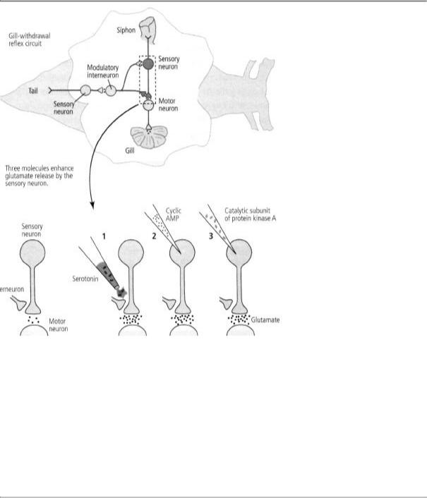
In 1978 Jimmy and I began to collaborate with Greengard. The three of us wanted to know whether cyclic AMP produces its effect on short-term memory through protein kinase A. We pulled the protein apart and injected directly into a sensory neuron only the catalytic unit, which normally carries out phosphorylation. We found that this unit does exactly what cyclic AMP does—it strengthens the synaptic connection by enhancing the release of glutamate. Then, just to make sure we were on the right track, we injected an inhibitor of protein kinase A into a sensory neuron and found that it indeed blocked the ability of serotonin to enhance glutamate release. In finding that cyclic AMP and protein kinase A are both necessary and sufficient for strengthening the connections between sensory and motor neurons, we were able to identify the first links in the chain of biochemical events leading to short-term memory storage (figure 16-4).
That did not tell us how serotonin and cyclic AMP bring about the slow synaptic potential or how this synaptic potential relates to the enhanced release of glutamate, however. In 1980 I met Steven Siegelbaum in Paris, where I was giving a series of seminars at the College of France. Steve was a technically gifted young biophysicist who specialized in studying the properties of single ion channels. We hit it off extremely well and, as fate would have it, he had recently accepted a position in the pharmacology department at Columbia. We therefore decided to join forces upon his arrival in New York and explore the biophysical nature of the slow synaptic potential.
Steve discovered one of the targets of cyclic AMP and protein kinase A: a potassium ion channel in sensory neurons that responds to serotonin. We called this channel the S channel because it responds to serotonin and because it was discovered by Steve Siegelbaum. The channel is open when the neuron is at rest and contributes to its resting membrane potential. Steve found that the channel is present in the presynaptic terminals and that he could cause it to close either by applying serotonin (the first messenger) to the outside of the cell membrane or by applying cyclic AMP (the second messenger) or protein kinase A to the inside. Closing the potassium ion channel causes
the slow synaptic potential that drew our attention to cyclic AMP in the first place.
Closing the channel also helps to enhance the release of glutamate. When the channel is open, it contributes, along with other potassium channels, to the resting membrane potential and to the outward movement of potassium during the descending stroke of the action potential. But when it is closed by serotonin, the ions move out of the cell less rapidly, slightly increasing the duration of the action potential by slowing the descending stroke. Steve showed that slowing the action potential allows more time for calcium to flow into the presynaptic terminals—and calcium, as Katz had shown in the squid's giant synapse, is essential for the release of glutamate. In addition, cyclic AMP and protein kinase A act directly on the machinery that releases the synaptic vesicles, thus stimulating the release of glutamate even further.
These exciting results regarding cyclic AMP were soon complemented by important genetic studies of learning in fruit flies, a research favorite for over half a century. In 1907 Thomas Hunt Morgan at Columbia began to use the fruit fly, Drosophila, as a model organism for genetic studies because of its small size and short reproductive cycle (twelve days). This proved to be a happy choice because Drosophila has only four pairs of chromosomes (compared with twenty-three pairs in humans), making it a relatively easy animal to study genetically. It had long been obvious that many physical characteristics of animals—the shape of the body, eye color, and speed, among others—are inherited. If external physical characteristics can be inherited, can mental characteristics produced by the brain also be inherited? Do genes have a role in a mental process such as memory?
The first person to address this question with modern techniques was Seymour Benzer, at the California Institute of Technology. In 1967 he began a brilliant series of experiments in which he treated flies with chemicals designed to produce random mutations, or changes, in single genes. He then examined the effects of these
mutations on learning and memory. To study memory in the fruit fly, Benzer's students Chip Quinn and Yadin Dudai used a classical conditioning procedure. They placed the flies in a small chamber and exposed them to
16-4 Molecules involved in short-term memory. Applying serotonin to the terminal of a sensory neuron (1), injecting cyclic AMP into the neuron (2), and injecting the catalytic part of protein kinase A (3) all lead to increased release of the neurotransmitter glutamate. This suggests that each of these three substances participates in the pathway for short-term memory.
two odors in sequence. The flies were then given an electric shock in the presence of odor 1, teaching them to avoid that odor. Later, the flies were placed in another chamber with the sources of the two odors at opposite ends. The conditioned flies avoided the end containing odor 1 and streamed to the end containing odor 2.
This training procedure gave Quinn and Dudai a way of identifying
the flies that lacked the ability to remember that odor 1 is accompanied by a shock. By 1974 they had screened thousands of flies and isolated the first mutant with a defect in short-term memory. Benzer called the mutant dunce. In 1981 Benzer's student Duncan Byers, following up on the work in Aplysia, began to examine the cyclic AMP pathway in dunce and found a mutation in the gene responsible for disposing of cyclic AMP. As a result, the fly accumulates too much of the substance; its synapses presumably become saturated, making them insensitive to further change and preventing them from functioning optimally. Other mutations in memory genes were subsequently identified. They, too, involve the cyclic AMP pathway.

THE MUTUALLY REINFORCING RESULTS IN APLYSIA AND
Drosophila—two very different experimental animals examined for different types of learning using different approaches—were vastly reassuring. Together, they made it clear that the cellular mechanisms underlying simple forms of implicit memory are likely to be the same in many animal species, including in people, and in many different forms of learning because those mechanisms have been conserved through evolution. Biochemistry and, later, molecular biology would be powerful tools for revealing common features in the biological machinery of different organisms.
The discoveries in Aplysia and Drosophila also reinforced an important biological principle: evolution does not require new, specialized molecules to produce a new adaptive mechanism. The cyclic AMP pathway is not unique to memory storage. As Sutherland had shown, it is not even unique to neurons: the gut, the kidney, and the liver all make use of the cyclic AMP pathway to produce persistent metabolic changes. In fact, of all the known second messengers, the cyclic AMP system is probably the most primitive. It is the most important, and in some cases the only second-messenger system found in single-celled organisms such as the bacterium E. coli, in which it signals hunger. Thus the biochemical actions underlying memory did not arise specifically to support memory. Rather, neurons simply recruited an efficient signaling system employed for other purposes in other cells and
used it to produce the changes in synaptic strength required for memory storage.
As the molecular geneticist François Jacob has pointed out, evolution is not an original designer that sets out to solve new problems with completely new sets of solutions. Evolution is a tinkerer. It uses the same collection of genes time and again in slightly different ways. It works by varying existing conditions, by sifting through random mutations in gene structure that give rise to slightly different variations of a protein or to variations in the way that protein is deployed in cells. Most mutations are neutral or even detrimental and do not survive the test of time. Only the rare mutation that enhances an individual's survival and reproductive capacities is likely to be retained. As Jacob writes:
The action of natural selection has often been compared to that of an engineer. This comparison, however, does not seem suitable. First . . . the engineer works according to a preconceived plan. Second, an engineer who prepares a new structure does not necessarily work from older ones. The electric bulb does not derive from the candle, nor does the jet engine descend from the internal combustion engine. . . . Finally, the objects thus produced de novo by the engineer, at least by the good engineer, reach the level of perfection made possible by the technology of the time.
In contrast to the engineer, evolution does not produce innovations from scratch. It works on what already exists, either transforming a system to give it a new function or combining several systems to produce a more complex one. If one wanted to use a comparison, however, one would have to say that this process resembles not engineering but tinkering, bricolage we say in French. While the engineer's work relies on his having the raw materials and the tools that exactly fit his project, the tinkerer manages with odds and ends. . . . He uses whatever he finds around him, old cardboards, pieces of string, fragments of wood or metal, to make some kind of workable object. The tinkerer
picks up an object that happens to be in his stock and gives it an unexpected function. Out of an old car wheel, he will make a fan; from a broken table a parasol.
In living organisms, new capabilities are achieved by modifying existing molecules slightly and adjusting their interaction with other existing molecules. Because human mental processes have long been thought to be unique, some early students of the brain expected to find many new classes of proteins lurking in our gray matter. Instead, science has found surprisingly few proteins that are truly unique to the human brain and no signaling systems that are unique to it. Almost all of the proteins in

the brain have relatives that serve similar purposes in other cells of the body. This is true even of proteins used in processes that are unique to the brain, such as the proteins that serve as receptors for neurotransmitters. All life, including the substrate of our thoughts and memories, is composed of the same building blocks.
I SUMMARIZED THE FIRST COHERENT INSIGHTS INTO THE CELL
biology of short-term memory in a book entitled Cellular Basis of Behavior, published in 1976. In it I spelled out my belief—almost as a manifesto—that to understand behavior, one had to apply to it the same type of radical reductionist approach that had proved so effective in other areas of biology. At about the same time, Steve Kuffler and John Nicholls published From Neuron to Brain, a book that emphasizes the power of the cellular approach. They used cell biology to explain how nerve cells work and how they form circuits in the brain, and I used cell biology to connect the brain to behavior. Steve also sensed that connection and saw that the field of neurobiology was poised to take another big step.
I was therefore particularly pleased that in August 1980 Steve and I had a chance to travel together. We were both invited to Vienna to be inducted as honorary members of the Austrian Physiological Society. Steve had fled Vienna in 1938. We were introduced to the medical faculty of the University of Vienna by Wilhelm Auerwald, a pretentious
academic who had accomplished little scientifically and who acted as if nothing out of the ordinary had caused these two sons of Vienna to flee the country. The professor blithely remarked that Kuffler had attended medical school in Vienna and that I had lived in Severingasse, literally around the corner from the university. His silence regarding our actual experiences in Vienna spoke volumes. Neither Steve nor I responded to his comments.
Two days later, we took a boat down the Danube from Vienna to Budapest, where we attended the International Meeting of Physiologists. It was the last important meeting Steve attended. He gave a superb lecture. Shortly thereafter, in October 1980, he died of a heart attack at his weekend home in Woods Hole, Massachusetts, having just come back from a long swim.
Like most of the neural science community, I was shattered when I heard the news. We were all indebted to and in some ways dependent upon him. Jack McMahan, one of Steve's most devoted students, described the reaction many of us felt: "How could he do this to us?"
I was president of the Society for Neuroscience that year and was responsible with the program committee for organizing the annual meeting in November. The meeting was held in Los Angeles just a few weeks after Steve's death, and about ten thousand neuroscien-tists attended. David Hubel delivered a remarkable eulogy. Accompanied by slides, he illustrated how prescient, insightful, and generous Steve had been and how much he had meant to us all. I don't think anyone on the American scene since then has been as influential or as beloved as Steve Kuffler. Jack McMahan organized a posthumous volume in his honor, and in my contribution I stated, "In writing this piece, I sense how much he is still here. Next to Alden Spencer, there is no colleague in science that I have lost that I think of and miss more."
The death of Steve Kuffler marked the end of an era, an era in which the neural science community was still relatively small and focused on the cell as the unit of brain organization. Steve's death coincided with the merger of molecular biology and neural science, a

step that expanded dramatically both the scope of the field and the number of scientists in it. My own work reflects this change: to a large degree I ended my cellular and biochemical studies of learning and memory in 1980. By that time it was becoming clear to me that the increase in cyclic AMP and the enhancement of transmitter release produced by serotonin in response to a single learning trial lasts only minutes. Long-term facilitation lasting days and weeks must involve something more, perhaps changes in the expression of genes as well as anatomical changes. So I turned to the study of genes.
I was ready for this step. Long-term memory was beginning to fire my imagination. How can one remember events from childhood for the whole of one's life? Denise's mother, Sara Bystryn, who imbued Denise and her brother, Jean-Claude, as well as their spouses and children, with her taste in decorative arts—art nouveau furniture, vases, and lamps— rarely spoke to me about my science. But she must have somehow sensed that I was ready to tackle genes and long-term memory.
On my fiftieth birthday, November 7, 1979, she bought me a beautiful Viennese vase by Teplist (figure 16-5) and gave it to me with the following note:
Dear Eric,
This vase by Teplist
The look of the Viennese forest
The nostalgia which emanates from
The trees
The flowers
The light
The sunset
Will bring memories to you
from other times
Reminiscences of your childhood.
And while you are jogging
along the trees of Riverdale forest,
the nostalgia of the Viennese
forest will envelop you. And for a short moment make you forget the events of your daily life.
Love, Sara
Sara Bystryn had defined my task.