

Erik_Kandel_-_V_poiskakh_pamyati_angl
.pdf16-5 The Teplist Vase. (From Eric Kandel's personal collection.)
17
LONG-TERM MEMORY
In reflecting on his genetic studies of bacteria, François Jacob distinguished between two categories of scientific investigation: day science and night science. Day science is rational, logical, and pragmatic, carried forward by precisely designed experiments. "Day science employs reasoning that meshes like gears, and achieves results with the force of certainty," Jacob wrote. Night science, on the other hand, "is a sort of workshop of the possible, where are elaborated what will become the building materials of science. Where hypotheses take the form of vague presentiments, of hazy sensations."
By the mid-1980s, I felt that our studies of short-term memory in Aplysia were edging toward the threshold of day science. We had succeeded in tracing a simple learned response in Aplysia to the neurons and synapses that mediate it and had found that learning gives rise to short-term memory by producing transient changes in the strength of existing synaptic connections between sensory and motor neurons. Those short-term changes are mediated by proteins and other molecules already present at the synapse. We had discovered that cyclic AMP and protein kinase A enhance the release of glutamate from the terminals of the sensory neurons, and that this enhanced release is a key element in short-term memory formation. In brief,
we had in Aplysia an experimental system whose molecular components we could manipulate experimentally in a logical way.
But a central mystery in the molecular biology of memory storage remained: How are short-term memories transformed into enduring, long-term memories? This mystery became for me a subject of night science: of romantic musings and unconnected ideas, of months of considering how we might pursue the solution through day science experiments.
Jimmy Schwartz and I had found that long-term memory formation depends upon the synthesis of new proteins. I had a hunch that long-term memory, which involves enduring changes in synaptic strength, could be tracked to changes in the genetic machinery of sensory neurons. Pursuing this vague idea

meant carrying our analysis of memory formation even deeper into the molecular labyrinth of the neuron: to the nucleus of the cell, where genes reside and where their activity is controlled.
In my late-night musings, I dreamed of taking the next step, of using the newly developed techniques of molecular biology to listen in on the dialogue between sensory neurons' genes and their synapses. This next step could not have come at a more opportune time. By 1980, molecular biology had become the dominant and unifying force within biology. It would soon extend its influence to neural science and help create a new science of mind.
HOW DID MOLECULAR BIOLOGY, PARTICULARLY MOLECULAR
genetics, get to be so important? The emergence of molecular biology and its initial influence can be traced to the 1850s, when Gregor Mendel first realized that hereditary information is passed from parent to offspring by means of discrete biological units we now call genes. In about 1915 Thomas Hunt Morgan discovered in fruit flies that each gene resides at a specific site, or locus, on the chromosomes. In flies and other higher organisms, the chromosomes are paired: one comes from the mother, the other from the father. The offspring thus receives one copy of each gene from each of its two parents. In 1942 the Austrian-born theoretical physicist Erwin Schrödinger gave a
series of lectures in Dublin that was later published in a little volume entitled What Is Life? In that book he noted that it is the differences in their genes that distinguish one animal species from another and human beings from other animals. Genes, Schrodinger wrote, endow organisms with their distinctive features; they code biological information in a stable form so that it can be copied and transmitted reliably from generation to generation. Thus when a paired chromosome separates, as it does during cell division, the genes on each chromosome must be copied exactly into genes on the new chromosome. Life's key processes—the storing and passing on of biological information from one generation to the next—are carried out through the replication of chromosomes and the expression of genes.
Schrodinger's ideas caught the attention of physicists and brought a number of them into biology. In addition, his ideas helped transform biochemistry, one of the core areas of biology, from a discipline concerned with enzymes and the transformation of energy (that is, with how energy is produced and utilized in the cell) to a discipline concerned with the transformation of information (how information is copied, transmitted, and modified within the cell). Viewed from this new perspective, the importance of chromosomes and genes is that they are carriers of biological information. By 1949 it was already clear that a number of neurological diseases, such as Huntington's and Parkinson's, as well as several mental illnesses, including schizophrenia and depression, had genetic components. The nature of the gene therefore became the central question for all of biology, including, ultimately, the biology of the brain.
What is the nature of the gene? Of what is it made? In 1944 Oswald Avery, Maclyn McCarty and Colin MacLeod at the Rockefeller Institute made the breakthrough discovery that genes are not proteins as many biologists had thought, but instead are made of deoxyribonucleic acid (DNA).
Nine years later, in the April 25, 1953, issue of Nature, James Watson and Francis Crick described their now historic model of the structure of DNA. With the help of X-ray photographs taken by the structural biologists Rosalind Franklin and Maurice Wilkins, Watson and Crick were able to infer that DNA is composed of two long
Strands wound around each other in the form of a spiral, or helix. Knowing that each strand in this double helix is made up of four small, repeating units called nucleotide bases—adenine, thymine, guanine, and cytosine—Watson and Crick assumed that the four nucleotides are the informationcarrying elements of the gene. This led them to the striking discovery that the two strands of DNA are complementary and that the nucleotide bases on one strand of DNA form pairs with specific nucleotide bases on the other strand: adenine (A) in one stand pairing and binding only to thymine (T)

in the other, and guanine (G) in one strand pairing and binding only to cytosine (C) in the other. The pairing of nucleotide bases at multiple points along their length holds the two strands together.
Watson and Crick's discovery put Schrödinger's ideas into a molecular framework, and molecular biology took off. The essential operation of genes, as Schrödinger pointed out, is replication. Watson and Crick ended their classic paper with the now famous sentence, "It has not escaped our notice that the specific pairing we have postulated immediately suggests a copying mechanism for genetic material."
The double helix model illustrates how gene replication works. When the two strands of DNA unwind during replication, each parent strand acts as a template for the formation of a complementary daughter strand. Since the sequence of the information-containing nucleotides on the parent strand is given, it follows that the sequence on the daughter strand will also be given: A will bind to T and G to C. The daughter strand can then serve as a template for the formation of still another strand. In this way, multiple copies of DNA can be replicated faithfully as a cell divides and the copies can be distributed to daughter cells. This pattern extends to all the cells of an organism, including the sperm and the egg, thus enabling the organism as a whole to be replicated from generation to generation.
Taking their cue from gene replication, Watson and Crick further suggested a mechanism for protein synthesis. Since each gene directs the production of a particular protein, they reasoned that the sequence of nucleotide bases in each gene carries the code for protein production. As in gene replication, the genetic code for proteins is "read out" by making a complementary copy of the nucleotide bases
in a strand of DNA. But in protein synthesis, later work showed, the code is carried by an intermediary molecule called messenger RNA (ribonucleic acid). Like DNA, messenger RNA is a nucleic acid made up of four nucleotides. Three of them—adenine, guanine, and cytosine—are identical to the nucleotides in DNA, but the fourth, uracil, is unique to RNA and replaces thymine. When the two strands of DNA in a gene separate, one of the strands is copied into messenger RNA. The sequence of nucleotides in messenger RNA is later translated into protein. Watson and Crick thus formulated the central dogma of molecular biology: DNA makes RNA, and RNA makes protein.
The next step was to crack the genetic code, the rules whereby the nucleotides in messenger RNA are translated into the amino acids of protein, including proteins important for memory storage. Attempts to do this began in earnest in 1956, when Crick and Sydney Brenner focused on how the four nucleotides in DNA could code for the twenty amino acids that combine to form proteins. A one-to- one system, with each nucleotide coding for a single amino acid, would yield only four amino acids. A code using different pairs of nucleotides would yield only sixteen amino acids. To produce twenty unique amino acids, Brenner argued, the system would have to be based on triplets—that is, on combinations of three nucleotides. However, triplets of nucleotides yield not twenty, but sixty-four combinations. Brenner therefore suggested that a code based on triplets is degenerate (redundant), meaning that more than one triplet of nucleotides encodes the same amino acid.
In 1961 Brenner and Crick proved that the genetic code consists of a series of nucleotide triplets, each of which contains the instructions for forming a unique amino acid. But they did not show which triplets code for which amino acids. That was revealed later in the same year by Marshall Nirenberg at NIH and by Har Gohind Khorana at the University of Wisconsin. They tested Brenner and Crick's idea biochemically and cracked the genetic code by describing the specific combinations of nucleotides that code for each amino acid.
In the late 1970s Walter Gilbert at Harvard and Frederick Sanger in Cambridge, England, developed a new biochemical technique that made it possible to sequence DNA rapidly, that is, to read segments of

the nucleotide sequences in DNA with relative ease and thus to determine what protein a given gene encodes. This proved a remarkable advance. It enabled scientists to observe that the same stretches of DNA occur in different genes and encode identical or similar regions in a variety of proteins. These recognizable regions, called domains, mediate the same biological function, regardless of the protein in which they occur. Thus, by merely looking at some of the nucleotide sequences that make up a gene, scientists could determine important aspects of how the protein encoded by that gene would work, whether the protein was a kinase, an ion channel, or a receptor, for example. Furthermore, by comparing the sequence of amino acids in different proteins, they could recognize similarities between proteins encountered in very different contexts, such as in different cells of the body or even in vastly different organisms.
From these sequences and comparisons of them, a blueprint emerged of how cells work and how they signal one another, forming a conceptual framework for understanding many of life's processes. In particular, these studies revealed once again that different cells—indeed, different organisms—are made out of the same material. All multicellular organisms have the enzyme that synthesizes cyclic AMP; they all have kinases, ion channels, and on and on. In fact, half of the genes expressed in the human genome are present in much simpler invertebrate animals such as the worm . elegans, the fly Drosophila, and the snail Aplysia. The mouse has more than 90 percent and the higher apes 98 percent of the coding sequences of the human genome.
A KEY ADVANCE IN MOLECULAR BIOLOGY THAT FOLLOWED DNA
sequencing, and the one that brought me into the field, was the emergence of recombinant DNA and gene cloning, techniques that make it possible to identify genes, including those expressed in the brain, and to determine their function. The first step is to isolate from a person, a mouse, or a snail the gene one wishes to study—that is, the segment of DNA that codes for a particular protein. One does this by locating the gene on the chromosome and then snipping it out with molecular scissors—enzymes that cut the DNA at appropriate spots.
The next step is to make many copies of the gene, a process known as cloning. In cloning, the ends of the excised gene are stitched to stretches of DNA from another organism, such as a bacterium, creating what is known as recombinant DNA—recombinant because a gene snipped from one organism's DNA is recombined with the genome from another organism. The genome of a bacterium divides every twenty minutes or so, producing large numbers of identical copies of the original gene. The final step is to decipher the protein that the gene encodes. This is done by reading the sequence of nucleotides or molecular building blocks, in the gene.
In 1972 Paul Berg of Stanford University succeeded in creating the first recombinant DNA molecule, and in 1973 Herbert Boyer of the University of California, San Francisco and Stanley Cohen of Stanford University elaborated on Berg's technique to develop gene cloning. By 1980 Boyer had spliced the human insulin gene into a bacterium, a feat that gave rise to an unlimited amount of human insulin and thereby created the biotechnology industry. Jim Watson, the co-discoverer of the structure of DNA, would write of these achievements as playing God:
We wanted to do the equivalent of what a word processor can now achieve: to cut, paste, and copy DNA . . .
after we cracked the genetic code. ... A number of discoveries made in the late sixties and seventies, however, serendipitously came together in 1973 to give us so-called "recombinant DNA' technology—the capacity to edit DNA. This was no ordinary advance in lab technique. Scientists were suddenly able to tailor DNA molecules, creating ones that had never before been seen in nature. We could "play God" with the molecular underpinning of all of life.
Before long, the remarkable tools and molecular insights that had been used to dissect gene and protein function in bacteria, yeast, and non-neuronal cells were eagerly seized upon by neuroscientists, especially by me, to study

the brain. I had no experience with any of these methods—it was all night science for me. But even at night I understood the power of molecular biology.
18
MEMORY GENES
Three events conspired to transform my plan to apply molecular biology to the study of memory from night science to day
science. The first was my move in 1974 to Columbia University's College of Physicians and Surgeons to replace my mentor Harry Grundfest, who was retiring. Columbia was attractive to me because it was a great university with a wonderful tradition in scientific medicine and was particularly strong in neurology and psychiatry.
Founded as King's College in 1754, it was the fifth oldest college in the United States and the first to grant a medical degree. The decisive factor was that Denise was on the faculty of the College of Physicians and Surgeons and we had bought our house in Riverdale because it was convenient to the campus. My move from NYU to Columbia therefore shortened my commute dramatically and made it possible for both of us to have independent careers yet participate in a common faculty
Moving to Columbia led to the second event, my collaboration with Richard Axel (figure 18-1). Just as Grundfest had been my mentor in the first stage of my biological career, spurring me to study brain functions at the cellular level, and Jimmy Schwartz had been my guide in the second stage, exploring the biochemistry of short-term memory, Richard Axel would prove to be the collaborator who guided
18-1 Richard Axel (b. 1946) and I became friends during our early years at Columbia University. Through our scientific interactions, I learned molecular biology and Richard began to work on the nervous system. In 2004 Richard and his colleague Linda Buck (b. 1947), who had been his postdoctoral fellow, won the Nobel Prize in Physiology or Medicine for their classic work on the sense of smell. (From Eric Kandel's personal collection.)
me into the third stage of my biological career, one centered on the dialogue between a neuron's genes and its synapses in the formation of long-term memory.
Richard and I met in 1977 at a tenure committee meeting. At the end of the meeting, he walked up to me and said, "I'm getting tired of all of this gene cloning. I want to do something on the nervous system. We should talk and maybe do something on the molecular biology of walking." This proposal was nowhere near as naive and

grandiose as my proposal to Harry Grundfest that I study the biological basis of the ego, superego, and id. Nevertheless, I felt obliged to tell Richard that as of the moment, walking was probably out of the reach of molecular biology. Perhaps a simple behavior in Aplysia, such as gill withdrawal, inking, or egg laying, might be more tractable.
As I got to know Richard, I quickly appreciated how remarkably interesting, intelligent, and generous he is. In his book on the origins of cancer, Robert Weinberg gives an excellent description of Richard's curiosity and his incisive intellect:
Tall, lanky, stoop-shouldered, Axel had an intense, angular face made even more intense by the shiny steelrimmed glasses he always wore. Axel . . . was the source of the "Axel syndrome,"
which I had discovered through careful observation and then described on occasion to members of my lab. I first recognized its existence at several scientific meetings where Axel was in attendance.
Axel would sit in the front row of a lecture audience, listening intently to every word from the podium. Afterwards he would ask penetrating, perceptive questions that came out in slow, well-measured words, each syllable pronounced with care and clarity. His questions invariably reached straight to the heart of the lecture, uncovering a weak point in the speaker's data or arguments. The prospect of a probing question from Axel was extremely unsettling for those not entirely comfortable with their own science.
Richard's glasses have actually always been gold-rimmed, but otherwise the description is right on target. Besides having added the 'Axel syndrome" to the annals of academic discomfort, Richard had made important contributions to recombinant DNA technology. He had developed a general method of transferring any gene into any cell in tissue culture. The method, called co-transfection, is widely used both by scientists in their research and by the pharmaceutical industry in generating drugs.
Richard was also an opera addict, and soon after we became friends, we went to the opera together on a number of occasions, always without tickets. The first time we went, we caught a performance of Wagner's Walküre. Richard insisted that we enter the opera house through the lower entrance that connects to the garage. The usher who collected tickets at this entrance recognized Richard immediately and let us in. We went into the orchestra and stood in the back until the lights were dimmed. Then another usher who had recognized Richard as we entered came to us and pointed to two empty seats. Richard slipped him money, the exact amount of which he refused to reveal to me. The performance was marvelous, but periodically I would break out in a cold sweat as I worried about reading a headline in the next day's New York Times: "Two Columbia Professors Discovered Sneaking into the Metropolitan Opera."
Shortly after we began our collaboration, Richard asked the people in his laboratory, "Does anyone want to learn neurobiology?" Only Richard Scheller stepped forward, and he became our joint postdoctoral student. Scheller proved a most fortunate addition—creative and bold, as his volunteering to explore the brain indicated. Scheller also knew a great deal about genetic engineering; he had contributed important technical innovations while still a graduate student, and he was generous in helping me learn molecular biology.
When Irving Kupfermann and I were investigating the behavioral function of various cells and cell clusters in Aplysia, we had found two symmetrical clusters of neurons, each containing about two hundred identical cells, which we called bag cells. Irving found that the bag cells release a hormone that initiates egg laying, an instinctive, fixed pattern of complex behavior. Aplysia s eggs are packaged in long gelatinous strings, each of which contains a million or more eggs. In response to the egg-laying hormone, the animal extrudes an egg string from an opening in its reproductive system, which is located near its head. As it does so, its heart rate increases and it breathes more rapidly. It then grabs the emerging egg string with its mouth and waves its head back and forth to draw the string out of the reproductive duct, kneads the egg string into a ball, and deposits it on a rock or an alga.

Scheller succeeded in isolating the gene that controls egg laying and showed that it encodes a peptide hormone, or short string of amino acids, that is expressed in the bag cells. He synthesized the peptide hormone, injected it into Aplysia, and watched as it set off the animal's whole egg-laying ritual. This was an extraordinary accomplishment for its day because it showed that a single short string of amino acids could trigger a complex sequence of behavioral actions. My work with Axel and with Scheller on the molecular biology of a complex behavior—egg laying—sparked both men's long-term interest in neurobiology and fueled my desire to move even further into the maze of molecular biology.
Our studies of learning and memory in the early 1970s had linked cellular neurobiology to learning in a simple behavior. My studies with Scheller and Axel, beginning in the late 1970s, convinced me, as they did
Axel, that molecular biology, brain biology, and psychology could be merged to create a new molecular science of behavior. We spelled this conviction out in the introduction to our first paper on the molecular biology of egg laying: "We describe a useful experimental system in Aplysia for examining the structure, expression, and modulation of genes that code for a peptide hormone of known behavioral function."
This shared project exposed me to the technique of recombinant DNA, which became crucial to my subsequent work on long-term memory. In addition, my collaboration with Axel laid the foundation for an important scientific and personal friendship. I therefore was delighted and not at all surprised when I learned on October 10, 2004, four years after I was recognized by the Nobel Prize committee, that Richard and one of his former postdoctoral fellows, Linda Buck, had been awarded the Nobel Prize in Physiology or Medicine for their extraordinary work in molecular neurobiology. Together, Richard and Linda made the astonishing discovery that there are about a thousand different receptors for smell in the nose of a mouse. This vast array of receptors— completely unpredicted—explains why we can detect thousands of specific odorants and indicates that a significant aspect of the brain's analysis of odors is carried out by receptors in the nose. Richard and Linda then used these receptors in independent studies to demonstrate the precision of connections between neurons in the olfactory system.
The third and final event that promoted my goal of learning molecular biology and using it to study memory occurred in 1983, when Donald Fredrickson, the newly appointed president of the Howard Hughes Medical Institute, asked Schwartz, Axel, and me to form the nucleus of a group devoted to this new science of mind— molecular cognition. Each group of scientists the medical institute supports at universities and other research institutions around the country is named by its location. We thus became the Howard Hughes Medical Institute at Columbia.
Howard Hughes was a creative and eccentric industrialist who also produced movies and designed and raced airplanes. He inherited from his father a major interest in the Hughes Tool Company and used it to build a large business empire. Within the tool company he established an aircraft division, the Hughes Aircraft Company, which became a
major defense contractor. In 1953 he gave the aircraft company in its entirety to the Howard Hughes Medical Institute, a medical research organization that he had just founded. By 1984, eight years after Hughes's death, the institute had become the largest private supporter of biomedical research in the United States. By 2004 the institute's endowment had risen to over $11 billion, and it supported 350 investigators in numerous universities in the United States. About 100 of those scientists belonged to the National Academy of Sciences, and 10 had Nobel Prizes.
The motto of the Howard Hughes Medical Institute is "People, not projects." It believes that science flourishes when outstanding researchers are provided both the resources and the intellectual flexibility to carry out bold, cutting-edge work. In 1983 the institute started three new initiatives—in neural science, in genetics, and in metabolic regulation. I was invited to be senior investigator of the neural science initiative, an opportunity that had an extraordinary impact on my career, as it did on Axel's.

The newly formed institute gave us the chance to recruit Tom Jessell and Gary Struhl from Harvard and to ask Steven Siegelbaum, who was about to leave Columbia, to remain. These were marvelous additions to the Hughes group at Columbia and to the Center for Neurobiology and Behavior. Jessell rapidly emerged as the leading scientist working on the development of the vertebrate nervous system. In a brilliant series of studies, he pinpointed the genes that endow different nerve cells in the spinal cord with their identity (the same cells that Sherrington and Eccles had studied). He went on to show that those genes also control the outgrowth of the axon and the formation of synapses. Siegelbaum brought his remarkable insights into ion channels to bear on how channels control the excitability of nerve cells and the strength of synaptic connections and how these are modulated by activity and by various modulatory neurotransmitters. Struhl developed an imaginative genetic approach in Drosophila to explore how the fruit fly develops its body form.
WITH THE TOOLS OF MOLECULAR BIOLOGY AND THE SUPPORT
of the Howard Hughes Medical Institute in hand, we could now
address questions about genes and memory. Since 1961 my experimental strategy had been to trap a simple form of memory in the smallest possible neural population and to use multiple microelectrodes to track the activity of participating cells. We could record signals from single sensory and motor cells for several hours in the intact animal, which was more than adequate for the study of short-term memory. But for long-term memory we needed to be able to record for one or more days. This required a new approach, so I turned to tissue cultures of the sensory and motor cells.
One cannot simply remove sensory and motor cells from adult animals and grow them, because adult cells do not survive well in culture. Instead, cells must be taken from the nervous system of very young animals and provided with an environment in which they can grow into adult cells. The crucial advance toward this goal was made by Arnold Kriegstein, an M.D.-Ph.D. student. Just before our lab moved to Columbia, Kriegstein succeeded in rearing Aplysia in the laboratory from the embryonic stage of the egg mass to adulthood, a feat that had eluded biologists for almost a century.
As it grows, Aplysia changes from a transparent, free-swimming larva that feeds on single-celled algae into a crawling, seaweed-eating juvenile slug, a small version of the adult. To achieve this radical change in body form, the larva must rest on a particular species of seaweed and be exposed to a specific chemical. No one had ever observed the metamorphosis in nature, so no one knew what the process entailed. Kriegstein observed immature Aplysia in the wild and noticed that they frequently rested on a particular species of seaweed. When he tested that seaweed by exposing larvae to it, he found that the larvae were transformed into juvenile slugs (figure 18-2). Most of us who were at Kriegstein's extraordinary seminar in December 1973 will not readily forget his description of how the larvae seek out a red seaweed called Laurencia pacifica, rest on it, and extract from it the chemicals needed to trigger metamorphosis. When Kriegstein showed the first pictures of the tiny juvenile snail, I remember saying to myself, "Babies are always so beautiful!"
After Kriegstein's discovery, we began to grow the seaweed and soon had all the juvenile animals we needed to culture cells of the
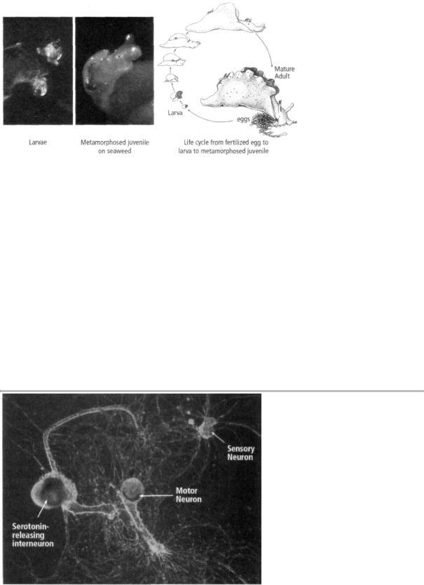
18-2 The life cycle of Aplysia. Aplysia larvae rest on a particular red seaweed (Laurencia pacifica) and extract from it the chemicals needed to trigger metamorphosis into a juvenile snail. (Drawing reprinted from Cellular Basis of Behavior, E. R. Kandel, W. H. Freeman and Company, 1976.)
nervous system. The next major task—how to grow individual nerve cells in culture and have them form synapses—was taken on by a former student of mine, Samuel Schacher, a cell biologist. With the help of two postdoctoral fellows, Schacher soon succeeded in culturing the individual sensory neurons, motor neurons, and interneurons involved in the gill-withdrawal reflex (figure 18-3).
We now had the elements of a learning circuit in tissue culture. This circuit enabled us to study a component of memory storage by focusing on a single sensory neuron and a single motor neuron. Our experiments showed that these isolated sensory and motor neurons form the same precise synaptic connections and exhibit the same physiological behavior in culture as they do in the intact animal. In nature, a shock to the tail activates modulatory interneurons that release serotonin, thereby strengthening the connections between sensory neurons and motor neurons. Since we already knew that these modulatory interneurons release serotonin, we found after a few experiments that we did not even need to culture them. We simply injected serotonin near the synapses between the sensory neuron and the motor neurons—that is, at the site in the intact animal where the modulatory interneurons terminate on the sensory neurons and
18-3 Using individual nerve cells grown in the lab to study long-term memory.
Single sensory neurons, motor neurons, and serotonin-releasing modulatory interneurons grown in culture form synapses that reproduce the simplest form of the circuit mediating and modulating the gill-withdrawal reflex. This simple learning circuit—

the first available in tissue culture—made it possible to investigate the molecular biology of long-term memory. (Courtesy of Sam Schacher.)
release serotonin. One of the great pleasures of working on a biological system over a long period is seeing today's discoveries become tomorrow's experimental tools. Our years of study of this neural circuit, our ability to isolate the key chemical signals being transmitted between and within its cells, enabled us to use these same signals to manipulate the system and probe more deeply.
We found that one brief pulse of serotonin strengthened the synaptic connection between the sensory and motor neuron for a few minutes by enhancing the release of glutamate from the sensory cell. As in the intact animal, this short-term enhancement of synaptic strength is a functional change: it does not require the synthesis of new proteins. In contrast, five separate pulses of serotonin, designed to simulate five shocks to the tail, strengthened the synaptic connection for
18-4 Changes underlying shortand long-term memory in a single sensory and motor neuron.
days and led to the growth of new synaptic connections, an anatomical change that did involve the synthesis of new protein (figure 18-4). This showed us that we could initiate new synaptic growth in the sensory neuron in tissue culture, but we still needed to find out what proteins are important for long-term memory.
My career in neurobiology now intersected with one of the great intellectual adventures of modern biology: the unraveling of the molecular machinery for regulating genes, the coded hereditary information at the heart of every life form on earth.
THIS ADVENTURE BEGAN IN 1961 WHEN FRANÇOIS JACOB AND
Jacques Monod of the Institut Pasteur in Paris published a paper entitled "Genetic Regulatory Mechanisms in the Synthesis of Protein." Using bacteria as a model system, they made the remarkable discovery that genes can be regulated—that is, they can be switched on and off like a water faucet.