

Erik_Kandel_-_V_poiskakh_pamyati_angl
.pdfThe ability to learn a drawing skill proved to be just one of a number of abilities that were intact in H.M. Moreover, this and other learning abilities described by Milner proved to be remarkably general and applied equally well to other people with damage to the hippocampus and the medial temporal lobe. Thus Milner's work revealed that we process and store information about the world in two fundamentally different ways (figure 8-6). It also revealed once again that, as with Broca and Wernicke, one learns a great deal from the careful study of clinical cases.
Larry Squire, a neuropsychologist working at the University of California, San Diego, expanded on Milner's finding. He did parallel experiments on memory storage in humans and in animals. These studies and those of Daniel Schacter, now at Harvard University, described the biology of two major classes of memory.
8-7 Despite his obvious loss of memory, H.M. could learn and retain new
skills. During his first attempt on Day 1 (left), H.M. made many errors tracing a star he could see only in a mirror. During his first attempt on Day 3 (right), H.M. retained what he had learned through practice—even though he had no recollection of the task.
What we usually think of as conscious memory we now call, following Squire and Schacter, explicit (or declarative) memory. It is the conscious recall of people, places, objects, facts, and events—the memory that H.M. lacked. Unconscious memory we now call implicit (or procedural) memory. It underlies habituation, sensitization, and classical conditioning, as well as perceptual and motor skills such as riding a bicycle or serving a tennis ball. This is the memory H.M. retained.
Implicit memory is not a single memory system but a collection of processes involving several different brain systems that lie deep within the cerebral cortex (figure 8-6). For example, the association of feelings (such as fear or happiness) with events involves a structure called the amygdala. The formation of new motor (and perhaps cognitive) habits requires the striatum, while learning new motor skills or coordinated activities depends on the cerebellum. In the simplest animals, including invertebrates, implicit memory for habituation, sensitization, and classical conditioning can be stored within the reflex pathways themselves.
Implicit memory often has an automatic quality It is recalled directly through performance, without any conscious effort or even awareness that we are drawing on memory. Although experiences change perceptual and motor abilities, those experiences are virtually inaccessible to conscious recollection. For example, once you learn to ride a bicycle, you simply do it. You do not consciously direct your body: "Now push with my left foot, now my right. . . ." If we paid that much attention to each movement, we would probably fall off the bicycle. When we speak, we do not consider where in the sentence to place the noun or the verb. We do it automatically, unconsciously. This is the type of reflexive learning studied by the behaviorists, notably Pavlov, Thorndike, and Skinner.
Many learning experiences recruit both explicit and implicit memory. Indeed, constant repetition can transform explicit memory into implicit memory. Learning to ride a bicycle initially involves conscious attention to one's body and the bicycle, but eventually riding becomes an automatic, unconscious motor activity.

Philosophers and psychologists had already anticipated the distinc-
tion between explicit and implicit memory. Hermann Helmholtz, the first person to measure the speed of conduction of the action potential, also worked on visual perception. In 1885 he pointed out that a great deal of mental processing for visual perception and for action occurs on an unconscious level. In 1890, in his classic work The Principles of Psychology, William James expanded on this idea, writing separate chapters on habit (unconscious, mechanical, reflexive action) and memory (conscious awareness of the past). In 1949 the British philosopher Gilbert Ryle distinguished between knowing how (the knowledge of skills), and knowing what (knowledge of facts and events). Indeed, a central premise of Freudian psychoanalytic theory, enunciated in 1900 in The Interpretation of Dreams, is an extension of Helmholtz's idea that experiences are recorded and recalled not only as conscious memories, but also as unconscious memories. Unconscious memories are ordinarily inaccessible to consciousness, but they nevertheless exert powerful effects on behavior.
While Freud's ideas were interesting and influential, many scientists were not convinced of their truth in the absence of experimental inquiry into how the brain actually stores information. Milner's star-tracing experiment with H.M. was the first time a scientist had uncovered the biological basis of a psychoanalytic hypothesis. By showing that a person who has no hippocampus (and therefore no ability to store conscious memories) can nonetheless remember an action, she validated Freud's theory that the majority of our actions are unconscious.
WHENEVER I RETURN TO BRENDA MILNER'S PAPERS ON H. M., I
am impressed yet again by how much these studies clarified our thinking about memory. Pierre Flourens in the nineteenth century and Karl Lashley well into the twentieth century thought of the cerebral cortex as a bowl of porridge, in which all regions were similar in how they worked. For them memory was not a discrete mental process that could be studied in isolation. But when other scientists began to track not only cognitive processes but also various memory processes to distinct regions of the brain, the theory of mass action was dismissed once and for all.
Thus by 1957, having read Milner's initial paper and having some
insight into where memory is stored in the brain, the question of how memory is stored in the brain had become the next meaningful scientific question for me. As I settled into Wade Marshall's laboratory, I began to think that this question would be an ideal challenge. Moreover, I thought that the answer would best be pursued by examining the cells involved in the storage of specific explicit memories. I would plant my flag midway between my interests in clinical psychoanalysis and the basic biology of nerve cells and proceed to investigate the territory of explicit memory "one cell at a time."
9
SEARCHING FORAN IDEAL
SYSTEM TO
STUDY MEMORY
Prior to Brenda Milner's discoveries, many behaviorists and some cognitive psychologists had followed the lead of Freud and Skinner and abandoned biology as a useful guide to the study of learning and memory They had

done so not because they were dualists, like Descartes, but because they thought that biology was unlikely to play a significant role in studies of learning in the near future. Indeed, Lashley's influential work made it seem that the biology of learning was essentially incomprehensible. In 1950, toward the end of his career, he wrote, "I sometimes feel, in reviewing the evidence on the localization of the memory trace, that the necessary conclusion is that learning is just not possible [italics added]."
Milner's work changed all that. Her discoveries that certain regions of the brain are necessary for some forms of memory provided the first evidence of where different memories are processed and stored. But the question of how memory is stored remained unanswered, and it fascinated me. Although I had only the most rudimentary preparation for researching how memory is stored in the nervous system, I was eager to give it a try—and the environment at NIH encouraged a certain boldness. All around me research on various problems in the spinal cord, first outlined by Sherrington, was being conducted at the
cellular level. Ultimately, cellular studies of memory had to answer a number of key questions: What changes occur in the brain when we learn? Do different types of learning involve different changes? What are the biochemical mechanisms of memory storage? These questions were swirling in my mind, but such questions do not translate easily into useful experiments.
I wanted to begin where Milner had left off. I wanted to tackle the most complex and interesting aspect of memory—the formation of long-term memory for people, places, and things that she found lacking in H.M. I therefore wanted to focus on the hippocampus, which Milner had shown was essential for the formation of new long-term memories. But my ideas about how to tackle the biology of memory in the hippocampus were not only vague, they were naive.
As a first step, I asked a simple question: Do nerve cells that participate in memory storage have easily recognizable distinguishing features? Were the nerve cells of the hippocampus—cells that are presumably critical for memory storage—physiologically different from motor neurons in the spinal cord, the only other well-studied neurons in the mammalian central nervous system? Possibly, I thought, the properties of hippocampal neurons would reveal something about how memory is recorded.
I was emboldened to try this technically demanding study because Karl Frank, who worked in the lab next door to me, and John Eccles in Australia were using microelectrodes to study individual motor neurons in the spinal cord of cats. Their electrodes were identical to the ones I had used to listen in on crayfish cells. Although Frank himself thought that studying the hippocampus was formidable and risky, he was not discouraging.
Marshall had only one laboratory and two postdoctoral fellows, Jack Brinley and me. Jack had obtained a medical degree from the University of Michigan and had started work on a Ph.D. in biophysics at Johns Hopkins University just before he came to NIH. His proposed thesis was on the movement of potassium ions through the membrane of neurons in the autonomic nervous system. Because Wade liked the cerebral cortex, Jack shifted his focus a bit and studied potassium movement through the cerebral cortex in response to spreading cortical
depression, a seizure process that Marshall had been interested in for several years. This was a perfectly good problem, but not one that interested me. Jack felt the same way about the hippocampus. So we worked out a compromise: we would share the lab. He would use it half of the time and I would help him, and I would use it the other half and he would help me.
This arrangement was working well when all of a sudden Marshall thrust upon us a third person, a new postdoctoral fellow, Alden Spencer, who had just graduated from the University of Oregon Medical School. The idea that now the lab would be shared by three independent projects with each of us having even less time in the lab to work on our own studies produced apprehension in both Jacks heart and mine. We each worked feverishly to persuade Alden to join our particular project.
To my delight, it took little effort to convince Alden that we should work together on the hippocampus. Part of my success, which I did not realize until later, was due to the fact that Alden never for a moment considered
working with Jack, whose project required using a radioactive form of potassium. Alden was a bit of a hypochondriac and was frightened to death of radioactivity.
MY RESEARCH TOOK AN EXTREMELY FORTUNATE TURN WITH
Alden's arrival. Born in Portland, he was a liberal in the best Oregonian tradition of independent thought based on moral rather than narrow political considerations (figure 9-1). Alden's father, a perpetual student, at once a freethinker and a religious man, was a conscientious objector during World War I and was recruited into the noncombatant corps. After the war, he went to divinity school in British Columbia and served for a while as pastor of a small church. He then went back to school at Stanford University, where he studied mathematics and statistics and later worked as a statistician for the civil service in Oregon.
Alden completely changed my narrow view of life outside the East Coast. He was strongly independent, with an original turn of mind, a great interest in music and art, and an enthusiasm for life that made him exciting to be with. He had novel insights about most things he
9-1 Alden Spencer (1931-1977), with whom I had the privilege of collaborating at the NIMH from 1958 to 1960 and who later joined me at NYU Medical School and at Columbia University. Alden made major contributions to the understanding of the hippocampus, the modification of simple reflex responses by learning, and the perception of touch. (From Eric Kandel's personal collection.)
experienced: a lecture, a concert, a tennis match. His creativity was so abundant and came to him so readily that he was always extending himself to something new, immersing himself in yet another problem. Alden had a considerable musical talent as well, having played the clarinet in the Portland Symphony Orchestra. His wife, Diane, was a fine pianist. Moreover, Alden was extremely modest and expressed these deep creative interests in an utterly unpretentious way. Denise and I soon became good friends with them, and the four of us routinely attended the Library of Congress's weekly chamber music recitals featuring the renowned Budapest String Quartet.
Among Alden's many talents were surgical skills, a good knowledge of the anatomical organization of the brain, and insights into what questions were scientifically important. Although he had no experience with intracellular recording, he had done some excellent electrophysiological research on the brain, studying how the pathways between the thalamus and the cortex contribute to the various brain rhythms displayed on EEGs (electroencephalograms). Alden was great company. We talked science incessantly and reinforced each

other's audacity. Once we decided it was important, we were not reluctant to tackle any difficult problem, such as trying to obtain recordings from individual cortical neurons in an intact brain.
Soon after we started we had our first successful experiment. I shall never forget it. I worked all morning and part of the afternoon to complete the surgery that exposed a cat's hippocampus. Late in the afternoon Alden took over and was advancing the recording electrode into the hippocampus. I was sitting in front of the oscilloscope, the instrument that displayed the electrical signals, and I also controlled the stimulators that could activate pathways into and out of the hippocampus. As I had done in Stanley Crain's laboratory, I connected the recording electrode to a loudspeaker so that any electrical signal we might obtain could be heard as well as seen. We were trying to record from pyramidal cells, the major class of neurons in the hippocampus. These cells receive and process the information coming into the hippocampus and send it on to the next relay point. We also set up a camera to photograph the display screen of the oscilloscope.
Suddenly we heard the loud bang! bang! bang! of action potentials, a sound I recognized immediately from my experiments on crayfish. Alden had penetrated a cell! We quickly realized it was a pyramidal cell because the axons of these neurons are bundled into a pathway (called the fornix) that leads out of the hippocampus, and I had positioned electrodes on that pathway. Every stimulus I applied elicited a beautiful, large action potential.
This method of stimulating the outgoing axon and causing pyramidal cells to fire proved to be a powerful way of identifying those cells. We also managed to excite pyramidal cells by stimulating the pathway that carries information into the hippocampus. We thus obtained a remarkable amount of information in the roughly ten minutes during which we recorded signals from pyramidal cells. We ran the camera continuously to ensure that every moment of the recording, every synaptic potential and every action potential in the pyramidal cells, was captured on film.
Alden and I were euphoric—we had obtained the first intracellular signals ever recorded from the region of the brain that stores our fondest memories! We almost danced around the lab. The mere accomplishment of recording from these cells successfully for several
minutes met our most optimistic expectations. In addition, our data looked fascinating and somewhat different from what Eccles and Frank had found in the motor neurons of the spinal cord.
This experiment and the ones that followed were physically exhausting, sometimes lasting twenty-four hours. It was a good thing we had both just finished a medical internship, where working twenty-four hours at a stretch was not uncommon. We carried out three experiments a week and used the intervening two days, often only partial days because Jack was using the laboratory to analyze data, to discuss the results, and just to talk. Many experiments were unsuccessful, but we eventually developed simple technical improvements that allowed us to obtain high-quality recordings once or twice a week.
By applying the powerful methodologies of cell biology to the hippocampus, Alden and I easily picked some low-hanging intellectual fruit. To begin with, we found that unlike motor neurons, a certain class of hippocampal neurons fires spontaneously, even without receiving instructions from sensory or other neurons. More interesting, we found that action potentials in the pyramidal cells of the hippocampus originate at more than one site within the cell. In the motor neuron, action potentials are initiated only at the base of the axon, where it emerges from the cell body. We had good evidence to suggest that action potentials in pyramidal cells of the hippocampus can also begin in the dendrites, and that they can be initiated in response to stimulation of the perforant pathway, an important direct synaptic input to the pyramidal cells from a part of the cortex called the entorrhinal cortex.
This proved to be an important discovery. Up to that time most neural scientists, including Dominick Purpura and Harry Grundfest, thought that dendrites could not be excited and therefore could not generate action potentials. Willifred Rall, a major theorist and model builder at NIH, had developed a mathematical model showing how the dendrites of motor neurons function. This model was based on

the fundamental assumption that the cell membrane of dendrites is passive: it does not contain voltagegated sodium channels and therefore cannot support an action potential. The intracellular signals we recorded were the first evidence to the contrary, and our finding later proved to be a general principle of neuronal function.
Our technical success and these intriguing findings brought enthusiastic encouragement and unstinting praise from our senior colleagues at NIH. John Eccles, who was emerging as the leading cellular physiologist in the mammalian brain, stopped by to see us when he visited NIH and was generous in his comments. Eccles invited Alden and me to join him in Australia to continue with him our work on the hippocampus, an offer we refused after much hesitation. Wade Marshall asked me to give a seminar at NIMH to summarize Alden's and my efforts; I did, to a packed conference room, and it was warmly received. But even in our headiest moments, we realized that ours was a typical NIH story. Young, inexperienced people were given the opportunity to try things on their own, knowing that wherever they turned, experienced people were available to help.
Not everything was wine and roses, however. Soon after I arrived, another young scientist, Felix Strumwasser, went to work at a neighboring laboratory. Unlike the rest of the young research associates, who were medical doctors, Felix had a Ph.D. in neurophysiology from the University of California, Los Angeles. While most of us knew relatively little about brain science, Felix was extremely knowledgeable. We became good friends and had dinner at each other's homes. I learned a great deal from him. In fact, in my conversations with Felix, I sharpened my thinking about how to tackle neurobiological studies of learning. Felix also got me to think about the hypothalamus, a region of the brain concerned with emotional expression and hormone secretions. The hypothalamus was becoming important in clinical discussions of how to treat stress and mental depression.
I was therefore taken aback—and hurt—when the day after I gave the seminar on our work, Felix stopped talking to me. I could not understand what had happened. Only with time did I realize that science is filled not simply with a passion for ideas but also with the ambition and strivings of people at different stages of their careers. Many years later, Felix renewed our friendship and explained that he had been chagrined when two relatively inexperienced scientists— incompetents, in his eyes—had been able to produce interesting and important experimental results.
As the afterglow of our beginner's luck began to fade, Alden and I
realized that as fascinating as our findings were, they were leading us in directions unrelated to memory. In fact, we found that the properties of hippocampal neurons were not sufficiently different from those of spinal motor neurons to account for the ability of the hippocampus to store memories. It took us a year to realize what should have been obvious from the start: the cellular mechanisms of learning and memory reside not in the special properties of the neuron itself, but in the connections it receives and makes with other cells in the neuronal circuit to which it belongs. As we learned through reading and discussions with each other to think more deeply about the biological mechanisms of learning and memory, we concluded that the role of the hippocampus in memory must arise in some other way, perhaps from the nature of the information it receives, the way its cells are interconnected, and from how that circuitry and the information it carries are affected by learning.
That change in our thinking led us to change our experimental approach. To understand how the neural circuitry of the hippocampus affects memory storage, we needed to know how sensory information reaches the hippocampus, what happens to it there, and where it goes after it leaves the hippocampus. This was a formidable challenge. Practically nothing was known then about how sensory stimuli reach the hippocampus or how the hippocampus sends information to other areas of the brain.
We therefore carried out a series of experiments to examine how various sensory stimuli—tactile, auditory, and visual—affect the firing pattern of the pyramidal neurons of the hippocampus. We saw

only occasional, sluggish responses—nothing comparable to the brisk responses reported by other investigators in the neural pathways of the somatosensory, auditory, and visual cortices. In a final attempt to understand how the hippocampus might participate in memory storage, we explored the properties of the synapses that incoming axons of the perforant pathway form with the nerve cells in the hippocampus. We stimulated those axons repetitively at the rate of 10 impulses per second and observed an increase in synaptic strength that lasted about 10 to 15 seconds. We then stimulated them at the rate of 60 to 100 impulses per second and produced an epileptic seizure. These were all interesting findings, but they were not what we were looking for!
As we became more familiar with the hippocampus, we realized that finding out how its neural networks process learned information and how learning and memory storage change those networks was an extraordinarily difficult task that would take a very long time.
I was initially drawn to the hippocampus because of my interest in psychoanalysis, which tempted me to tackle the biology of memory in its most complex and intriguing form. But it became clear to me that the reductionist strategy used by Hodgkin, Katz, and Kuffler to study the action potential and synaptic transmission also applied to research on learning. To make any reasonable progress toward understanding how memory storage occurs, it would be desirable, at least initially, to study the simplest instance of memory storage and to study it in an animal with the simplest possible nervous system, so I could trace the flow of information from sensory input to motor output. I therefore searched for an experimental animal—perhaps an invertebrate such as a worm, a fly, or a snail—in which simple but modifiable behaviors were controlled by simple neuronal circuits made up of a small number of nerve cells.
But what animal? Here Alden and I parted intellectual company. He was committed to mammalian neurophysiology and wanted to stay with the mammalian brain. He felt that although invertebrates are instructive, the organization of the invertebrate brain is so fundamentally different from that of the vertebrate brain that he did not want to work on them. Moreover, the components of the vertebrate brain were already well described. Biological solutions valid for the rest of the animal kingdom would draw his interest and his admiration, but unless they were true for the vertebrate brain, the human brain, they would not draw his effort. Alden therefore turned to one of the simple subsystems of the spinal cord of the cat and examined spinal reflexes, which are modified through learning. Over the next five years Alden made important contributions in this area of research in collaboration with the psychologist Richard Thompson. However, even the relatively simple reflex circuits in the spinal cord proved too difficult for a detailed cellular analysis of learning, and by 1965 Alden had turned from the spinal cord and the study of learning to other areas of research.
EVEN THOUGH IT MEANT SWIMMING AGAINST THE TIDE OF
current thinking, I yearned for a more radical, reductionist approach to the biology of learning and memory storage. I was convinced that the biological basis of learning should be studied first at the level of individual cells and, moreover, that the approach was most likely to succeed if it focused on the simplest behavior of a simple animal. Many years later, Sydney Brenner, a pioneer of molecular genetics who introduced the worm
Caenorhabditis elegans to biology, was to write:
What you need to do is find which is the best system to experimentally solve the problem, and as long as it [the problem] is general enough you will find the solution there.
The choice of an experimental object remains one of the most important things to do in biology and is, I think, one of the great ways to do innovative work. . . . The diversity in the living world is so large, and since everything is connected in some way, let's find the best one.
In the 1950s and 1960s, however, most biologists shared Alden's reluctance to apply a strictly reductionist strategy to the study of behavior because they thought it would have no relevance for human behavior. People have mental abilities not found in simpler animals, and these biologists believed that the functional organization

of the human brain must be quite different from that of simpler animals. Although this view holds some truth, I thought it overlooked the fact—amply demonstrated in the fieldwork of ethologists like Konrad Lorenz, Niko Tinbergen, and Karl von Frisch—that certain elementary forms of learning are common to all animals. It seemed likely to me that, in the course of evolution, humans had retained some of the cellular mechanisms of learning and memory storage found in simpler animals.
Not surprisingly, I was discouraged from pursuing this research strategy by a number of senior scientists in neurobiology, including Eccles. His concern reflected in part the hierarchy of acceptable research questions in neurobiology at that time. Although some scientists were studying behavior in invertebrates, that work was not con-
sidered important—indeed, it was largely ignored—by most people working on the mammalian brain. Of even greater concern to me was the skepticism of knowledgeable psychologists and psychoanalysts that anything interesting about higher-order mental processes Uke learning and memory could be found by focusing on individual nerve cells—particularly the cells of an invertebrate. I had made up my mind, however. The only remaining question was which invertebrate would best suit the cellular study of learning and memory.
Besides being a good place to do research, NIH was also a good place to learn about new developments in biology. During the course of any given year, most good scientists working on the brain visit the NIH campus. As a result, I was able to speak with many people and to attend seminars in which I learned about the experimental advantages of various invertebrate animals, such as crayfish, lobsters, honeybees, flies, land snails, and the nematode worm Ascaris.
I remembered vividly Kuffler's description of the advantages of the crayfish's sensory neuron for studying the properties of dendrites. But I ruled out crayfish: although they have a few very large axons, their nerve cell bodies are not very large. I wanted an animal with a simple reflex that could be modified by learning and that was controlled by a small number of large nerve cells whose pathway from input to output could be identified. In that way, I could relate changes in the reflex to changes in the cells.
After about six months of careful consideration, I settled on the giant marine snail Aplysia as a suitable animal for my studies. I had been greatly impressed with two lectures I had heard about the snail, one given by Angelique Arvanitaki-Chalazonitis, a senior, highly accomplished scientist who had discovered the usefulness of Aplysia for studying the signaling characteristics of nerve cells, and the other by Ladislav Tauc, a younger person who brought a new biophysical perspective to the study of how nerve cells function.
Aplysia was first mentioned by Pliny the Elder in his encyclopedic study Historia Naturalis, written during the first century a.D. It was mentioned again in the second century by Galen. These ancient scholars called it lepus marinus, or sea hare, because, when sitting still and contracted, it resembles a rabbit. When I began to examine
Aplysia
myself, I found, as others had before me, that it releases copious amounts of purple ink when disturbed. That ink was once erroneously thought to be the royal purple used to dye the stripe on the togas of the Roman emperors. (The royal purple dye is actually secreted by the clam Murex.) Because of Aplysia's tendency to ink so profusely, some ancient naturalists also thought it was holy.
The American species of Aplysia that lives off the California coast (A. californica), and which I have spent most of my career studying, measures more than one foot in length and weighs several pounds (figure 9-2). It assumes the reddish brown coloration of the seaweed it feeds on. It is a large, proud, attractive, and obviously highly intelligent beast—-just the sort of animal one would select for studies of learning!
What drew my attention to Aplysia was not its natural history or physical beauty but several other features outlined by Arvanitaki-Chalazonitis and by Taue in their lectures on the European species (A. depilans). They both emphasized that the brain of Aplysia has a small number of cells, about 20,000, compared with about 100 billion in the
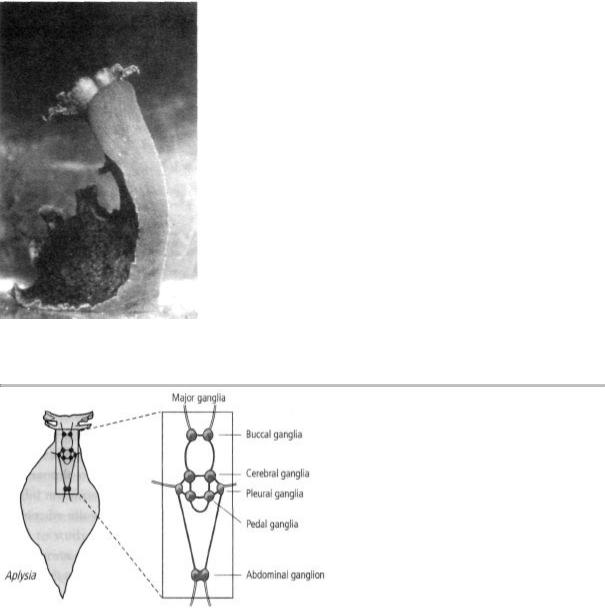
9-2 Aplysia californica: the giant marine snail. (Courtesy of Thomas Teyke.)
9-3 The brain of Aplysia is very simple. It has 20,000 neurons grouped into nine separate clusters, or ganglia. Since each ganglion has a small number of cells, researchers can isolate simple behaviors that are controlled by it. They can then study changes in particular cells as a behavior is altered by learning.
mammalian brain. Most of these cells are grouped into nine clusters, or ganglia (figure 9-3). Since individual ganglia were thought to control several simple reflex responses, it seemed to me that the number of cells committed to a single simple behavior was likely to be small. In addition, some of Aplysia's cells are the largest in the animal kingdom, making it relatively easy to insert microelectrodes into them to record electrical activity. The pyramidal cells of the cat hippocampus, whose activity Alden and I had recorded, are among the largest
nerve cells in the mammalian brain, yet they are only 20 micrometers in diameter (1/1250 of an inch) and can be seen only under a high-powered microscope. Some cells in Aplysia's nervous system are fifty times that size and
can be seen with the naked eye.
Arvanitaki-Chalazonitis had found that a few nerve cells in Aplysia are uniquely identifiable—that is, the same cells can easily be recognized by sight under the microscope in every single snail. In time I realized that the same thing is true of most other cells in its nervous system, heightening the prospect of mapping the entire neural circuitry controlling a behavior. As it turned out, the circuitry controlling Aplysia s most elementary reflexes was quite simple. Moreover, I later

found that stimulating a single neuron often produced a large synaptic potential in its target cells, a clear sign and measure of the strength of the synaptic connection between the two cells. These large synaptic potentials made it possible to map neural connections cell by cell and eventually enabled me to work out for the first time the precise wiring diagram of a behavior.
Many years later, Chip Quinn, one of the first scientists to conduct genetic studies of learning in the fruit fly noted that the ideal experimental animal for biological studies of learning must have "no more than three genes, be able to play the cello or at least recite classical Greek, and learn these tasks with a nervous system containing only ten large, differently colored and therefore easily recognizable neurons." I have often thought that Aplysia meets these criteria to a surprising degree.
At the time I decided to work on Aplysia, I had never dissected the snail or recorded the electrical activity of its neurons. Moreover, no one in the United States was working on Aplysia. The only two people in the world who were studying it in 1959 were Tauc and Arvanitaki-Chalazonitis. Both of them were in France, Tauc in Paris and Arvanitaki-Chalazonitis in Marseilles. Denise, ever the Parisian chauvinist, thought Paris the better choice. Living in Marseilles, she said, would be like living in Albany instead of New York City. So the decision was for Tauc. Before leaving NIH in May 1960, I visited Tauc and we arranged that I would join him in September 1962, as soon as I had completed my residency in psychiatry at Harvard Medical School.
AS I LEFT NIH IN JUNE 1960, I FELT A DEEP SADNESS, SOMEWHAT
similar to that which I experienced when I graduated from Erasmus Hall High School. I had come as a novice, and I left as a limited but nonetheless working scientist. At NIH I had walked the walk. I found that I liked it and that I was quite successful at what I tried. But I was genuinely surprised by my success. For a long time I felt that it was due to pure chance, my good fortune, my enjoyable and productive collaboration with Alden, the generous psychological support of Wade Marshall, and the youth-oriented scientific culture of NIH. I had had a number of ideas that proved to be useful, but I thought they
were beginner's luck. I was very much afraid that I would run out of ideas and would not last in science.
This insecurity about my ability to come up with new ideas was not helped by the fact that John Eccles and several other senior scientists whom I respected thought I was making a grave error in switching from a highly promising start in the mammalian hippocampus to a new beginning with an invertebrate whose behavior had not been well studied. But three factors drove me onward. First, there was the Kuffler-Grundfest principle of biological research: for every biological problem there is a suitable organism in which to study it. Second, I was now a cell biologist. I wanted to think about how cells functioned during learning, and I wanted to spend my time reading, thinking, and discussing ideas with others. I did not want to spend hours simply setting up an experiment time and again as Alden and I had done on the hippocampus, only to find an occasional good cell to study. I liked the idea of big cells and, despite the risks involved, I was convinced that Aplysia was the right system and that I had the tools to study behavior in this snail effectively.
Finally, I had learned something in marrying Denise. I had been reluctant and fearful of marriage, even to Denise, whom I loved much more than any other woman I had ever thought of marrying. But Denise was confident that our marriage would work, so I took a leap of faith and went ahead. I learned from that experience that there are many situations in which one cannot decide on the basis of cold facts alone—because facts are often insufficient. One ultimately has to trust one's unconscious, one's instincts, one's creative urge. I did this again in choosing Aplysia.