

Erik_Kandel_-_V_poiskakh_pamyati_angl
.pdfTogether, the work of Hodgkin, Huxley, and Katz showed that there are two fundamentally different types of ion channels. Voltage-gated channels generate action potentials that carry information within neurons, while chemical transmitter-gated channels transmit information between neurons (or between neurons and muscle cells) by generating synaptic potentials in postsynaptic cells. Thus Katz discovered that by producing the synaptic potential, transmitter-gated ion channels in effect translate chemical signals from motor neurons into electrical signals in muscle cells.
Just as there are diseases of voltage-gated ion channels, so there are diseases of transmitter-gated channels. For instance, myasthenia gravis, a serious autoimmune disease that occurs primarily in men, produces antibodies that destroy the acetylcholine receptors in muscle cells and thus weaken muscle action. Muscular weakness can become so severe that patients cannot keep their eyes open.
SYNAPTIC TRANSMISSION IN THE SPINAL CORD AND BRAIN IS
decidedly more complex than signaling between motor neurons and muscle. Eccles had spent the years 1925 to 1935 working directly with Sherrington on the spinal cord. He returned to those studies full-time in 1945, and by 1951 had obtained intracellular recordings from motor neurons. Eccles confirmed Sherrington's finding that motor neurons receive both excitatory and inhibitory signals and that these signals are produced by distinctive neurotransmitters acting on distinctive receptors. In the motor neuron, excitatory neurotransmitters released by the presynaptic neurons lower the resting membrane potential of the postsynaptic cell from -70 millivolts to -55 millivolts, the threshold for firing an action potential, while inhibitory neurotransmitters increase the membrane potential from -70 millivolts to -75 millivolts, making it much more difficult for the cell to fire an action potential.
6-2 The propagated action potential.
We now know that the major excitatory neurotransmitter in the brain is the amino acid glutamate, while the main inhibitory transmitter is the amino acid GABA (gamma-aminobutyric acid). A variety of tranquilizing drugs—benzodiazepines, barbiturates, alcohol, and general anesthetics—bind to GABA receptors and produce a calming effect on behavior by enhancing the receptors' inhibitory function.
Eccles thus confirmed Katz's finding that excitatory synaptic transmission is chemically mediated, and he showed that inhibitory synaptic transmission is also chemically mediated. When he described these findings in later years, Eccles wrote, "I had been encouraged by Karl Popper to make my hypothesis as precise as possible, so that it would call for experimental attack and falsification. It turned out that it
was I who was to succeed in this falsification." Eccles celebrated his discoveries by abandoning the electrical hypothesis he had so vigorously championed and converting wholeheartedly to the chemical hypothesis, arguing with equal enthusiasm and vigor for its universality.
It was at this point, in October 1954, that Paul Fatt, one of Katz's outstanding collaborators, wrote a masterly review of synaptic trans-
mission. Fatt took a farsighted view, pointing out that it was premature to conclude that all synaptic transmission is chemical. He concluded, "Although there is every indication that chemical transmission occurs across those junctions ... which are most familiar to the physiologist, it is probable that electrical transmission occurs at certain other junctions [emphasis added]."
Three years later, Fatt's prediction was convincingly demonstrated by Edwin Furshpan and David Potter, two postdoctoral fellows in Katz's laboratory who found an actual case of electrical transmission between two cells of the nervous system in the crayfish. Thus, as sometimes happens in scientific controversies, both sides of the argument had merit. We now know that most synapses, including those under scrutiny at the time of the controversy, are chemical in nature. But some neurons form electrical synapses with other nerve cells. At such synapses there are small bridges between the two cells that allow electrical current to pass from one cell to the other, very much as Golgi had predicted.
The existence of two forms of synaptic transmission raised questions in my mind that would reemerge in my thinking later. Why do chemical synapses predominate in the brain? Do chemical and electrical transmission have different roles in behavior?
IN THE FINAL PHASE OF AN EXTRAORDINARY CAREER, KATZ
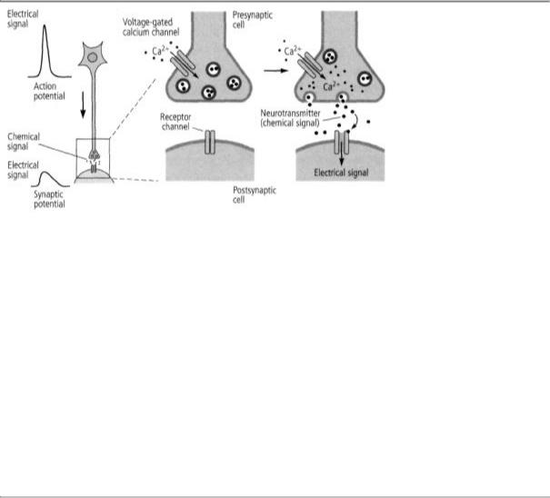
turned his attention from the synaptic potential in the target cell to the release of neurotransmitters from the signaling cell. He wanted to know how an electrical event in the presynaptic terminal, the action potential, leads to the release of a chemical transmitter. Here, he made two more remarkable discoveries. First, as an action potential propagates along the axon into the presynaptic terminal, it leads to the opening of voltage-gated channels that admit calcium ions. The influx of calcium ions into the presynaptic terminals sets off a series of molecular steps that lead to the release of the neurotransmitter. Thus, in the signaling cell, voltage-gated calcium channels opened by the action potential start the process of translating an electrical signal into a chemical signal, just as in the receiving cell, transmitter-gated channels translate chemical signals back into electrical signals.
Second, Katz discovered that transmitters such as acetylcholine are
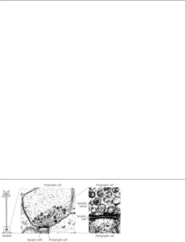
6-3 How signals travel from cell to cell. The first images of a synapse showed that the presynaptic terminal contains synaptic vesicles, which were later found to enclose about 5,000 molecules of neurotransmitter. These vesicles cluster near the membrane of the presynaptic terminal, where they prepare to release the transmitter into the space between the two cells, the synaptic cleft. After crossing the synaptic cleft, the neurotransmitters bind to receptors on dendrites of the postsynaptic cell. (Reprinted from Cell, vol. 10, 1993, page 2, Jessell and Kandel. Used with permission from Elsevier. Center image courtesy of C. Bailey and M. Chen.)
not released from the axon terminal as single molecules but in small, discrete packets containing about five thousand molecules each. Katz called these packets quanta and postulated that each one is packaged in a membrane-bound sac that he called the synaptic vesicle. In 1955, images of the synapse taken by Sanford Palay and George Palade with an electron microscope confirmed Katz's prediction, showing that the presynaptic terminal is packed with vesicles that were later shown to contain neurotransmitters (figure 6-3).
To test this idea further, Katz made a brilliant strategic decision. He shifted from studying the nerve-muscle synapse of the frog to the giant synapse of the squid. Using this advantageous system, Katz was able to infer what calcium ions do when they flow into the presynaptic terminal: they cause the synaptic vesicles to fuse with the surface membrane of the presynaptic terminal and open a pore in the membrane through which the vesicles release their transmitter into the synaptic cleft (figure 6-4).
THE REALIZATION THAT THE WORKINGS OF THE BRAIN —THE
ability not only to perceive, but to think, learn, and store informa-
6-4 From electrical to chemical signals and back again. Bernard Katz discovered that when an action potential enters a presynaptic terminal, it causes calcium channels to open, letting calcium ions flow into the cell. This leads to the release of neurotransmitters into the synaptic cleft. The neurotransmitter binds to receptors on the surface of the postsynaptic cell, and the chemical signals are reconverted into electrical signals.
tion—may occur through chemical as well as electrical signals expanded the appeal of brain science from anatomists and electro-physiologists to biochemists. In addition, since biochemistry is a universal language of biology, synaptic transmission piqued the interest of the biological science community as a whole, not to mention students of behavior and mind, like me.
How fortunate for brain science throughout the world that England, Australia, New Zealand, and the United States opened their doors to the remarkable scholars of the synapse cast out by Austria and Germany, including Loewi, Feldberg, Kuffler, and Katz. I am reminded of a story told about Sigmund Freud when he arrived in England and was shown the beautiful house on the outskirts of London that he was to live in. On seeing the tranquility and civility that his forced emigration had brought him to, he was moved to whisper with typical Viennese irony, "Heil Hitler!"
7
SIMPLE AND COMPLEX NEURONAL SYSTEMS
Soon after I arrived at Columbia in 1955, Grundrest suggested that I work alongside Dominick Purpura, a young physician whom he had encouraged to change careers from neurosurgery to basic research on the brain (figure 7-1). When I met Dom, he had just made the decision to focus his research on the cerebral cortex, the most highly developed region of the brain. Dom was interested in mind-altering drugs, and the first experiments I helped him with concerned the role of the psychedelic agent LSD (lysergic acid diethylamide) in producing visual hallucinations.
LSD was discovered in the 1940s. By the mid-1950s it had become extremely well-known because of its widespread recreational use. Aldous Huxley had publicized its mind-altering properties in his book The Doors of Perception, in which he described how LSD enhanced his own awareness of visual experiences, giving rise to powerful, brightly colored images and a greater sense of clarity. The ability of LSD and related psychedelic drugs to alter perception, thought, and feeling in ways that are not ordinarily experienced except in dreams and exalted religious states makes them markedly different from other classes of drugs. People taking LSD often have the sense that their mind has expanded and split in two: one part is organized, experiencing the
7-1 Dominick Purpura (b. 1927) trained as a neurosurgeon but switched to full-time research and became a major contributor to the physiology of the cortex. I worked with him in 1955-56 during my first stay in the Grundfest laboratory. He later became an academic leader at Stanford University and then at the Albert Einstein School of Medicine. (From Eric Kandel's personal collection.)
enhanced perceptual effects; the other part is passive, observing the events as a neutral outsider. Attention is typically turned inward, and the clear distinction between self and non-self is lost, giving the user of LSD a mystical sense of being part of the cosmos. In many people the perceptual distortions take the form of visual hallucinations; in some people LSD can even cause a psychotic reaction resembling schizophrenia. Because of these remarkable properties, Dom wanted to know how LSD worked.
A year earlier D. W Woolley and E. N. Shaw, two pharmacologists at the Rockefeller Institute, had found that LSD binds to the same receptor as serotonin, a substance that had recently been discovered in the brain and was thought to be a neurotransmitter. For their studies they used a preparation favored by experimental pharmacologists, the smooth muscle of the rat uterus, which they found would undergo spontaneous contractions in response to serotonin. LSD counteracted this effect of serotonin, and it did so by displacing serotonin from its

receptor. This led Woolley and Shaw to suggest that LSD might counteract serotonin in the brain. They further suggested that since LSD can cause psychotic reactions, it might do so by preventing the normal action of serotonin in the brain. If that were so,
they argued, serotonin might well be required for our sanity—for normal mental functioning.
Although Dom had no problem with the idea of using smooth uterine muscle to test ideas about chemicals in the brain, he thought a more relevant test about brain functioning in mental health and illness would be to look at the brain directly to see how psychedelic drugs act. Specifically, he wanted to know whether LSD affects synaptic activity in the visual cortex, the area of the cortex concerned with visual perception, where presumably the dramatic visual distortions and hallucinations occur. He asked me to help him explore the action of serotonin on a neural pathway in cats that ends in the visual cortex.
We anesthetized the animals, opened their skulls to expose the brain, and placed electrodes on the surface of the visual cortex. We found that in the visual cortex, serotonin and LSD did not act in opposition to each other, as they did in the smooth muscle of the uterus. Not only did both have the same action, inhibiting synaptic signaling, but each enhanced the other's inhibitory activity. Thus our studies, and subsequent studies from other laboratories, seemed to disprove Woolley and Shaw's notion that the disorienting visual effects of LSD were due to the drug's blocking the action of serotonin in the visual system. (We now know that serotonin acts on as many as eighteen different types of receptors throughout the brain and that LSD seems to produce its hallucinatory action by stimulating one of these receptors, located in the frontal lobe of the brain.)
This was quite a nice result. In the course of these studies I learned from Dom how to set up experiments with cats and how to operate electrical recording and stimulating equipment. To my surprise, I found my first laboratory experiences to be absorbing, quite unlike the rather dry science I had been taught in college and medical school classrooms. In the laboratory, science is a means for formulating interesting questions about nature, discussing whether those questions are important and well formulated, and then designing a series of experiments to explore possible answers to a particular question.
The questions Grundfest and Purpura were asking were not immediately related to the ego, superego, or id, but they made me realize that neural science was beginning to be able to test ideas about aspects
of major mental illnesses, such as the perceptual distortions and hallucinations of schizophrenia.
More important, I found discussions with Grundfest and Purpura fascinating—they were penetrating and sometimes marvelously gossipy about other scientists' work, their careers, their sex lives. Dom was extremely bright, technically strong, and highly entertaining (I later called him the Woody Allen of neurobiology). I began to realize that what makes science so distinctive, particularly in an American laboratory, is not just the experiments themselves, but also the social context, the sense of equality between student and teacher, and the open, ongoing, and brutally frank exchange of ideas and criticism. Grundfest and Purpura admired each other and were involved together in the design of the experiment, but Grundfest would criticize Dom's data as if he were a rival from another laboratory. Grundfest was at least as demanding about the experiments from his and Dom's laboratory as he was about other people's experiments.
In addition to learning about the important new ideas emerging from biological studies of the brain, I learned methodology and strategy from Grundfest and Purpura, and later from Stanley Crain, a young colleague of Grundfest's. In a larger sense, much as the painful memories of my youth in Vienna in 1938 were to obsess me in later years, these early positive research experiences and the ideas to which I was exposed when I was twenty-five years old had a major impact on my thinking and my life's work.

The findings regarding serotonin and LSD encouraged Dom to carry his analysis to the edge of what was technically possible in the mammalian cortex. We had used flashes of light to activate the visual cortex. Those stimuli activated a pathway that ended on the dendrites of neurons in the visual cortex. Very little was known about dendrites. In particular, it was not known whether they could generate action potentials like those in the axon. Based on their studies, Purpura and Grundfest proposed that dendrites have limited electrical properties: they can produce synaptic potentials, but they cannot generate action potentials.
In reaching this conclusion Grundfest and Purpura were tentative, however, because they were uncertain that the experimental methods they were using were adequate to the task of studying dendrites. To
detect changes in synaptic transmission produced by LSD, Grundfest and Purpura ideally needed to obtain intracellular recordings from the dendrites of the neurons in the visual cortex one dendrite at a time. This required using small glass electrodes of the sort used by Katz in single muscle fibers and by Eccles in single motor neurons. After some discussion they concluded that intracellular recordings were unlikely to succeed, because the neurons in the visual cortex were much smaller than the cells studied by Katz and Eccles. The slender dendrites, which are only one-twentieth the size of the cell body, seemed impossibly difficult recording targets.
IT WAS IN THE CONTEXT OF THESE DISCUSSIONS THAT I ONCE
again encountered Stephen Kuffler. One evening Grundfest threw into my lap an issue of the Journal of General Physiology that contained three papers by Kuffler based on his work with single nerve cells and their dendrites in the crayfish. I found the idea of a contemporary neuro-physiologist working on crayfish simply remarkable: one of Freud's first scientific papers, published in 1882, when he was only twenty-six, was on the nerve cells of the crayfish! It was in the course of this study that Freud almost discovered, independently of Cajal, that the nerve cell body and all of its processes are a single unit, the signaling unit of the brain.
I read Kuffler's papers as best I could. Even though I did not understand them fully, one thing popped out immediately: Kuffler was doing what Purpura and Grundfest aspired to do but could not achieve in the mammalian brain. He was studying the dendrites of a single, isolated nerve cell. Here, without any other nerve cells present, Kuffler could actually see the individual branches of the dendrites and could record the conseqences of electrical changes in them.
Kuffler's papers drove home the point that selecting an anatomically simple system is crucial to the success of an experiment and that invertebrate animals are a rich source of simple systems. The papers also reminded me that the choice of an experimental system is one of the most important decisions a biologist makes, a lesson I had learned earlier from Hodgkin and Huxley's work on the squid's giant axon and Katz's work on the squid's giant synapse.
These insights had a great impact on me, and I was eager to test the new research strategies directly for myself. I had no specific idea in mind, but I was beginning to think like a biologist. I appreciated that all animals have some form of mental life that reflects the architecture of their nervous system, and I knew I wanted to study nervous system function at the cellular level. All I knew at this point is that someday I might want to test an idea with an invertebrate animal.
AFTER GRADUATING FROM MEDICAL SCHOOL IN 1956, I SPENT A
year as a medical intern at Montefiore Hospital in New York City. In the spring of 1957, during a brief elective period in my internship, I returned to Grundfest 's lab and spent six weeks with Stanley Crain, a master of simple systems. I sought out Crain because he was a cell biologist who had searched for

appropriate experimental systems to solve important problems. He was one of the first to study the properties of single, isolated nerve cells removed from the brain and grown in tissue culture apart from all other cells. It doesn't get much simpler than that!
Knowing of my growing curiosity about invertebrate animals, particularly about the crayfish, Grundfest suggested that I set up an electrophysical recording system with Crain's help. I could use the system to replicate Hodgkin and Huxley's experiment by recording from the large axon of the crayfish, which controls the animal's tail and thus its escape from predators. This crayfish axon is smaller than that of the squid but nonetheless very large.
Crain showed me how to manufacture glass microelectrodes for insertion into individual axons and how to obtain and interpret electrical recordings from them. It was in the course of those experiments— which were almost laboratory exercises, since I was not exploring new ground scientifically or conceptually—that I first began to feel the excitement of working on my own. I connected the output from the amplifier I was using to record the electrical signal to a loudspeaker, as Adrian had done thirty years earlier. Whenever I penetrated a cell, I, too, could hear the crack of an action potential. I am not fond of the sound of gunshots, but I found the bang! bang! bang! of action potentials intoxicating. The idea that I had successfully impaled an axon and was actually listening in on the brain of the crayfish as it conveyed mes-
sages seemed marvelously intimate. I was becoming a true psychoanalyst: I was listening to the deep, hidden thoughts of my crayfish!
The beautifully straightforward results I obtained from early experiments on the simple nervous system of the crayfish—measurements of the resting membrane and action potentials, confirmation that the action potential is all-or-none and that it does not simply nullify the resting membrane potential but overshoots it—made a profound impression on me and confirmed the importance of selecting just the right animal for my studies. My results were completely unoriginal, but to me they were wonderful.
Based on my two brief periods in his laboratory, Grundfest offered to nominate me for a research position at the National Institute of Mental Health (NIMH), the psychiatric component of NIH, as an alternative to being drafted into the armed forces. During the years following the Korean War, physicians were drafted to provide medical care for members of the armed services and their families. The Public Health Service, then part of the Coast Guard, was an alternative form of active duty for those who were deemed eligible, and NIH was one of the installations that belonged to the Public Health Service. With Grundfest's recommendation, I was accepted by Wade Marshall, chief of the Laboratory of Neurophysiology at NIMH, and was slated to arrive in July 1957.
IN THE LATE 1930s WADE MARSHALL WAS PROBABLY THE MOST
promising and accomplished young scientist working on the brain in the United States (figure 7-2). In a now classic series of studies he asked: How are the touch receptors of the body surface—the hands, face, chest, back—represented in the brain of cats and monkeys? Marshall and his colleagues discovered that the internal representation of touch is organized spatially: neighboring areas on the body surface are preserved in the brain.
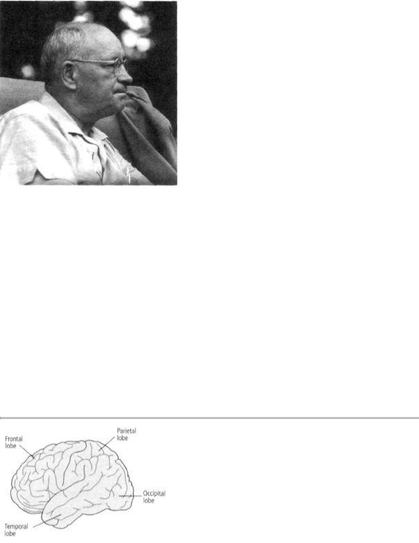
By the time Marshall began his research, a great deal was already known about the anatomy of the cerebral cortex. The cortex is a convoluted structure that covers the two symmetrical hemispheres of the forebrain and is divided into four parts, or lobes (frontal, parietal, temporal, and occipital) (figure 7-3). Unfolded, the human cerebral cortex is about the size of a large cloth dinner napkin, only somewhat thicker. It
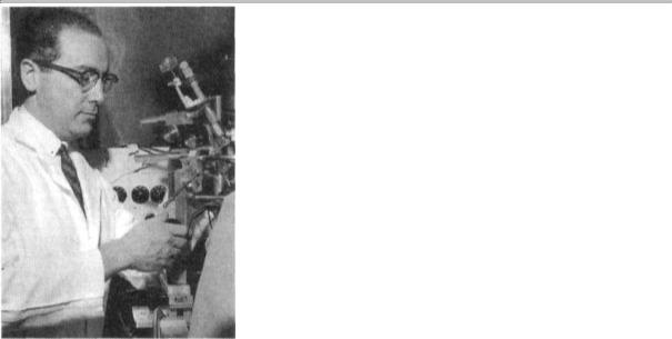
7-2 Wade Marshall (1907-1972) was the first scientist to map the detailed sensory representation of touch and vision in the cerebral cortex. He moved to the NIH in 1947 and became head of the Laboratory of Neurophysiology at NIMH in 1950, where I worked for him from 1957 to 1960. (Courtesy of Louise Marshall.)
contains about 100 billion neurons, each with about a thousand synapses, making a total of about 1 quadrillion synaptic connections.
Marshall began his studies of touch sensation as a graduate student at the University of Chicago in 1936. He discovered that moving the hairs on a cat's leg or touching its skin produces an electrical response in specific groups of neurons in the somatosensory cortex, a region of the parietal lobe that governs the sense of touch. These studies showed only that the sense of touch is represented in the brain, but Marshall immediately realized that he could advance his analysis much further. He wanted to know whether neighboring areas of the skin are represented in neighboring areas of the somatosensory cortex or scattered at random across it.
For guidance in answering that question, Marshall sought postdoctoral training under Philip Bard, chairman of the department of physi-
7-3 The four lobes of the cerebral cortex. The frontal lobe is part of the neural circuit governing social judgments, planning and organization of activities, aspects of language, control of movement, and a form of short-term memory called working memory. The parietal lobe receives sensory information about touch, pressure, and space around the body and helps integrate that information into coherent perceptions. The occipital lobe is involved with vision. The temporal lobe is involved with auditory processing and aspects of language and memory.
ology at the Johns Hopkins Medical School and a major figure in American biology. Marshall joined Bard in carrying out studies on monkeys in which they discovered that the entire body surface is
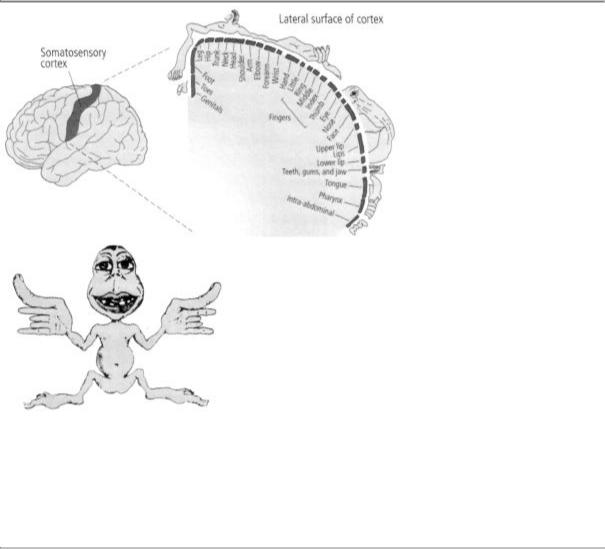
represented in the somatosensory cortex in the form of a point-for-point neural map. Parts of the body surface that are adjacent to one another, such as the fingers, are represented near each other in the somatosensory cortex. A few years later a remarkably gifted Canadian neurosurgeon, Wilder Penfield, extended the study from monkeys to people, revealing that the parts of the body surface most sensitive to touch are represented by the largest areas of the somatosensory cortex (figure 7-4).
Next, Marshall found that the light receptors in the retina of the eye are also represented in an orderly way in the primary visual cortex, a region of the occipital lobe. Finally, Marshall showed that the temporal lobe has a sensory map for sound frequencies, with different pitches represented systematically in the brain.
These studies revolutionized our understanding of how sensory
7-4 A sensory map of the body, as represented in the brain. The somatosensory cortex—a strip in the parietal lobe of the cerebral cortex—receives sensations of touch. Each part of the body is represented separately. Fingers, mouth, and other particularly sensitive areas take up most space. Wilder Penfield called this cross-sectional map a sensory homunculus. A composite representation of the sensory homunculus as a figurine drawing (below) is based on the cross-sectional map. It shows a human with large hands, fingers, and mouth. (From Mechanics of the Mind, Colin Blakemore. © Cambridge University Press, 1977.)
information is organized and represented in the brain. Marshall showed that, even though the different sensory systems carry different types of information and end up in different regions of the cerebral cortex, they share a common logic in their organization: all sensory information is organized topographically in the brain in the form of precise maps of the body's sensory receptors, such as, the retina of the eye, the basilar membrane in the ear, or the skin on the body surface.

These sensory maps are most easily understood by the representation of touch in the somatosensory cortex. Touch begins with receptors in the skin that translate the energy of a stimulus—for example, the energy transmitted by a pinch—into electrical signals in sensory neurons. The signals then travel along precise pathways to the brain, passing through several processing, or relay, stages in the brain stem and thalamus before terminating in the somatosensory cortex. At each stage the signals traveling from adjacent points on the skin are carried by nerve fibers that run alongside each other. In this way stimulation of two adjacent fingers, for instance, activates adjacent populations of nerve cells in the brain.
Knowledge of the brain's sensory maps and an understanding of how they are organized topographically is exceedingly helpful in treating patients. Because these maps are incredibly precise, clinical neurology has long been an accurate diagnostic discipline, even though it has relied, until the recent development of brain imaging, only on the simplest, most primitive tools—a wad of cotton to test for touch, a safety pin to test for pain, a tuning fork to test for vibration, and a hammer to test reflex actions. Disturbances in the sensory and motor systems can be located with remarkable accuracy because of the one-to-one relationship between sites on the body and areas of the brain.
A dramatic example of this relationship is the Jacksonian sensory march, which characterizes a certain type of epileptic seizure first described by British neurologist John Hughlings Jackson in 1878. In such a seizure, numbness and sensations such as burning or prickling begin in one place and spread throughout the body. For example, numbness might begin at the fingertips and spread over the next minute to the hand, up the arm, across the shoulder, into the back,
and down the leg on the same side. This sequence of sensations is explained by the arrangement of the sensory map of the body: the seizure, which is a wave of abnormal electrical activity in the brain, starts in the lateral area of the somatosensory cortex, where the hand is represented, and propagates across the cortex toward the midline, where the leg is represented.
Marshall's marvelous scientific achievements came at a price, however. The experiments were physically demanding, often lasting more than twenty-four hours at a time. Frequently deprived of sleep, he became exhausted. In addition, there was tension with Bard. In 1942 Marshall collapsed with an acute psychotic paranoid episode after having actually threatened Bard physically. This episode of mental illness required that Marshall be hospitalized for eighteen months.
When Marshall returned to neuroscience in the late 1940s, he moved to a completely new set of problems: spreading cortical depression, an experimentally induced, reversible silencing of electrical activity in the cerebral cortex. By the time I arrived at NIH, he had passed the peak of his brilliant career. He still enjoyed doing occasional experiments, but he had lost his scientific drive and the clarity of his vision, and he focused much of his energy and interest on administrative matters, which he did well.
Although eccentric, moody, and somewhat suspicious in unpredictable ways, Marshall was a generous lab chief who was extremely supportive of the young people for whom he was responsible. I learned a great deal from him about the modesty and rigor that belong in a scientific laboratory. He had high standards of scientific conduct and a fine sense of humor about himself, which was reflected in the wonderful aphorisms he would call up at appropriate moments. One of his favorites, used whenever one of his findings was challenged, was, "We were confused and they were confused, but we were more accustomed to being confused." On other occasions he would mutter, "Things will go along like this for a while and then they'll get worse!"
In addition to humility I learned from Marshall that with great personal strength and the passage of time, one might substantially recover from a severe mental illness (therapeutic drugs were not yet