

Учебник по анатомии (для англ.яз)
.pdfThorax
Visceral afferents in the vagus nerves relay information to the central nervous system about normal physiological
processes and reflex activities. They do not transmit pain sensation.
Right vagus nerve
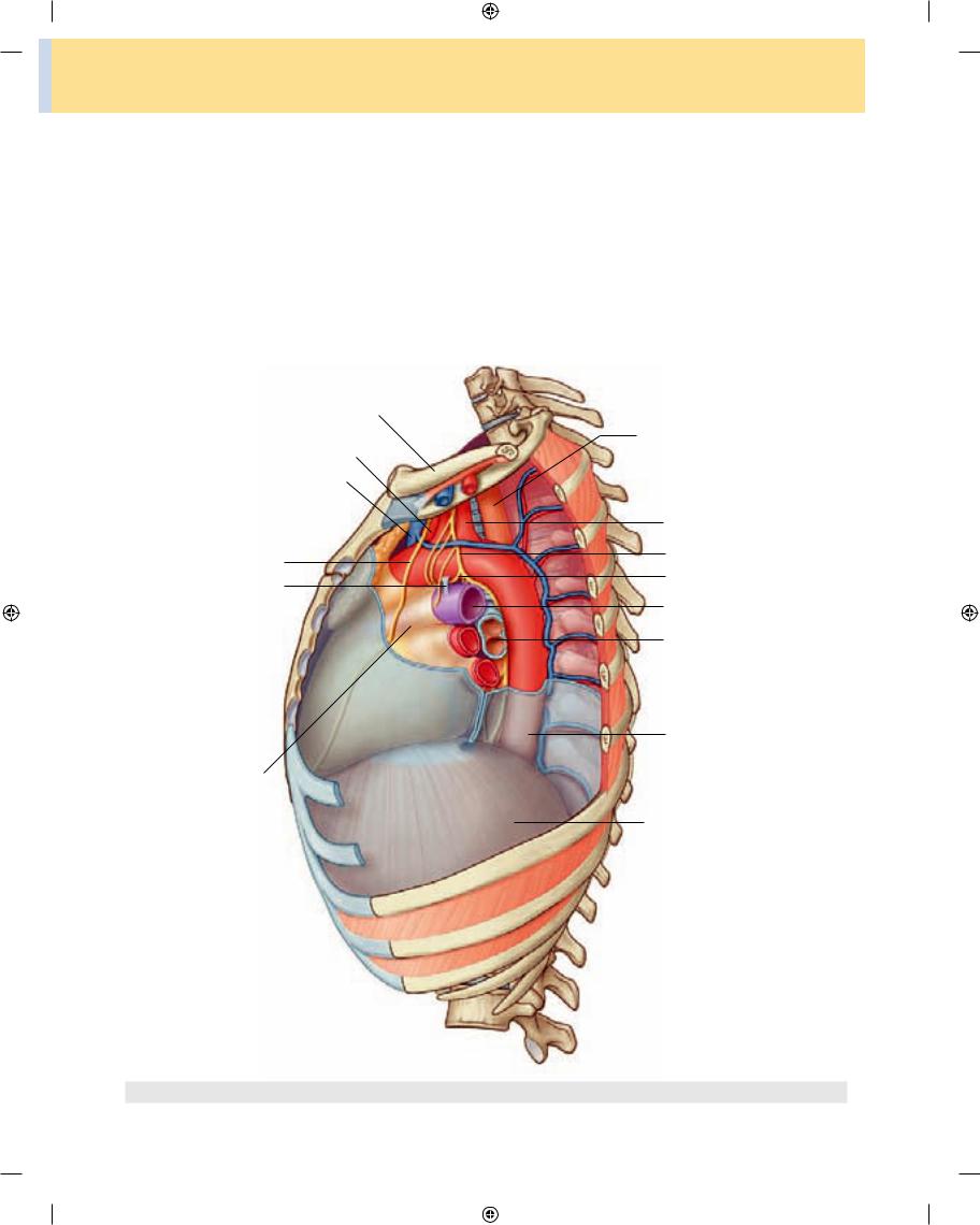
The right vagus nerve enters the superior mediastinum and lies between the right brachiocephalic vein and the brachiocephalic trunk. It descends in a posterior direction toward the trachea (Fig. 3.86), crosses the lateral surface of the trachea and passes posteriorly to the root of the right lung to reach the esophagus. Just before the esophagus, it is crossed by the arch of the azygos vein.
As the right vagus nerve passes through the superior mediastinum, it gives branches to the esophagus, cardiac plexus, and pulmonary plexus.
Esophagus
Trachea
Right vagus nerve
Azygos vein
Bronchus
Esophagus
Esophageal plexus
3-92 |
Fig. 3.86 Right vagus nerve passing through the superior mediastinum. |
|
Trachea
Brachiocephalic |
Left brachiocephalic |
|
trunk |
||
vein |
||
|
||
Superior vena |
Arch of aorta |
|
cava |
|
|
TIV/V vertebral |
|
|
level |
|
|
|
Left main |
|
|
bronchus |
|
Right main bronchus |
Pulmonary trunk |
Fig. 3.85 Trachea in the superior mediastinum.
Brachiocephalic trunk
Right brachiocephalic vein
Left brachiocephalic vein
Superior vena cava
Right phrenic nerve
Diaphragm
Drake_ch03_main.indd 92 |
|
|
8/25/2008 4:14:06 PM |
|
|
||
|
|
|
|
Left vagus nerve
The left vagus nerve enters the superior mediastinum posterior to the left brachiocephalic vein and between the left common carotid and left subclavian arteries (Fig. 3.87). As it passes into the superior mediastinum, it lies just deep to the mediastinal part of the parietal pleura and crosses the left side of the arch of aorta. It continues to descend in a posterior direction and passes posterior to the root of the left lung to reach the esophagus in the posterior mediastinum.
Regional anatomy • Mediastinum |
3 |
|
|
As the left vagus nerve passes through the superior mediastinum, it gives branches to the esophagus, the cardiac plexus, and the pulmonary plexus.
The left vagus nerve also gives rise to the left recurrent laryngealnerve,whicharisesfromitattheinferiormargin of the arch of aorta just lateral to the ligamentum arterio sum. The left recurrent laryngeal nerve passes inferior to the arch of aorta before ascending on its medial surface. Entering a groove between the trachea and esophagus, the left recurrent laryngeal nerve continues superiorly to enter the neck and terminate in the larynx (Fig. 3.88).
Rib I
Left common carotid artery
Left brachiocephalic vein
Left phrenic nerve
Ligamentum arteriosum
Pericardial sac
Fig. 3.87 Left vagus nerve passing through the superior mediastinum.
Esophagus
Left subclavian artery
Left vagus nerve
Left recurrent laryngeal nerve
Left pulmonary artery
Bronchus
Thoracic aorta
Diaphragm
3-93
Drake_ch03_main.indd 93 |
|
|
8/25/2008 4:14:16 PM |
|
|
||
|
|
|
|

Thorax
Phrenic nerves
The phrenic nerves arise in the cervical region mainly from the fourth, but also from the third and fifth cervical spinal cord segments.
The phrenic nerves descend through the thorax to supply motor and sensory innervation to the diaphragm and its associated membranes. As they pass through the thorax, they provide innervation through somatic afferent
fibers to the mediastinal pleura, fibrous pericardium, and parietal layer of serous pericardium.
Right phrenic nerve
The right phrenic nerve enters the superior mediasti num lateral to the right vagus nerve and lateral and slightly posterior to the beginning of the right brachiocephalic vein (see Fig. 3.86). It continues inferiorly along the right side of this vein and the right side of the superior vena cava.
Esophagus |
Left recurrent |
|
laryngeal nerve |
|
Left subclavian |
Trachea |
artery |
On entering the middle mediastinum, the right phrenic nerve descends along the right side of the pericardial sac, within the fibrous pericardium, anterior to the root of the right lung. The pericardiacophrenic vessels accompany it through most of its course in the thorax (see Fig. 3.54). It leaves the thorax by passing through the diaphragm with the inferior vena cava.
Left phrenic nerve
The left phrenic nerve enters the superior mediastinum in a position similar to the path taken by the right phrenic nerve. It lies lateral to the left vagus nerve and lateral and slightly posterior to the beginning of the left brachioce phalic vein (see Fig. 3.82), and continues to descend across the left lateral surface of the arch of aorta, passing superfi cially to the left vagus nerve and the left superior intercos tal vein.
On entering the middle mediastinum, the left phrenic nerve follows the left side of the pericardial sac, within the fibrous pericardium, anterior to the root of the left lung, and is accompanied by the pericardiacophrenic vessels (see Fig. 3.54). It leaves the thorax by piercing the diaphragm near the apex of the heart.
|
Left vagus nerve |
|
Right main |
Arch of aorta |
|
|
||
bronchus |
Ligamentum |
|
TIV/V |
arteriosum |
|
Left |
||
vertebral |
||
pulmonary |
||
level |
||
artery |
||
|
|
Left main |
Pulmonary trunk |
bronchus |
Esophagus |
Thoracic aorta |
|
Fig. 3.88 Left recurrent laryngeal nerve passing through the
superior mediastinum.
3-94
In the clinic
The vagus nerves, recurrent laryngeal nerves, and hoarseness
The left recurrent laryngeal nerve is a branch of the left vagus nerve. It passes between the pulmonary artery and the aorta, a region known clinically as the aortopulmonary window and may be compressed in any patient with a pathological mass in this region. This compression results in vocal cord paralysis and hoarseness of the voice. Lymph node enlargement, often associated with the spread of lung cancer, is a common condition that may produce compression. Chest radiography is therefore usually carried out for all patients whose symptoms include a hoarse voice.
More superiorly, the right vagus nerve gives off the right recurrent laryngeal nerve which “hooks” around the right subclavian artery at the superior sulcus of the right lung. If a patient has a hoarse voice and a right vocal cord palsy is demonstrated at laryngoscopy, chest radiography with an apical lordotic view should be obtained to assess for cancer in the right lung apex (Pancoast’s tumor).
Drake_ch03_main.indd 94 |
|
|
8/25/2008 4:14:20 PM |
|
|
||
|
|
|
|

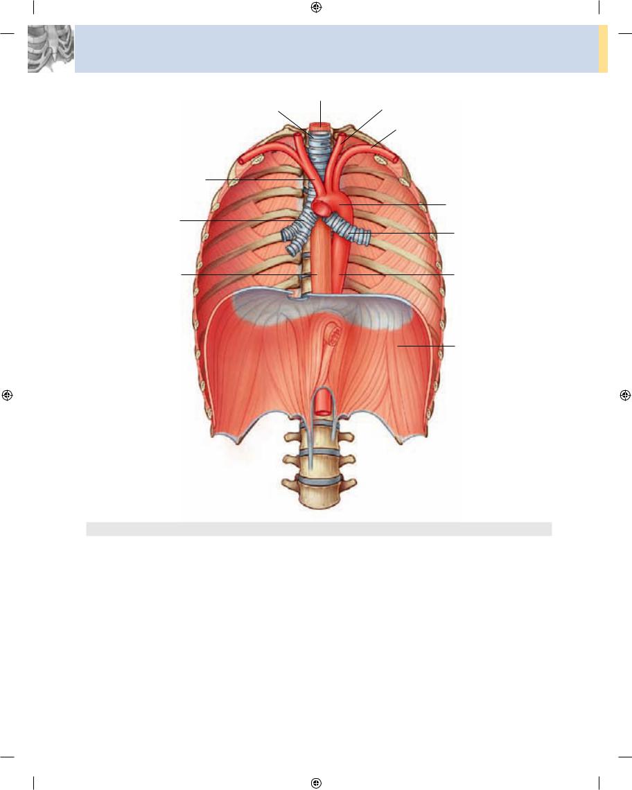
Thoracic duct in the superior mediastinum
The thoracic duct, which is the major lymphatic vessel in the body, passes through the posterior portion of the superior mediastinum (see Figs. 3.79 and 3.84). It:
■enters the superior mediastinum inferiorly, slightly to the left of the midline, having moved to this position just before leaving the posterior mediastinum opposite ver tebral level TIV/V; and
■continues through the superior mediastinum, posterior to the arch of aorta, and the initial portion of the left subclavian artery, between the esophagus and the left mediastinal part of the parietal pleura.
Posterior mediastinum
The posterior mediastinum is posterior to the pericar dial sac and diaphragm and anterior to the bodies of the mid and lower thoracic vertebrae (see Fig. 3.52).
■Its superior boundary is a transverse plane passing from the sternal angle to the intervertebral disc between ver tebrae TIV and TV.
■Its inferior boundary is the diaphragm.
■Laterally, it is bordered by the mediastinal part of pari etal pleura on either side.
■Superiorly, it is continuous with the superior mediastinum.
Major structures in the posterior mediastinum include the:
■esophagus and its associated nerve plexus;
■thoracic aorta and its branches;
■azygos system of veins;
■thoracic duct and associated lymph nodes;
■sympathetic trunks; and
■thoracic splanchnic nerves.
Esophagus
The esophagus is a muscular tube passing between the pharynx in the neck and the stomach in the abdomen. It begins at the inferior border of the cricoid cartilage, oppo site vertebra CVI, and ends at the cardiac opening of the stomach, opposite vertebra TXI.
The esophagus descends on the anterior aspect of the bodies of the vertebrae, generally in a midline position as it moves through the thorax (Fig. 3.89). As it approaches
Regional anatomy • Mediastinum |
3 |
|
|
the diaphragm, it moves anteriorly and to the left, crossing from the right side of the thoracic aorta to eventually assume a position anterior to it. It then passes through the esophageal hiatus, an opening in the muscular part of the diaphragm, at vertebral level TX.
The esophagus has a slight anterior-to-posterior curva ture that parallels the thoracic portion of the vertebral column, and is secured superiorly by its attachment to the pharynx and inferiorly by its attachment to the diaphragm.
Relationships to important structures in the posterior mediastinum
In the posterior mediastinum, the esophagus is related to a number of important structures. The right side is covered by the mediastinal part of the parietal pleura.
Posterior to the esophagus, the thoracic duct is on the right side inferiorly, but crosses to the left more superiorly. Also on the left side of the esophagus is the thoracic aorta.
Anterior to the esophagus, below the level of the tra cheal bifurcation, are the right pulmonary artery and the left main bronchus. The esophagus then passes immedi ately posteriorly to the left atrium, separated from it only by pericardium. Inferior to the left atrium, the esophagus is related to the diaphragm.
Structures other than the thoracic duct posterior to the esophagus include portions of the hemiazygos veins, the right posterior intercostal vessels, and, near the diaphragm, the thoracic aorta.
The esophagus is a flexible, muscular tube that can be compressed or narrowed by surrounding structures at four locations (Fig. 3.90):
■the junction of the esophagus with the pharynx in the neck;
■in the superior mediastinum where the esophagus is crossed by the arch of aorta;
■in the posterior mediastinum where the esophagus is compressed by the left main bronchus;
■in the posterior mediastinum at the esophageal hiatus in the diaphragm.
These constrictions have important clinical conse quences. For example, a swallowed object is most likely to lodge at a constricted area. An ingested corrosive sub stance would move more slowly through a narrowed region, causing more damage at this site than elsewhere along the esophagus. Also, constrictions present problems during the passage of instruments.
3-95
Drake_ch03_main.indd 95 |
|
|
8/25/2008 4:14:20 PM |
|
|
||
|
|
|
|
Thorax
Brachiocephalic trunk
Right main bronchus
Esophagus
Fig. 3.89 Esophagus.
|
Esophagus |
Trachea |
Left common carotid artery |
|
Left subclavian artery |
Arch of aorta
Left main bronchus
Thoracic aorta
Diaphragm
Arterial supply and venous and lymphatic drainage
The arterial supply and venous drainage of the esophagus in the posterior mediastinum involves many vessels. Esophageal arteries arise from the thoracic aorta, bron chial arteries, and ascending branches of the left gastric artery in the abdomen.
Venous drainage involves small vessels returning to the azygos vein, hemiazygos vein, and esophageal branches to the left gastric vein in the abdomen.
Lymphatic drainage of the esophagus in the posterior mediastinum returns to posterior mediastinal and left
3-96 gastric nodes.
Innervation
Innervation of the esophagus, in general, is complex. Esophageal branches arise from the vagus nerves and sym pathetic trunks.
Striated muscle fibers in the superior portion of the esophagus originate from the branchial arches and are innervated by branchial efferents from the vagus nerves.
Smooth muscle fibers are innervated by components of the parasympathetic part of the autonomic division of the peripheral nervous system, visceral efferents from the vagus nerves. These are preganglionic fibers that synapse in the myenteric and submucosal plexuses of the enteric nervous system in the esophageal wall.
Drake_ch03_main.indd 96 |
|
|
8/25/2008 4:14:38 PM |
|
|
||
|
|
|
|

Pharynx
Junction of esophagus
with pharynx
Esophagus
Trachea
|
Where esophagus is |
|
|
crossed by arch of |
|
|
aorta |
|
|
Where esophagus |
|
|
is compressed by |
|
|
left main bronchus |
|
Position of |
At the esophageal |
|
esophagus |
||
hiatus |
||
posterior to |
||
|
||
left atrium |
Diaphragm |
|
|
Fig. 3.90 Sites of normal esophageal constrictions.
Sensory innervation of the esophagus involves visceral afferent fibers originating in the vagus nerves, sympathetic trunks, and splanchnic nerves.
The visceral afferents from the vagus nerves are involved in relaying information back to the central nervous system
about normal physiological processes and reflex activities. They are not involved in the relay of pain recognition.
The visceral afferents that pass through the sympathetic trunks and the splanchnic nerves are the primary partici pants in detection of esophageal pain and transmission of this information to various levels of the central nervous system.
Esophageal plexus
After passing posteriorly to the root of the lungs, the right and left vagus nerves approach the esophagus. As they reach the esophagus, each nerve divides into several branches that spread over this structure, forming the esophageal plexus (Fig. 3.91). There is some mixing of fibers from the two vagus nerves as the plexus continues inferiorly on the esophagus toward the diaphragm. Just above the diaphragm, fibers of the plexus converge to form two trunks:
Regional anatomy • Mediastinum |
3 |
|
|
In the clinic
Esophageal cancer
When patients present with esophageal cancer, it is important to note which portion of the esophagus contains the tumor because tumor location determines the sites to which the disease will spread.
Esophageal cancer spreads quickly to lymphatics, draining to lymph nodes in the neck and around the celiac artery. Endoscopy or barium swallow is used to assess the site. CT and MRI may be necessary to stage the disease.
Once the extent of the disease has been assessed, treatment can be planned.
In the clinic
Esophageal rupture
The first case of esophageal rupture was described by Herman Boerhaave in 1724. This case was fatal, but early diagnosis has increased the survival rate up to 65%. If the disease is left untreated, mortality is 100%.
Typically, the rupture occurs in the lower third of the esophagus with a sudden rise in intraluminal esophageal pressure produced by vomiting secondary
to an incoordination and failure of the cricopharyngeus 7 muscle to relax. Because the tears typically occur on the
left, they are often associated with a large left pleural effusion that contains the gastric contents. In some patients, subcutaneous emphysema may be demonstrated.
Treatment is optimal with urgent surgical repair.
■the anterior vagal trunk on the anterior surface of the esophagus, mainly from fibers originally in the left vagus nerve;
■the posterior vagal trunk on the posterior surface of the esophagus, mainly from fibers originally in the right vagus nerve.
The vagal trunks continue on the surface of the eso phagus as it passes through the diaphragm into the abdomen.
Thoracic aorta
The thoracic portion of the descending aorta (thoracic aorta) begins at the lower edge of vertebra TIV, where it
is continuous with the arch of aorta. It ends anterior to the 3-97
Drake_ch03_main.indd 97 |
|
|
8/25/2008 4:14:50 PM |
|
|
||
|
|
|
|

Thorax
Right vagus nerve
Esophageal
plexus
Posterior vagal trunk
|
Trachea |
Left subclavian artery |
|
|
|
|
|||
Esophagus |
Supreme |
|
|
|
|
Esophagus |
Arch of aorta |
|
|
|
intercostal artery |
|
||
|
|
|
|
|
|
Right |
|
Left |
|
Left vagus nerve |
bronchial |
|
bronchial |
|
|
artery |
|
artery |
|
Anterior vagal trunk
Stomach
Posterior |
Esophageal |
Mediastinal |
intercostal |
branches |
branches |
arteries |
|
|
|
Esophagus |
|
Fig. 3.92 Thoracic aorta and branches.
Fig. 3.91 Esophageal plexus.
lower edge of vertebrae TXII, where it passes through the aortic hiatus posterior to the diaphragm. Situated to the left of the vertebral column superiorly, it approaches the midline inferiorly, lying directly anterior to the lower thoracic vertebral bodies (Fig. 3.92). Throughout its course, it gives off a number of branches, which are sum marized in Table 3.3.
Azygos system of veins
The azygos system of veins consists of a series of longitudi nal vessels on each side of the body that drain blood from the body wall and move it superiorly to empty into the superior vena cava. Blood from some of the thoracic viscera may also enter the system, and there are anastomotic con nections with abdominal veins.
The longitudinal vessels may or may not be continuous and are connected to each other from side to side at various
3-98 points throughout their course (Fig. 3.93).
The azygos system of veins serves as an important anas tomotic pathway capable of returning venous blood from the lower part of the body to the heart if the inferior vena cava is blocked.
The major veins in the system are:
■the azygos vein, on the right; and
■the hemiazygos vein and the accessory hemiazygos vein, on the left.
There is significant variation in the origin, course, tribu taries, anastomoses, and termination of these vessels.
Azygos vein
The azygos vein arises opposite vertebra LI or LII at the junction between the right ascending lumbar vein and the right subcostal vein (Fig. 3.93). It may also arise as a direct branch of the inferior vena cava, which is joined by a common trunk from the junction of the right ascend ing lumbar vein and the right subcostal vein.
Drake_ch03_main.indd 98 |
|
|
8/25/2008 4:15:00 PM |
|
|
||
|
|
|
|

|
|
|
|
Regional anatomy • Mediastinum |
3 |
||
|
|
|
|
|
|
|
|
|
|
|
|
|
|
||
|
|
|
Table 3.3 Branches of the thoracic aorta |
|
|
||
|
|
|
|
|
|
|
|
|
|
|
Branches |
Origin and course |
|
|
|
|
|
|
Pericardial branches |
A few small vessels to the posterior surface of the pericardial sac |
|
|
|
|
|
|
Bronchial branches |
Vary in number, size, and origin—usually, two left bronchial arteries from the thoracic aorta and one right |
|
|
|
|
|
|
|
bronchial artery from the third posterior intercostal artery or the upper left bronchial artery |
|
|
|
|
|
|
Esophageal branches |
Four or five vessels from the anterior aspect of the thoracic aorta, which form a continuous anastomotic |
|
|
|
|
|
|
|
chain—anastomotic connections include esophageal branches of the inferior thyroid artery superiorly, and |
|
|
|
|
|
|
|
esophageal branches of the left inferior phrenic and the left gastric arteries inferiorly |
|
|
|
|
|
|
Mediastinal branches |
Several small branches supplying lymph nodes, vessels, nerves, and areolar tissue in the posterior mediastinum |
|
|
|
|
|
|
Posterior intercostal arteries |
Usually nine pairs of vessels branching from the posterior surface of the thoracic aorta—usually supply lower |
|
|
|
|
|
|
|
nine intercostal spaces (first two spaces are supplied by the supreme intercostal artery—a branch of the |
|
|
|
|
|
|
|
costocervical trunk) |
|
|
|
|
|
|
Superior phrenic arteries |
Small vessels from the lower part of the thoracic aorta supplying the posterior part of the superior surface of |
|
|
|
|
|
|
|
the diaphragm—they anastomose with the musculophrenic and pericardiacophrenic arteries |
|
|
|
|
|
|
Subcostal artery |
The lowest pair of branches from the thoracic aorta located inferior to rib XII |
|
|
|
|
|
|
|
|
|
|
|
Right superior intercostal vein |
|
|
|
Left superior intercostal vein |
|
|
|
||
|
|
|
|
Opening of azygos vein into superior vena cava
|
|
|
|
|
Accessory hemiazygos |
|
|
|
|
|
|
||
Azygos vein |
|
|
|
|
vein |
|
|
|
|
|
Posterior intercostal vein |
||
|
|
|
|
|
|
|
|
|
|
|
|
|
|
Right subcostal vein |
|
|
|
|
|
|
|
|
|
Hemiazygos vein |
|
|
|
|
|
|
|
|
|
||
|
|
|
|
|
|
|
|
Ascending lumbar vein |
||
|
|
|
|
|
|
|
|
|||
Right ascending lumbar vein |
|
|
|
|
|
|
|
|||
|
|
|
|
|
|
|
||||
|
|
|
|
|
|
Inferior vena cava |
||||
|
|
|
|
|
|
|
|
|||
|
|
|
|
|
|
|
|
|||
3-99
Fig. 3.93 Azygos system of veins.
Drake_ch03_main.indd 99 |
|
|
8/25/2008 4:15:18 PM |
|
|
||
|
|
|
|

Thorax
The azygos vein enters the thorax through the aortic hiatus of the diaphragm, or it enters through or posterior to the right crus of the diaphragm. It ascends through the posterior mediastinum, usually to the right of the thoracic duct. At approximately vertebral level TIV, it arches anteri orly, over the root of the right lung, to join the superior vena cava before the superior vena cava enters the pericardial sac.
Tributaries of the azygos vein include:
■the right superior intercostal vein (a single vessel formed by the junction of the second, third, and fourth intercostal veins);
■fifth to eleventh right posterior intercostal veins;
■the hemiazygos vein;
■the accessory hemiazygos vein;
■esophageal veins;
■mediastinal veins;
■pericardial veins; and
■right bronchial veins.
Hemiazygos vein
The hemiazygos vein (inferior hemiazygos vein) usually arises at the junction between the left ascending lumbar vein and the left subcostal vein (Fig. 3.93). It may also arise from either of these veins alone and often has a connection to the left renal vein.
The hemiazygos vein usually enters the thorax through the left crus of the diaphragm, but may enter through the aortic hiatus. It ascends through the posterior mediasti num, on the left side, to approximately vertebral level TIX. At this point, it crosses the vertebral column, posterior to the thoracic aorta, esophagus, and thoracic duct, to enter the azygos vein.
Tributaries joining the hemiazygos vein include:
■the lowest four or five left posterior intercostal veins;
■esophageal veins; and
■mediastinal veins.
Accessory hemiazygos vein
The accessory hemiazygos vein (superior hemiazygos vein) descends on the left side from the superior portion of the posterior mediastinum to approximately vertebral level TVIII (Fig. 3.93). At this point, it crosses the vertebral column to join the azygos vein, or ends in the hemiazygos vein, or has a connection to both veins. Usually, it also has a connection superiorly to the left superior intercostal vein.
Vessels that drain into the accessory hemiazygos vein include:
■the fourth to eighth left posterior intercostal veins; and
■sometimes, the left bronchial veins.
Thoracic duct in the posterior mediastinum
The thoracic duct is the principal channel through which lymph from most of the body is returned to the venous system. It begins as a confluence of lymph trunks in the abdomen, sometimes forming a saccular dilation referred to as the cisterna chyli (chyle cistern), which drains the abdominal viscera and walls, pelvis, perineum, and lower limbs.
The thoracic duct extends from vertebra LII to the root of the neck.
Entering the thorax, posterior to the aorta, through the aortic hiatus of the diaphragm, the thoracic duct ascends through the posterior mediastinum to the right of midline between the thoracic aorta on the left and the azygos vein on the right (Fig. 3.94). It lies posterior to the diaphragm and the esophagus and anterior to the bodies of the vertebra.
At vertebral level TV, the thoracic duct moves to the left of midline and enters the superior mediastinum. It con tinues through the superior mediastinum and into the neck.
3-100
Drake_ch03_main.indd 100 |
|
|
8/25/2008 4:15:20 PM |
|
|
||
|
|
|
|

Right common carotid artery
Superior vena cava
Azygos vein
Thoracic duct
Cisterna chyli
Fig. 3.94 Thoracic duct.
Regional anatomy • Mediastinum |
3 |
|
|
Esophagus
Thoracic duct
Left brachiocephalic vein
Accessory hemiazygos vein
Hemiazygos vein
After being joined, in most cases, by the left jugular trunk, which drains the left side of the head and neck, and the left subclavian trunk, which drains the left upper limb, the thoracic duct empties into the junction of the left subclavian and left internal jugular veins.
The thoracic duct usually receives the contents from:
■the confluence of lymph trunks in the abdomen;
■descending thoracic lymph trunks draining the lower six or seven intercostal spaces on both sides;
■upper intercostal lymph trunks draining the upper left five or six intercostal spaces;
■ducts from posterior mediastinal nodes; and
■ducts from posterior diaphragmatic nodes.
Sympathetic trunks
The sympathetic trunks are an important component of |
|
the sympathetic part of the autonomic division of the PNS |
|
and are usually considered a component of the posterior |
|
mediastinum as they pass through the thorax. |
3-101 |
Drake_ch03_main.indd 101 |
|
|
8/25/2008 4:15:34 PM |
|
|
||
|
|
|
|