

Учебник по анатомии (для англ.яз)
.pdfThorax
Pleural recesses
The lungs do not completely fill the anterior or posterior inferior regions of the pleural cavities (Fig. 3.38). This results in recesses in which two layers of parietal pleura
become opposed. Expansion of the lungs into these spaces usually occurs only during forced inspiration; the recesses also provide potential spaces in which fluids can collect and from which fluids can be aspirated.
Costomediastinal recesses
Anteriorly, a costomediastinal recess occurs on each side where costal pleura is opposed to mediastinal pleura. The largest is on the left side in the region overlying the heart.
Costodiaphragmatic recesses
The largest and clinically most important recesses are the costodiaphragmatic recesses, which occur in each
Midclavicular line
Midaxillary
Vertebra TX (posterior)
Rib VIII (lateral)
Costodiaphragmatic recess
3-42 |
Fig. 3.38 Parietal pleural reflections and recesses. |
|
pleural cavity between the costal pleura and diaphrag matic pleura (Fig. 3.38). The costodiaphragmatic recesses are the regions between the inferior margin of the lungs and inferior margin of the pleural cavities. They are deepest after forced expiration and shallowest after forced inspiration.
During quiet respiration, the inferior margin of the lung crosses rib VI in the midclavicular line, rib VIII in the midaxillary line, and then courses somewhat horizontally to reach the vertebral column at vertebral level TX. From the midclavicular line and around the thoracic wall to the vertebral column, the inferior margin of the lung can be approximated by a line running between rib VI, rib VIII, and vertebra TX. The inferior margin of the pleural cavity at the same points is rib VIII, rib X, and vertebra TXII. The costodiaphragmatic recess is the region between the two margins.
During expiration, the inferior margin of the lung rises and the costodiaphragmatic recess becomes larger.
Costomediastinal recess
Rib VI (anterior)
Drake_ch03_main.indd 42 |
|
|
8/25/2008 4:11:56 PM |
|
|
||
|
|
|
|

Lungs
The two lungs are organs of respiration and lie on either side of the mediastinum surrounded by the right and left pleural cavities. Air enters and leaves the lungs via main bronchi, which are branches of the trachea.
The pulmonary arteries deliver deoxygenated blood to the lungs from the right ventricle of the heart. Oxygen ated blood returns to the left atrium via the pulmonary veins.
The right lung is normally a little larger than the left lung because the middle mediastinum, containing the heart, bulges more to the left than to the right.
Each lung has a half-cone shape, with a base, apex, two surfaces, and three borders (Fig. 3.39).
■The base sits on the diaphragm.
■The apex projects above rib I and into the root of the neck.
■The two surfaces—the costal surface lies immediately adjacent to the ribs and intercostal spaces of the tho racic wall. The mediastinal surface lies against the
Right lung
Anterior border
Regional anatomy • Pleural cavities |
3 |
|
|
mediastinum anteriorly and the vertebral column pos teriorly and contains the comma-shaped hilum of the lung through which structures enter and leave.
■The three borders—the inferior border of the lung is sharp and separates the base from the costal surface. The anterior and posterior borders separate the costal surface from the medial surface. Unlike the ante rior and inferior borders, which are sharp, the posterior border is smooth and rounded.
The lungs lie directly adjacent to, and are indented by, structures contained in the overlying area. The heart and major vessels form bulges in the mediastinum that indent the medial surfaces of the lung; the ribs indent the costal surfaces. Pathology, such as tumors, or abnormalities in one structure can affect the related structure.
Root and hilum
The root of each lung is a short tubular collection of struc tures that together attach the lung to structures in the mediastinum (Fig. 3.40). It is covered by a sleeve of medi
Left lung
Apex
Hilum
Bronchus
Pulmonary
artery
Pulmonary
veins
Posterior
border
Costal surface |
Mediastinal |
|
|
surface |
|
|
Inferior border |
|
|
Base (diaphragmatic surface) |
|
|
|
|
Fig. 3.39 Lungs. |
|
3-43 |
|
|
|
Drake_ch03_main.indd 43 |
|
|
8/25/2008 4:11:59 PM |
|
|
||
|
|
|
|

Thorax
astinal pleura that reflects onto the surface of the lung as visceral pleura. The region outlined by this pleural reflec tion on the medial surface of the lung is the hilum, where structures enter and leave.
A thin blade-like fold of pleura projects inferiorly from the root of the lung and extends from the hilum to the mediastinum. This structure is the pulmonary ligament. It may stabilize the position of the inferior lobe and may also accommodate the down-and-up translocation of structures in the root during breathing.
In the mediastinum, the vagus nerves pass immediately posterior to the roots of the lungs, while the phrenic nerves pass immediately anterior to them.
Within each root and located in the hilum are:
■a pulmonary artery;
■two pulmonary veins;
■a main bronchus;
■bronchial vessels;
■nerves; and
■lymphatics.
Generally, the pulmonary artery is superior at the hilum, the pulmonary veins are inferior, and the bronchi are somewhat posterior in position.
On the right side, the lobar bronchus to the superior lobe branches from the main bronchus in the root, unlike on the left where it branches within the lung itself, and is superior to the pulmonary artery.
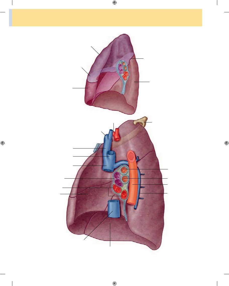
Right lung
The right lung has three lobes and two fissures (Fig. 3.41A). Normally, the lobes are freely movable against each other because they are separated, almost to the hilum, by invaginations of visceral pleura. These invaginations form the fissures:
■the oblique fissure separates the inferior lobe (lower lobe) from the superior lobe and the middle lobe of the right lung;
■the horizontal fissure separates the superior lobe
(upper lobe) from the middle lobe.
Root |
Pulmonary artery |
|
Hilum |
(deoxygenated blood) |
|
Pulmonary veins |
||
|
||
Bronchus |
(oxygenated blood) |
|
|
Pulmonary
artery
Pulmonary
veins
|
Pulmonary ligament |
Right lung |
Left lung |
Fig. 3.40 Roots and hila of the lungs.
3-44
Drake_ch03_main.indd 44 |
|
|
8/25/2008 4:12:02 PM |
|
|
||
|
|
|
|
A
Superior lobe
Horizontal fissure
Middle lobe
B
Anterior
Subclavian artery
Subclavian vein
Right brachiocephalic vein
Left brachiocephalic vein
Superior vena cava
Pulmonary artery
Pulmonary veins
Heart
Regional anatomy • Pleural cavities |
3 |
|
|
Oblique fissure
Inferior lobe
Posterior
Rib I
Bronchus to superior lobe
Bronchus
Esophagus
Azygos vein
Inferior vena cava
Diaphragm
Fig. 3.41 A. Right lung. B. Major structures related to the right lung. |
3-45 |
|
|
Drake_ch03_main.indd 45 |
|
|
8/25/2008 4:12:07 PM |
|
|
||
|
|
|
|

Thorax
|
|
The approximate position of the oblique fissure on a |
|
|||
|
patient, in quiet respiration, can be marked by a curved |
|
||||
|
line on the thoracic wall that begins roughly at the spinous |
|
||||
|
process of vertebra TIV level of the spine, crosses the fifth |
|
||||
2 |
interspace laterally, and then follows the contour of rib VI |
|
||||
anteriorly (see p. 000). |
|
|
||||
|
|
|||||
|
|
The horizontal fissure follows the fourth intercostal |
||||
|
space from the sternum until it meets the oblique fissure as |
|||||
|
it crosses rib V. |
|||||
|
|
|
|
|
||
|
|
The orientations of the oblique and horizontal fissures |
|
|||
|
|
|
|
|
|
|
|
determine where clinicians should listen for lung sounds |
|
||||
|
from each lobe. |
|
|
|
||
|
|
The largest |
surface of the superior lobe is in contact with |
|||
|
the upper part of the anterolateral wall and the apex of this |
|||||
|
lobe projects into the root of the neck. The surface of the |
|||||
|
middle lobe lies mainly adjacent to the lower anterior and |
|||||
|
lateral wall. The costal surface of the inferior lobe is in |
|||||
|
contact with the posterior and inferior walls. |
|||||
|
|
|
|
|||
|
|
When listening to lung sounds from each of the lobes, |
|
|||
|
|
|
|
|
||
|
it is important to position the stethoscope on those areas of |
|
||||
|
the thoracic wall related to the underlying positions of the |
|
||||
3 |
lobes (see p. 000). |
|
|
|||
|
|
The medial surface of the right lung lies adjacent to a |
||||
|
number of important structures in the mediastinum and |
|||||
|
the root of the neck (Fig. 3.41B). These include the: |
|||||
|
■ |
heart, |
||||
|
■ |
inferior vena cava, |
||||
|
■ |
superior vena cava, |
||||
|
■ |
azygos vein, and |
||||
|
■ |
esophagus. |
||||
The right subclavian artery and vein arch over and are related to the superior lobe of the right lung as they pass over the dome of cervical pleura and into the axilla.
Left lung
The left lung is smaller than the right lung and has two lobes separated by an oblique fissure (Fig. 3.42A). The
3-46
oblique fissure of the left lung is slightly more oblique than the corresponding fissure of the right lung.
During quiet respiration, the approximate position of the left oblique fissure can be marked by a curved line on the thoracic wall that begins between the spinous pro
cesses of vertebrae TIII and TIV, crosses the fifth interspace laterally, and follows the contour of rib VI anteriorly (see p. 000).
As with the right lung, the orientation of the oblique fissure determines where to listen for lung sounds from each lobe.
The largest surface of the superior lobe is in contact with the upper part of the anterolateral wall, and the apex of this lobe projects into the root of the neck. The costal surface of the inferior lobe is in contact with the posterior and inferior walls.
When listening to lung sounds from each of the lobes, the stethoscope should be placed on those areas of the tho
racic wall related to the underlying positions of the lobes (see p. 000).
The inferior portion of the medial surface of the left lung, unlike the right lung, is notched because of the heart’s projection into the left pleural cavity from the middle mediastinum.
From the anterior border of the lower part of the supe rior lobe a tongue-like extension (the lingula of left lung) projects over the heart bulge.
The medial surface of the left lung lies adjacent to a number of important structures in the mediastinum and root of the neck (Fig. 3.42B). These include the:
■heart,
■aortic arch,
■thoracic aorta, and
■esophagus.
The left subclavian artery and vein arch over and are related to the superior lobe of the left lung as they pass over the dome of cervical pleura and into the axilla.
4
5
Drake_ch03_main.indd 46 |
|
|
8/25/2008 4:12:07 PM |
|
|
||
|
|
|
|

A
Oblique fissure
Inferior lobe
B
Posterior
Rib I
Bronchus
Esophagus
Thoracic aorta
Fig. 3.42 A. Left lung. B. Major structures related to the left lung.
Regional anatomy • Pleural cavities |
3 |
|
|
Superior lobe
Lingula
Anterior
Left subclavian artery
Left brachiocephalic vein
Aortic arch
Pulmonary artery
Pulmonary veins
Heart
Diaphragm
3-47
Drake_ch03_main.indd 47 |
|
|
8/25/2008 4:12:12 PM |
|
|
||
|
|
|
|

Thorax
Bronchial tree
The trachea is a flexible tube that extends from vertebral level CVI in the lower neck to vertebral level TIV/V in the mediastinum where it bifurcates into a right and a left
main bronchus (Fig. 3.43). The trachea is held open by C- shaped transverse cartilage rings embedded in its wall— the open part of the C facing posteriorly. The lowest tracheal ring has a hook-shaped structure, the carina, that projects
A
Right main bronchus
Lobar bronchi
Segmental bronchi of middle lobe
B
Lateral bronchopulmonary segment of middle lobe of right lung
3-48
Fig. 3.43 A. Bronchial tree. B. Bronchopulmonary segments.
Trachea
Left main bronchus
Lobar bronchi
Branch of pulmonary artery
Medial bronchopulmonary segment of middle lobe of right lung
Drake_ch03_main.indd 48 |
|
|
8/25/2008 4:12:17 PM |
|
|
||
|
|
|
|

backwards in the midline between the origins of the two main bronchi. The posterior wall of the trachea is com posed mainly of smooth muscle.
Each main bronchus enters the root of a lung and passes through the hilum into the lung itself. The right main bronchus is wider and takes a more vertical course through the root and hilum than the left main bronchus (Fig. 3.43A). Therefore, inhaled foreign bodies tend to lodge more frequently on the right side than on the left.
The main bronchus divides within the lung into lobar bronchi (secondary bronchi), each of which supplies a lobe. On the right side, the lobar bronchus to the superior lobe originates within the root of the lung.
The lobar bronchi further divide into segmental bronchi (tertiary bronchi), which supply bronchopulmo nary segments (Fig. 3.43B).
Within each bronchopulmonary segment, the segmen tal bronchi give rise to multiple generations of divisions and, ultimately, to bronchioles, which further subdivide and supply the respiratory surfaces. The walls of the bronchi are held open by discontinuous elongated plates of cartilage, but these are not present in bronchioles.
Bronchopulmonary segments
A bronchopulmonary segment is the area of lung sup plied by a segmental bronchus and its accompanying pul monary artery branch.
Tributaries of the pulmonary vein tend to pass interseg mentally between and around the margins of segments.
Each bronchopulmonary segment is shaped like an irregular cone with the apex at the origin of the segmental bronchus and the base projected peripherally onto the surface of the lung.
A bronchopulmonary segment is the smallest, function ally independent region of a lung and the smallest area of lung that can be isolated and removed without affecting adjacent regions.
There are ten bronchopulmonary segments in each lung (Fig. 3.44); some of them fuse in the left lung.
Pulmonary arteries
The right and left pulmonary arteries originate from the pulmonary trunk and carry deoxygenated blood to the lungs from the right ventricle of the heart (Fig. 3.45).
The bifurcation of the pulmonary trunk occurs to the left of the midline just inferior to vertebral level TIV/V, and anteroinferiorly to the left of the bifurcation of the trachea.
Right pulmonary artery
The right pulmonary artery is longer than the left and passes horizontally across the mediastinum (Fig. 3.45). It passes:
Regional anatomy • Pleural cavities |
3 |
|
|
■anteriorly and slightly inferiorly to the tracheal bifur cation and anteriorly to the right main bronchus; and
■posteriorly to the ascending aorta, superior vena cava, and upper right pulmonary vein.
The right pulmonary artery enters the root of the lung and gives off a large branch to the superior lobe of the lung. The main vessel continues through the hilum of the lung, gives off a second (recurrent) branch to the superior lobe, and then divides to supply the middle and inferior lobes.
Left pulmonary artery
The left pulmonary artery is shorter than the right and lies anterior to the descending aorta and posterior to the superior pulmonary vein (Fig. 3.45). It passes through the root and hilum and branches within the lung.
Pulmonary veins
On each side a superior pulmonary vein and an inferior pulmonary vein carry oxygenated blood from the lungs back to the heart (Fig. 3.45). The veins begin at the hilum of the lung, pass through the root of the lung, and immediately drain into the left atrium.
Bronchial arteries and veins
The bronchial arteries (Fig. 3.45) and veins constitute the “nutritive” vascular system of the pulmonary tissues (bronchial walls and glands, walls of large vessels, and visceral pleura). They interconnect within the lung with branches of the pulmonary arteries and veins.
The bronchial arteries originate from the thoracic aorta or one of its branches:
■a single right bronchial artery normally arises from the third posterior intercostal artery (but occasion ally, it originates from the upper left bronchial artery);
■two left bronchial arteries arise directly from the anterior surface of the thoracic aorta—the superior left bronchial artery arises at vertebral level TV, and the inferior one inferior to the left bronchus.
The bronchial arteries run on the posterior surfaces of the bronchi and ramify in the lungs to supply pulmonary tissues.
The bronchial veins drain into:
■either the pulmonary veins or the left atrium; and
■into the azygos vein on the right or into the
superior intercostal vein or hemiazygos vein on the 3-49 left.
Drake_ch03_main.indd 49 |
|
|
8/25/2008 4:12:17 PM |
|
|
||
|
|
|
|

Thorax
A |
Medial view |
Apical segment (S I)
Superior lobe
Anterior segment (S III)
Medial segment (S V)
Middle lobe
Anterior basal segment (S VIII)
Posterior segment (S II)
Superior segment (S VI)
Inferior lobe
Medial basal segment (S VII)
Posterior basal segment (S X)
Lateral basal segment (S IX)
Lateral view
Apical segment (S I)
Anterior segment (S III)
Medial segment (S V)
Lateral segment (S IV)
Anterior basal segment (S VIII)
B
Superior segment (S VI)
Inferior lobe
Posterior basal segment (S X)
Medial basal segment (S VII)
Apicoposterior segment (S I & II)
Superior lobe
Anterior segment (S III)
Superior lingular segment (S IV)
Inferior lingular segment (S V)
Anterior basal segment (S VIII)
Lateral basal segment (S IX)
Superior segment (S VI)
Posterior basal segment (S X)
Fig. 3.44 Bronchopulmonary segments. A. Right lung. B. Left lung. (Bronchopulmonary segments are numbered and named.)
3-50
Drake_ch03_main.indd 50 |
|
|
8/25/2008 4:12:18 PM |
|
|
||
|
|
|
|

|
Regional anatomy • Pleural cavities |
3 |
|
|
|
A
Right bronchial artery |
Aortic arch |
|
(branch from right third |
||
|
||
posterior intercostal artery) |
|
Superior left bronchial artery
Right pulmonary artery
Bronchial vessels |
|
Left pulmonary artery |
|
|
|
|
|
on posterior surface |
|
|
|
of bronchi |
|
|
|
|
|
Left pulmonary veins |
|
Right pulmonary |
|
|
|
veins |
|
|
|
Pulmonary trunk |
|
Pulmonary ligament |
|
|
|
Thoracic aorta |
|
Esophagus |
|
|
|
B |
|
C |
|
Superior vena cava Ascending aorta |
Pulmonary trunk |
Superior vena cava Ascending aorta |
Pulmonary trunk |
|
|
|
|
|
|
|
|
|
|
|
|
|
|
|
|
|
|
|
|
|
|
|
|
|
|
|
|
|
|
|
|
|
|
|
|
|
|
|
|
|
|
|
|
|
|
|
|
Right main bronchus |
Esophagus |
Left pulmonary artery |
Right pulmonary artery |
Esophagus |
Thoracic aorta |
||||||||||
|
|
|
|
|
|
|
|
|
|
|
|
|
|
|
|
|
|
|
|
|
Thoracic aorta |
|
|
|
|
|
|
|
|||
Fig. 3.45 Pulmonary vessels. A. Diagram of an anterior view. B. Axial computed tomography image showing the left pulmonary artery branching from the pulmonary trunk. C. Axial computed tomography image (just inferior to the image in B) showing the right pulmonary
artery branching from the pulmonary trunk.
3-51
Drake_ch03_main.indd 51 |
|
|
8/25/2008 4:12:20 PM |
|
|
||
|
|
|
|