

Учебник по анатомии (для англ.яз)
.pdfConceptual overview
General description Functions
Breathing
Protection of vital organs Conduit
Component parts
Thoracic wall
Superior thoracic aperture Inferior thoracic aperture Diaphragm
Mediastinum Pleural cavities
Relationship to other regions
Neck Upper limb Abdomen Breast
Key features
Vertebral level TIV/V
Venous shunts from left to right Segmental neurovascular supply of
thoracic wall Sympathetic system
Flexible wall and inferior thoracic aperture Innervation of the diaphragm
Regional anatomy
Pectoral region
Breast
Venous drainage
Muscles of the pectoral region
Thoracic wall
Skeletal framework
Intercostal spaces
124 |
Diaphragm |
156 |
|
Venous drainage |
160 |
124 |
Innervation |
160 |
125 |
Movements of the thoracic wall and |
|
125 |
diaphragm during breathing |
160 |
125 |
Pleural cavities |
160 |
125 |
Pleura |
160 |
125 |
Lungs |
165 |
125 |
Mediastinum |
178 |
126 |
Middle mediastinum |
178 |
126 |
Superior mediastinum |
206 |
127 |
Posterior mediastinum |
217 |
127 |
Anterior mediastinum |
225 |
127 |
|
|
130 |
Surface anatomy |
226 |
130 |
||
130 |
|
|
131 |
Thorax surface anatomy |
226 |
131 |
How to count ribs |
226 |
132 |
Surface anatomy of the breast in women |
227 |
132 |
Visualizing structures at the TIV/V |
|
132 |
vertebral level |
228 |
|
Visualizing structures in the superior |
|
132 |
mediastinum |
228 |
135 |
Visualizing the margins of the heart |
228 |
135 |
Where to listen for heart sounds |
229 |
137 |
Visualizing the pleural cavities and lungs, |
|
|
pleural recesses, and lung lobes and |
|
|
fissures |
229 |
139 |
Where to listen for lung sounds |
230 |
139 |
Clinical cases |
235 |
139 |
||
139 |
|
|
141
142
142
149
Drake_ch03_main.indd 2 |
|
|
8/25/2008 4:10:06 PM |
|
|
||
|
|
|
|

3
Thorax
Drake_ch03_main.indd 3 |
|
|
8/25/2008 4:10:13 PM |
|
|
||
|
|
|
|

Thorax
Conceptual overview
GENERAL DESCRIPTION
The thorax is an irregularly shaped cylinder with a narrow opening (superior thoracic aperture) superiorly and a rela tively large opening (inferior thoracic aperture) inferiorly (Fig. 3.1). The superior thoracic aperture is open, allowing continuity with the neck; the inferior thoracic aperture is closed by the diaphragm.
The musculoskeletal wall of the thorax is flexible and consists of segmentally arranged vertebrae, ribs, muscles, and the sternum.
The thoracic cavity enclosed by the thoracic wall and the diaphragm is subdivided into three major compartments:
|
Superior thoracic aperture |
|
Vertebral column |
|
Mediastinum |
Right pleural cavity |
Left pleural cavity |
|
Rib I |
Manubrium of sternum
Sternal angle
Body of sternum
Ribs
Xiphoid process
Diaphragm
Inferior thoracic aperture
3- |
Fig. 3.1 Thoracic wall and cavity. |
|
Drake_ch03_main.indd 4 |
|
|
8/25/2008 4:10:18 PM |
|
|
||
|
|
|
|

■a left and a right pleural cavity, each surrounding a lung;
■the mediastinum.
The mediastinum is a thick, flexible soft tissue partition oriented longitudinally in a median sagittal position. It contains the heart, esophagus, trachea, major nerves, and major systemic blood vessels.
The pleural cavities are completely separated from each other by the mediastinum. Therefore, abnormal events in one pleural cavity do not necessarily affect the other cavity. This also means that the mediastinum can be entered sur gically without opening the pleural cavities.
Another important feature of the pleural cavities is that they extend above the level of rib I. The apex of each lung actually extends into the root of the neck. As a conse
quence, abnormal events in the root of the neck can involve the adjacent pleura and lung, and events in the adjacent pleura and lung can involve the root of the neck.
FUNCTIONS
Breathing
One of the most important functions of the thorax is breath ing. The thorax not only contains the lungs but also pro vides the machinery necessary—the diaphragm, thoracic wall, and the ribs—for effectively moving air into and out of the lungs.
Up and down movements of the diaphragm and changes in the lateral and anterior dimensions of the thoracic wall, caused by movements of the ribs, alter the volume of the thoracic cavity and are key elements in breathing.
Protection of vital organs
The thorax houses and protects the heart, lungs, and great vessels. Because of the domed shape of the diaphragm, the thoracic wall also offers protection to some important abdominal viscera.
Much of the liver lies under the right dome of the dia phragm, and the stomach and spleen lie under the left. The posterior aspects of the superior poles of the kidneys lie on the diaphragm and are anterior to rib XII, on the right, and to ribs XI and XII, on the left.
Conduit
The mediastinum acts as a conduit for structures that pass completely through the thorax from one body region to another and for structures that connect organs in the thorax to other body regions.
Conceptual overview • component parts |
3 |
|
|
The esophagus, vagus nerves, and thoracic duct pass through the mediastinum as they course between the abdomen and neck.
The phrenic nerves, which originate in the neck, also pass through the mediastinum to penetrate and supply the diaphragm.
Other structures such as the trachea, thoracic aorta, and superior vena cava course within the mediastinum en route to and from major visceral organs in the thorax.
COMPONENT PARTS
Thoracic wall
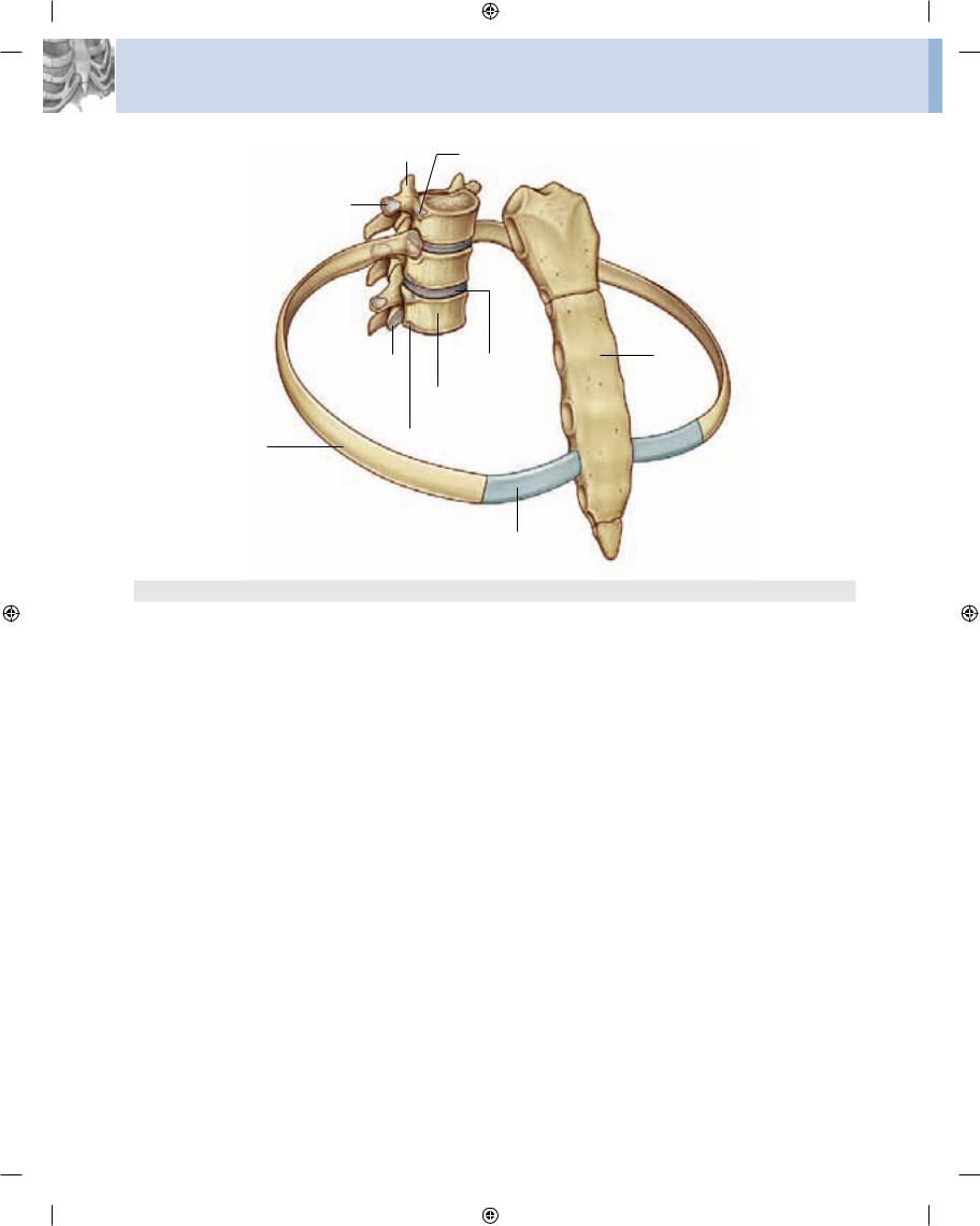
The thoracic wall consists of skeletal elements and muscles (Fig. 3.1):
■posteriorly, it is made up of twelve thoracic vertebrae and their intervening intervertebral discs;
■laterally, the wall is formed by ribs (twelve on each side) and three layers of flat muscles, which span the intercostal spaces between adjacent ribs, move the ribs and provide support for the intercostal spaces;
■anteriorly, the wall is made up of the sternum, which consists of the manubrium of sternum, body of sternum, and xiphoid process.
The manubrium of sternum, angled posteriorly on the |
|
body of sternum at the manubriosternal joint, forms the |
|
sternal angle, which is a major surface landmark used |
|
by clinicians in performing physical examinations of the |
|
thorax. |
|
The anterior (distal) end of each rib is composed of costal |
|
cartilage, which contributes to the mobility and elasticity |
|
of the wall. |
|
All ribs articulate with thoracic vertebrae posteriorly. |
|
Most ribs (from rib II to IX) have three articulations |
|
with the vertebral column. The head of each rib articulates |
|
with the body of its own vertebra and with the body of the |
|
vertebra above (Fig. 3.2). As these ribs curve posteriorly, |
|
each also articulates with the transverse process of its |
|
vertebra. |
|
Anteriorly, the costal cartilages of ribs I to VII articulate |
|
with the sternum. |
|
The costal cartilages of ribs VIII to X articulate with the |
|
inferior margins of the costal cartilages above them. Ribs |
|
XI and XII are called floating ribs because they do not |
|
articulate with other ribs, costal cartilages, or the sternum. |
|
Their costal cartilages are small, only covering their tips. |
|
The skeletal framework of the thoracic wall provides |
|
extensive attachment sites for muscles of the neck, |
|
abdomen, back, and upper limbs. |
3- |
Drake_ch03_main.indd 5 |
|
|
8/25/2008 4:10:18 PM |
|
|
||
|
|
|
|
Thorax
Superior articular process |
Superior costal facet |
Costal facet of transverse process
Sternum
Inferior articular Intervertebral disc process
Vertebral body
Inferior costal facet
Rib V
Costal cartilage
Fig. 3.2 Joints between ribs and vertebrae.
A number of these muscles attach to ribs and function as accessory respiratory muscles; some of them also stabi lize the position of the first and last ribs.
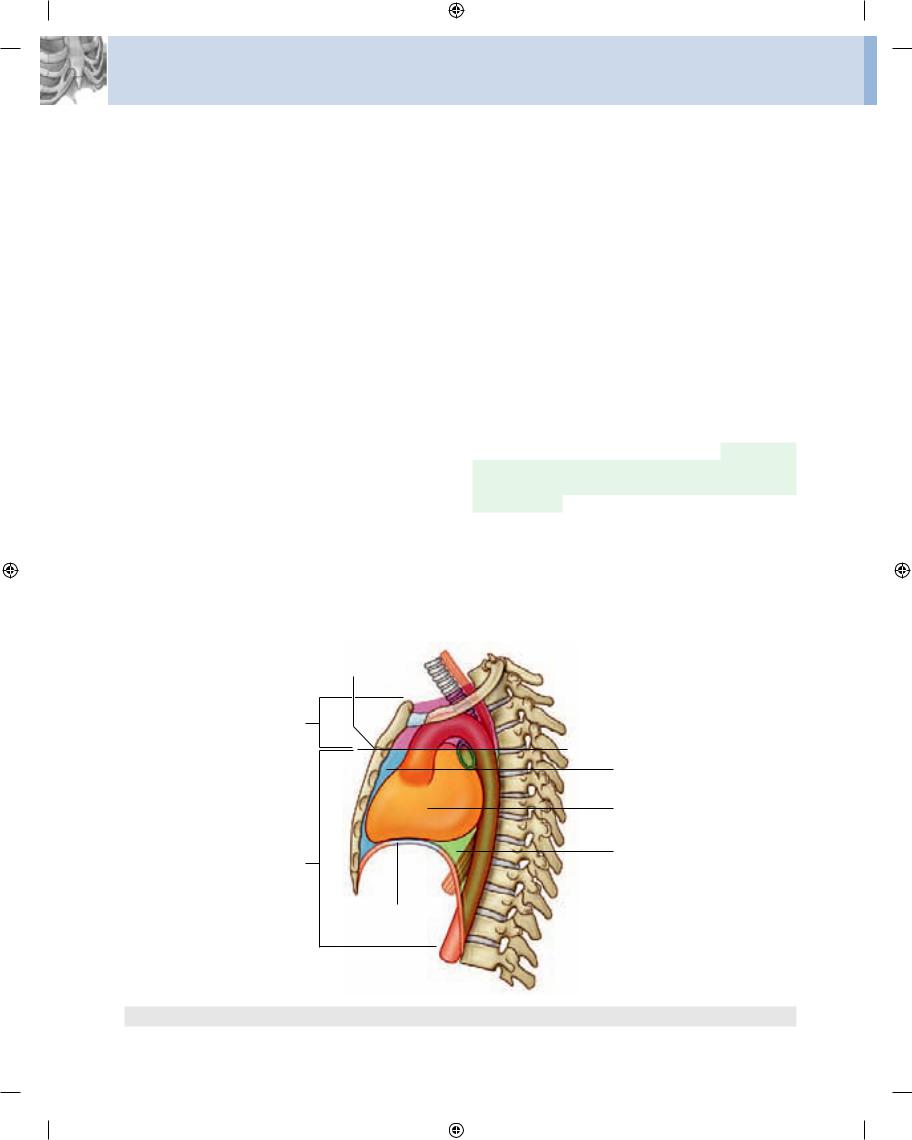
Superior thoracic aperture
Completely surrounded by skeletal elements, the superior thoracic aperture consists of the body of vertebra TI pos teriorly, the medial margin of rib I on each side, and the manubrium anteriorly.
The superior margin of the manubrium is in approxi mately the same horizontal plane as the intervertebral disc between vertebrae TII and TIII.
The first ribs slope inferiorly from their posterior articu lation with vertebra TI to their anterior attachment to the manubrium. Consequently, the plane of the superior tho racic aperture is at an oblique angle, facing somewhat anteriorly.
At the superior thoracic aperture, the superior aspects of the pleural cavities, which surround the lungs, lie on either side of the entrance to the mediastinum (Fig. 3.3).
Structures that pass between the upper limb and thorax 3- pass over rib I and the superior part of the pleural cavity as they enter and leave the mediastinum. Structures that pass
between the neck and head and the thorax pass more verti cally through the superior thoracic aperture.
Inferior thoracic aperture
The inferior thoracic aperture is large and expand able. Bone, cartilage, and ligaments form its margin (Fig. 3.4A).
The inferior thoracic aperture is closed by the dia phragm, and structures passing between the abdomen and thorax pierce or pass posteriorly to the diaphragm.
Skeletal elements of the inferior thoracic aperture are:
■the body of vertebra TXII posteriorly;
■rib XII and the distal end of rib XI posterolaterally;
■the distal cartilaginous ends of ribs VII to X, which unite to form the costal margin anterolaterally; and
■the xiphoid process anteriorly.
The joint between the costal margin and sternum lies roughly in the same horizontal plane as the intervertebral disc between vertebrae TIX and TX. In other words, the post erior margin of the inferior thoracic aperture is inferior to the anterior margin.
When viewed anteriorly, the inferior thoracic aperture is tilted superiorly.
Drake_ch03_main.indd 6 |
|
|
8/25/2008 4:10:20 PM |
|
|
||
|
|
|
|

|
|
Conceptual overview • component parts |
3 |
|
|
|
|
|
|
Esophagus |
|
|
|
Common carotid artery |
|
|
|
Vertebra TI |
|
|
|
Trachea |
|
|
Superior thoracic aperture |
|
|
|
Rib I |
Internal |
|
|
|
jugular vein |
|
Apex of right lung
Subclavian
artery and vein Manubrium of sternum
Rib II
Fig. 3.3 Superior thoracic aperture.
A |
B |
|
|
Right dome |
|
|
Central |
|
|
tendon |
|
Xiphoid process |
|
|
Inferior thoracic |
Left dome |
|
aperture |
||
|
||
Distal cartilaginous |
|
|
ends of ribs VII to X; |
|
|
costal margins |
Esophageal |
|
|
||
Rib XI |
hiatus |
|
Aortic |
||
Rib XII |
||
hiatus |
||
Vertebra TXII |
||
|
Fig. 3.4 A. Inferior thoracic aperture. B. Diaphragm.
Diaphragm
The musculotendinous diaphragm seals the inferior tho racic aperture (Fig. 3.4B).
Generally, muscle fibers of the diaphragm arise radially, from the margins of the inferior thoracic aperture, and converge into a large central tendon.
Because of the oblique angle of the inferior thoracic aperture, the posterior attachment of the diaphragm is inferior to the anterior attachment.
The diaphragm is not flat; rather, it “balloons” superi
orly, on both the right and left sides, to form domes. The 3- right dome is higher than the left, reaching as far as rib V.
Drake_ch03_main.indd 7 |
|
|
8/25/2008 4:10:24 PM |
|
|
||
|
|
|
|
Thorax
As the diaphragm contracts, the height of the domes decreases and the volume of the thorax increases.
The esophagus and inferior vena cava penetrate the dia phragm; the aorta passes posterior to the diaphragm.
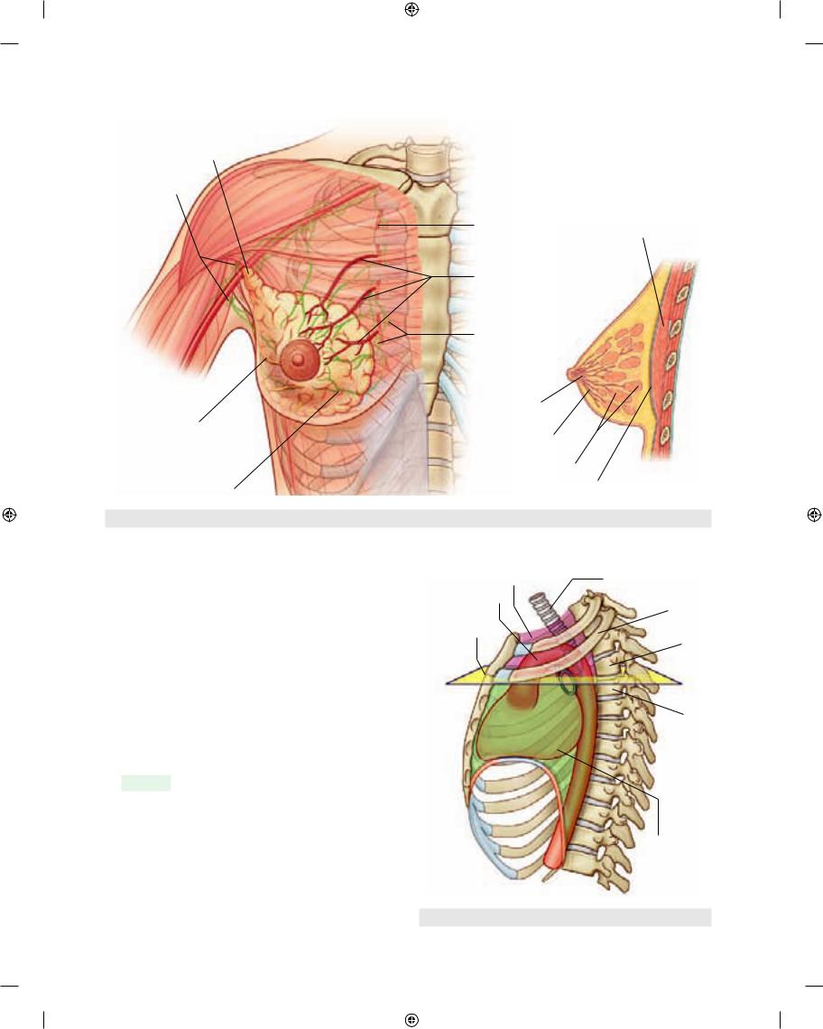
Mediastinum
The mediastinum is a thick midline partition that extends from the sternum anteriorly to the thoracic vertebrae posteriorly, and from the superior thoracic aperture to the inferior thoracic aperture.
A horizontal plane passing through the sternal angle and the intervertebral disc between vertebrae TIV and TV separates the mediastinum into superior and inferior parts (Fig. 3.5). The inferior part is further subdivided by the pericardium, which encloses the pericardial cavity sur rounding the heart. The pericardium and heart constitute the middle mediastinum.
The anterior mediastinum lies between the sternum and the pericardium; the posterior mediastinum lies be tween the pericardium and thoracic vertebrae.
Pleural cavities
The two pleural cavities are situated on either side of the mediastinum (Fig. 3.6).
Sternal angle
Superior mediastinum
Inferior mediastinum
Diaphragm
3- |
Fig. 3.5 Subdivisions of the mediastinum. |
|
Each pleural cavity is completely lined by a mesothe lial membrane called the pleura.
During development, the lungs grow out of the medias tinum, becoming surrounded by the pleural cavities. As a result, the outer surface of each organ is covered by pleura.
Each lung remains attached to the mediastinum by a root formed by the airway, pulmonary blood vessels, lym phatic tissues, and nerves.
The pleura lining the walls of the cavity is the parietal pleura, whereas that reflected from the mediastinum at the roots and onto the surfaces of the lungs is the visceral pleura. Only a potential space normally exists between the visceral pleura covering lung and the parietal pleura lining the wall of the thoracic cavity.
The lung does not completely fill the potential space of the pleural cavity, resulting in recesses, which do not contain lung and are important for accommodating changes in lung volume during breathing. The costodia phragmatic recess, which is the largest and clinically most important recess, lies inferiorly between the thoracic wall and diaphragm.
I |
|
|
Rib I |
|
|
||
|
|
|
|
|
|
IV |
|
|
|
V |
Anterior mediastinum |
|
|
|
|
|
|
|
Middle mediastinum |
|
|
|
Posterior mediastinum |
|
X |
|
|
XII |
|
|
|
Drake_ch03_main.indd 8 |
|
|
8/25/2008 4:10:25 PM |
|
|
||
|
|
|
|

|
|
Conceptual overview • Relationship to other regions |
3 |
|
|
|
|
|
Apex of right lung |
Trachea |
|
|
|
Left pleural cavity |
|
Right main bronchus
Parietal pleura |
|
Visceral pleura |
Mediastinum |
|
Costodiaphragmatic
recess
Diaphragm
Fig. 3.6 Pleural cavities.
RELATIONSHIP TO
OTHER REGIONS
Neck
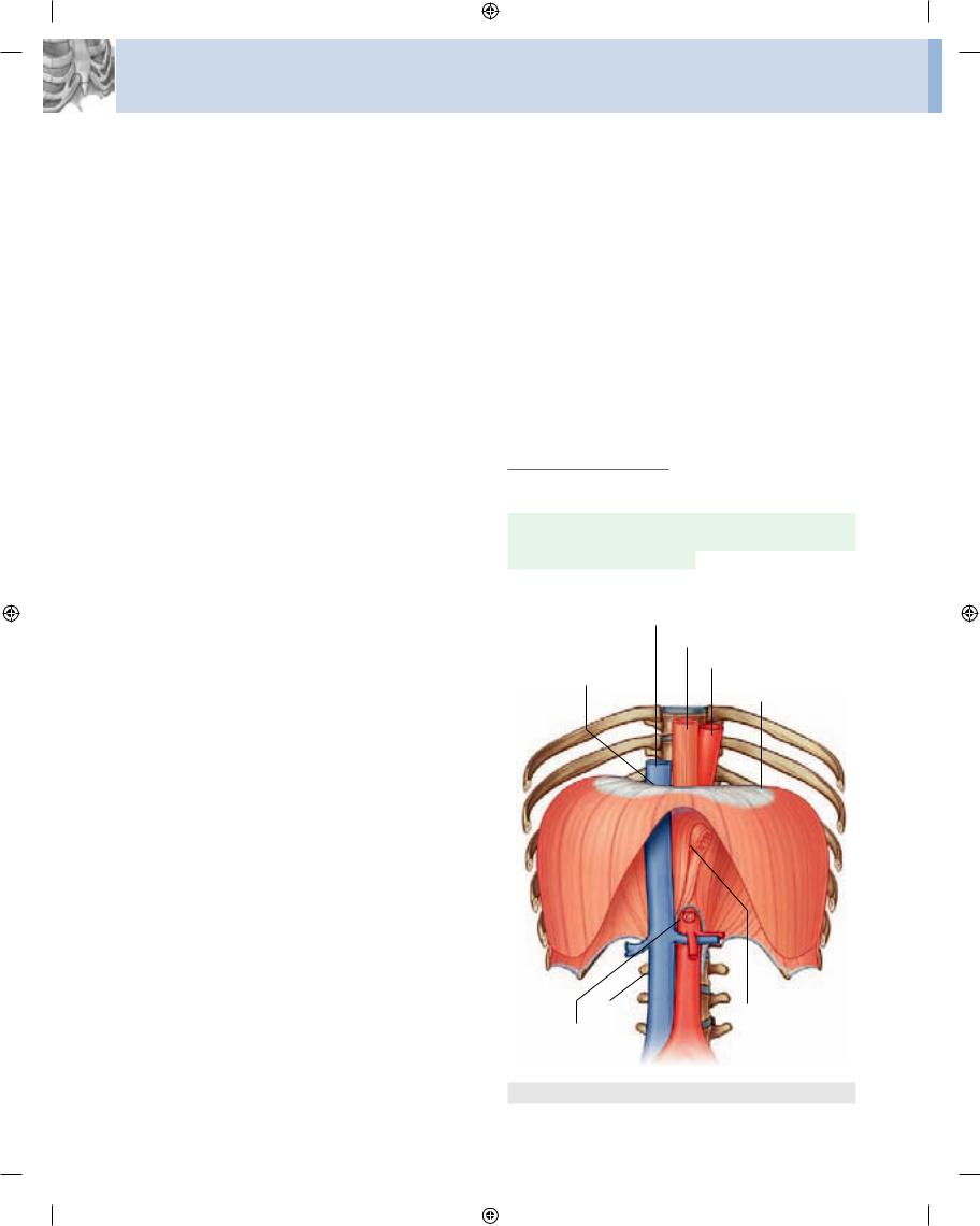
The superior thoracic aperture opens directly into the root of the neck (Fig. 3.7).
The superior aspect of each pleural cavity extends approximately 2–3 cm above rib I and the costal cartilage into the neck. Between these pleural extensions, major visceral structures pass between the neck and superior mediastinum. In the midline, the trachea lies immediately anterior to the esophagus. Major blood vessels and nerves pass in and out of the thorax at the superior thoracic aper ture anteriorly and laterally to these structures.
Superior thoracic aperture |
Rib I |
||||||
Esophagus |
|
|
|
Scapula |
|||
|
|
|
|||||
Brachial plexus |
|
|
|
Axillary inlet |
|||
|
|
|
|
|
|
|
|
|
|
|
|
|
|
|
|
|
|
|
|
|
|
|
|
|
|
|
|
|
|
|
|
|
|
|
|
|
|
|
|
|
|
|
|
|
|
|
|
|
|
|
|
|
|
|
|
|
|
|
|
|
|
|
|
|
|
|
|
|
|
|
|
|
|
|
|
|
|
|
|
|
|
|
|
|
|
|
|
|
|
|
|
|
|
|
|
|
|
|
|
|
|
|
|
|
|
|
|
Subclavian |
Trachea |
|
|
Coracoid |
|||||
artery and vein |
|
|
|
|
process |
||||
|
|
Clavicle |
|||||||
|
|
|
|
|
|
|
|
||
Fig. 3.7 Superior thoracic aperture and axillary inlet. |
3- |
|
Drake_ch03_main.indd 9 |
|
|
8/25/2008 4:10:29 PM |
|
|
||
|
|
|
|
Thorax
Upper limb
An axillary inlet, or gateway to the upper limb, lies on each side of the superior thoracic aperture. These two axil lary inlets and the superior thoracic aperture communi cate superiorly with the root of the neck (Fig. 3.7).
Each axillary inlet is formed by:
■the superior margin of the scapula posteriorly;
■the clavicle anteriorly; and
■the lateral margin of rib I medially.
The apex of each triangular inlet is directed laterally and is formed by the medial margin of the coracoid process, which extends anteriorly from the superior margin of the scapula.
The base of the axillary inlet’s triangular opening is the lateral margin of rib I.
Large blood vessels passing between the axillary inlet and superior thoracic aperture do so by passing over rib I.
Proximal parts of the brachial plexus also pass between the neck and upper limb by passing through the axillary inlet.
Abdomen
The diaphragm separates the thorax from the abdomen. Structures that pass between the thorax and abdomen either penetrate the diaphragm or pass posteriorly to it (Fig. 3.8):
■the inferior vena cava pierces the central tendon of the diaphragm to enter the right side of the mediasti num near vertebral level TVIII;
■the esophagus penetrates the muscular part of the diaphragm to leave the mediastinum and enter the abdomen just to the left of the midline at vertebral level TX;
■the aorta passes posteriorly to the diaphragm at the midline at vertebral level TXII;
■numerous other structures that pass between the thorax and abdomen pass through or posterior to the diaphragm.
Breast
The breasts, consisting of secretory glands, superficial fascia, and overlying skin, are in the pectoral region on each side of the anterior thoracic wall (Fig. 3.9).
Branches from the internal thoracic arteries and veins 3-10 perforate the anterior chest wall on each side of the ster
num to supply anterior aspects of the thoracic wall. Those branches associated mainly with the second to fourth intercostal spaces also supply the anteromedial parts of each breast.
Lymphatic vessels from the medial part of the breast accompany the perforating arteries and drain into the parasternal nodes on the deep surface of the thoracic wall:
■vessels and lymphatics associated with lateral parts of the breast emerge from or drain into the axillary region of the upper limb;
■lateral and anterior branches of the fourth to sixth inter costal nerves carry general sensation from the skin of the breast.
KEY FEATURES
Vertebral level TIV/V
When working with patients, physicians use vertebral levels to determine the position of important anatomical structures within body regions.
Inferior vena cava
|
Esophagus |
Caval opening |
Aorta |
(vertebral level TVIII) |
Central tendon |
|
|
|
of diaphragm |
LI |
Esophageal hiatus |
|
|
Aortic hiatus |
(vertebral level TX) |
(vertebral level TXII) |
|
Fig. 3.8 Major structures passing between abdomen and thorax.
Drake_ch03_main.indd 10 |
|
|
8/25/2008 4:10:31 PM |
|
|
||
|
|
|
|
|
Conceptual overview • Key features |
3 |
|
|
|
A
Axillary process
Axillary
lymph nodes
B
Internal |
|
thoracic artery |
Pectoralis major |
Second, third, and fourth perforating branches of internal thoracic artery
Parasternal lymph nodes
Lactiferous
sinuses
Fourth thoracic
intercostal nerve Lactiferous ducts
Secretory glands
Lymphatic vessel |
Deep (pectoral) fascia |
Fig. 3.9 Right breast.
|
|
|
Superior mediastinum |
Trachea |
|
|
|
Aortic arch |
|
|
|
|
|
Rib II |
|
|
|
Sternal angle |
|
The horizontal plane passing through the disc that sepa |
TIV |
|||
rates thoracic vertebrae TIV and TV is one of the most sig |
|
|||
nificant planes in the body (Fig. 3.10) because it: |
|
|||
■ passes through the sternal angle anteriorly, marking |
TV |
|||
|
the position of the anterior articulation of the costal |
|||
|
|
|||
|
|
|
||
|
cartilage of rib II with the sternum. |
The sternal angle is |
|
|
|
|
|
|
|
|
used to find the position of rib II as a |
reference for count |
|
|
|
ing ribs (because of the overlying clavicle, rib I is not |
|
|
|
|
palpable); |
|
|
|
■separates the superior mediastinum from the inferior mediastinum and marks the position of the superior limit of the pericardium;
■ |
marks where the arch of the aorta begins and ends; |
Inferior |
■ |
passes through the site where the superior vena cava |
mediastinum |
|
||
|
penetrates the pericardium to enter the heart; |
|
■is the level at which the trachea bifurcates into right and left main bronchi; and
■ marks the superior limit of the pulmonary trunk. |
Fig. 3.10 Vertebral level TIV/V. |
3-11 |
|
Drake_ch03_main.indd 11 |
|
|
8/25/2008 4:10:36 PM |
|
|
||
|
|
|
|