

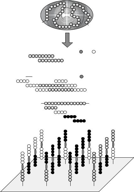
Micro-Nano Technology for Genomics and Proteomics BioMEMs - Ozkan
.pdfPEPTIDE ARRAYS IN PROTEOMICS AND DRUG DISCOVERY |
203 |
to eject extremely small droplets without possibly clogging a nozzle is unique. However, acoustic jets enabling to handle many different solutions in parallel have yet to be developed.
The prototype of a piezoelectric device is a glass capillary surrounded by a cylindrical piezoelectric device [3, 66, 509]. A control circuit sends a short pulse (typically 5 µs at 100 V) to the piezoelectric element, which causes compression of the piezoelectric device, generating shock pulses in the fluid chamber and forcing droplets out of the nozzle (Figure 7.37B). The size of the droplet depends upon the diameter of the nozzle, the magnitude of the pulse and the physical properties of the peptide solution. Micro-machined piezo or thermal jets [15, 251, 296, 302, 404] generate precisely sized droplets due to the highly defined orifice geometry, but unlike piezoelectric capillary jets these devices cannot be loaded directly from a microtitre plate.
One disadvantage of the piezoelectric ink-jets is the relatively large amount of peptide solution needed to generate reproducible droplets (typically 1–4 microliters). Therefore, these jets are used if one wants to deposit a large number of spots of the same peptide solution. Nevertheless, there are special piezoelectric micropipettes with extremely small dead volume available (GeSIMmbH, Großerkmannsdorf, Germany; [148]).
A major advantage of the ink-jet approach is the speed of printing. In the case of contact printing using pin tools the required close proximity of the reagent delivery tip and the surface makes a vertical motion necessary. During this vertical motion, touching the surface to deposit a droplet of the peptide solution, horizontal motion is stopped. Since motion must be stopped for each contact event, the time required to generate a microarray batch increases linearly with the number of peptide arrays in the batch. In contrast, with non-contact printing in the “on the fly” mode (no stop of horizontal motion necessary for delivering aliquots of peptide solution) total printing time increases much less rapidly with batch size.
Special cases of non-contact printing are the parallel direct displacement of liquids using an elastomer stamp [101] and the highly parallel top-spot-printing technology (Figure 7.37C; [124]) developed by IMTEK (University of Freiburg, Germany) and the HSG-IMIT Institute (Villingen-Schwenningen, Germany; www.hsg-imit.de). A pneumatic pulse to a specially designed mini-microtitre plate ejects up to 96 different sample droplets simultaneously onto a surface within one second. This high speed and the parallel mode makes top-spot- printing perfectly suited for applications that require an extremely high number of similar microarrays.
7.3. LIBRARY TYPES
A great variety of different library types has evolved, from the origin of multiple peptide synthesis through to the huge peptide collections generated and applied nowadays either in solution or immobilized as peptide arrays. In general, the design principles can be classified into protein sequence-based approaches dissecting or modifying the primary structure of a protein or peptide and de novo approaches exploring the entire or at least a significant and sometimes biased part of the potential sequence space. Only in exceptional cases are aspects of sequence-based and de novo strategies combined within one library (see 7.3.1.2. and 7.3.1.8.). So far, most investigations applying peptide arrays (Table 7.4) are based on the SPOT technology since it was the first generally amenable method suitable for even
204 |
ULRICH REINEKE, JENS SCHNEIDER-MERGENER AND MIKE SCHUTKOWSKI |
non-specialized laboratories. However, although many examples refer to publications using this technology, all library types described here can also be prepared by other newly developed peptide array production technologies.
7.3.1. Protein Sequence-Derived Libraries
Protein sequence-derived libraries provide the basic tools to elucidate interactions between a protein and a ligand, such as other proteins (enzymes), DNA, metal ions, cofactors and lipids. This provides detailed information about the ligand binding site. In addition, peptides derived from the binding site are often valuable starting compounds for peptide inhibitor or substrate development.
7.3.1.1.Scans of Overlapping Peptides The standard library to identify a protein’s ligand binding site is a scan of overlapping peptides, also called a peptide scan or simply pepscan (Figure 7.38A; [176, 178]). The entire protein sequence, or a certain part of it corresponding to a particular domain perhaps, is synthesized as short, overlapping, linear peptides that are subsequently tested for ligand binding. Usually, this involves 6- to 15-mer peptides since most linear binding sites (see 7.5.1.1.) do not exceed this range ([113, 415, 458, 562]). Furthermore, longer peptides result in raw products with rather limited purities, which cannot be used for array preparation without expensive purification procedures. However, to identify discontinuous binding sites (see 7.5.1.2.) longer peptides are considered an advantage if the peptide covers a folding motif comparable to the native protein structure (“domain scan” http://www.pepscan.nl/html/outframeset.html). In addition to the peptide length, another important parameter of peptide scans is the number of overlapping amino acids between two consecutive peptides. Usually, the peptides are shifted by one to three positions along the protein sequence. With shorter overlaps important peptides may be overlooked. In peptide scans derived from proteins containing disulfide bonds or free cysteine residues, these residues are commonly exchanged by similar amino acids, such as serine, to avoid dimerization and oligomerization of the peptides or covalent linkage to thiols in the ligands [417].
7.3.1.2.Hybritope Scans The mapping of discontinuous (conformational) binding sites (see 7.5.1.2.) necessitates analyzing peptide-ligand interactions with very low affinities. This led to the introduction of the hybritope and duotope scan (see 7.3.1.3.). Discontinuous epitopes are composed of two or more binding regions separated in the primary structure. Upon folding they are brought together on the protein surface to form a composite epitope. In the hybritope scan (Figure 7.38B) the peptides of a peptide scan (see 7.3.1.1.) are N-, C- or N- and C-terminally flanked by randomized positions ([445]; for synthesis procedures of randomized positions refer to [269]). The rationale is that a juxtaposed binding region within the discontinuous binding site can be mimicked by the randomized positions or a subset of peptides with the appropriate amino acid composition in the randomized sequence. Deconvolution libraries (see 7.3.2.1.) must then be used to identify single peptides from these peptide mixtures.
7.3.1.3.Duotope Scans and Matrix Scans Flanking randomized positions of the hybritope scan may either contribute via sequence motifs homologous to binding regions
PEPTIDE ARRAYS IN PROTEOMICS AND DRUG DISCOVERY |
205 |
||||||
|
|
|
|
|
|
|
|
H2N
COOH
AH2N 


 .......

 COOH
COOH


 .......
BH2N 


 .......

 COOH
COOH
X X X X


 X X X X
|
X X X X |
X X X X |
|
X X X X |
X X X X ....... |
C |
H2N |
COOH |
FIGURE 7.38. Protein sequence-derived peptide libraries. The amino acid sequence of the protein under investigation is used to generate short linear overlapping peptides. (A) Scan of overlapping peptides (peptide scan) (see 7.3.1.1.), (B) hybritope scan (see 7.3.1.2.), and (C) duotope scan or matrix scan (see 7.3.1.3.).
206 |
ULRICH REINEKE, JENS SCHNEIDER-MERGENER AND MIKE SCHUTKOWSKI |
of the discontinuous binding site, mimicking sequences according to the “mimotope concept” [177, 363] or detract via adverse effects, e.g., by unfavorable charged amino acid side chains. The duotope scan (Figure 7.38C) was therefore introduced to provide a rational tool to identify peptide mimics for discontinuous binding sites. The concept is that these binding sites can only be mimicked adequately if two or more binding regions are connected in one molecule by a linker moiety resembling their spacing in the protein’s three-dimensional structure. This means synthesizing all possible combinations of two overlapping peptides from a conventional peptide scan as one linear peptide for each combination, i.e., combinatorial chemistry with peptides as second level building blocks [451, 452]. This concept was validated by mapping the discontinuous epitope of the anti-hen-egg white lysozyme (HEL) antibody D1.3. A complete lysozyme duotope scan comprising all combinations of HEL-derived 10-mer overlapping peptides (offset by thee amino acids) combined via two β-alanine residues as a spacer moiety, synthesized by the SPOT method, resulted in an array of 41 by 41 (1681) 22-mer peptides, a number recommending array-based techniques for cost-effective experiments. Probing the array with mab D1.3 revealed a duotope peptide composed of two binding regions that exactly matched the structural epitope known from X-ray crystallography of the HEL-Fab D1.3 complex [40, 41]. The dissociation constant of the duotope scan peptide in a complex with mab D1.3 was determined by ELISA as 27 µM, a value significantly higher than that of the native HEL-D1.3 complex due to complete loss of the conformational stability conferred by the protein fold. Nevertheless, the duotope peptide clearly has a higher affinity compared to peptides spanning the single binding regions [451, 452].
If information about the binding region is available, e.g., from site-directed mutagenesis studies, one can carry out a partial duotope scan covering only the protein-derived sequences of interest. This strategy was applied to identify a mimic for a discontinuous epitope recognized by a neutralizing anti-interleukin-10 (IL-10) antibody [448, 451, 452]. Disadvantages of the duotope scan include having to assess a relatively large number of long peptides and possibly identifying false positives since hydrophobic residues that are often buried in the three-dimensional structure of the protein become exposed by dissecting the protein into duotope peptides and can cause unspecific interactions.
A concept analogous to the duotope approach called “matrix scan” has been developed (http://www.pepscan.nl/html/outframeset.html; [364]). However, experimental details were not available at the time of preparing this manuscript.
7.3.1.4. Amino Acid Substitution Scans The interaction of a peptide with a binding partner usually relies upon a limited number of amino acid residues that are effectively in contact with the binding partner. These amino acids contribute either to the binding free energy or to the specificity of the interaction and are referred to as key residues. If the peptide has to adopt a certain conformation upon or prior to binding, amino acids facilitating these conformations can also be regarded as critical. The concept of “alanine scanning” introduced to map protein-protein interactions by site-directed mutagenesis has been used to identify these residues (Figure 7.39A; [95]). Here, residues that cannot be exchanged by alanine without loss of binding and/or biological activity are regarded as key residues for the interaction. Scans with other amino acids are similarly used for alanine residues in the starting peptide itself or to explore the effect of charged residues, for example. It has to be considered that this only reveals effects that depend on the amino acid side
PEPTIDE ARRAYS IN PROTEOMICS AND DRUG DISCOVERY |
207 |
AH2N 


 COOH
COOH
A
 A
A


 A
A


 A
A


 A
A


 A
A


 A
A


 A
A


 A
A
wt
B
wt
wt
FIGURE 7.39. Libraries of substitution analogs. These peptide arrays of substitution analogs are used to identify the key residues required for the interaction with a binding partner or a certain biological activity. (A) Amino acid substitution scan (alanine scan) of a 9-mer peptide (see 7.3.1.4) and (B) complete substitutional analysis of a 3-mer peptide (see 7.3.1.5).
chains unless one incorporates building blocks that lead to a modified backbone. Among the naturally occurring amino acids proline plays a special role in amino acids substitution scans (synonym: replacement scan) and is therefore often used as a substitute. It can influence the pre-binding conformation by inducing a turn structure or preventing helical structures, providing indirect information about binding modes.
7.3.1.5. Substitutional Analyses If, for example, the amino acid substitution scanning approach employs all genetically encoded amino acids it is called (complete) substitutional analysis (synonyms: mutational analysis as referred to in some former publications, replacement analysis, analoguing). These experiments explore the effects of all possible single site substitutions of the starting peptide (Figure 7.39B; [415]; Frank and Overwin, 1996; [273, 447]). This identifies key residues that are those that cannot be substituted at all or only by physicochemically similar amino acids (e.g., leucine/isoleucine). Usually, other positions are not sensitive to substitutions and some may even lead to increased binding activity. Complete substitutional analyses are a rapid and effective way to delineate the structureactivity relationship of peptides and to simultaneously optimize the starting sequences with respect to the activity (binding, enzyme substrate properties, etc.) being screened for in the assay. In addition to substitutional analyses using the genetically encoded amino acids, D-amino acids [274], other unnatural amino acids [172, 265], or peptoidic building blocks [16, 456] are used to increase the diversity of side chain functionalities or backbone modifications. This often results in identifying substitution analogs that are stabilized against proteolytic degradation.
208 |
ULRICH REINEKE, JENS SCHNEIDER-MERGENER AND MIKE SCHUTKOWSKI |
|||
|
A |
B |
C |
D |
OOOOOOOOOOOOOOOOOOOO OOOOOOOOOO OOOOOOOOOO OOOOOOOOO -OOOOOOOOO --OOOOOOOO AOOOOOOOOO OOOOOOOO O-OOOOOOOO -O-OOOOOOO AAOOOOOOOO
OOOOOOO |
OO-OOOOOOO |
-OO-OOOOOO |
AAAOOOOOOO |
.... |
OOO-OOOOOO |
-OOO-OOOOO |
.... |
OOOOOOOOO |
OOOO-OOOOO |
-OOOO-OOOO |
|
OOOOO-OOOO |
-OOOOO-OOO |
OOOOOOOOO |
|
OOOOOOOO |
OOOOOO-OOO |
.... |
OOOOOOOOA |
OOOOOOO |
OOOOOOO-OO |
|
OOOOOOOAA |
OOOOOOOOOOOOOO-O O--OOOOOOO OOOOOOAAA OOOOOOOOO- O-O-OOOOOO
.... |
--OOOOOOOO |
O-OO-OOOOO |
.... |
OOOOOOOO |
O-OOO-OOOO |
AOOOOOOOOA |
|
O--OOOOOOO |
O-OOOO-OOO |
||
OOOOOO |
OO--OOOOOO |
O-OOOOO-OO |
AAOOOOOOAA |
OOOO |
OOO--OOOOO |
.... |
AAAOOOOAAA |
.... |
.... |
|
.... |
FIGURE 7.40. Analysis and optimization of peptide length (see 7.3.1.6.). (A) Truncation library with N- terminal, C-terminal and bi-directional stepwise truncations; (B) deletion library (one or more consecutive amino acids deleted at all possible positions), (C) combinatorial deletion library comprising all peptides with two or more positions omitted independently all over the starting sequence, and (D) progressive alanine substitution library.
7.3.1.6. Truncation, Deletion, and Combinatorial Deletion Libraries Biologically active peptides identified, for example using a peptide scan or by other types of peptide libraries, including chemical and biological approaches, often contain a well-defined core of key residues. In addition, these peptides include other dispensable positions resulting from the predefined peptide length used in the library design. In order to narrow down the peptide to the “active principle” or to minimize the molecular weight to facilitate peptidebased drug design, three different types of libraries are useful: (1) Truncation libraries (synonyms: size scan, window scan) comprise peptides omitting one or more N-, C- or N- and C-terminal amino acids (Figure 7.40A; Frank and Overwin, 1996). (2) Peptides from libraries of deletion analogs (Figure 7.40B) have one or more consecutive amino acids deleted at all possible positions. (3) Compared to deletion libraries, combinatorial deletion libraries additionally cover peptides with two or more positions omitted independently all over the sequence (Figure 7.40C). It should be noted that the number of peptide analogs covered by a combinatorial deletion library rapidly increases depending on the number of deleted positions and the peptide length.
As an alternative to truncation analyses, a few authors used progressive substitutions by alanine while retaining the overall peptide length (Figure 7.40D). Such library types were called progressive alanine substitution or progressive alanine fill-up libraries [75, 139, 140].
7.3.1.7. Cyclization Scans A widespread strategy to optimize the binding free energy of a peptide interacting with a binding partner is to stabilize the binding conformation. This is often achieved by cyclization, for example via disulfide bonds [191, 448]. However, the binding conformation is usually unknown since structure determination experiments by X-ray crystallography or NMR are time-consuming and laborious. Furthermore, docking of peptides to binding partners in silico is one of the most complex modeling problems due to the tremendous intrinsic flexibility. Therefore, a large number of cyclic peptide analogs have to be synthesized and screened to seek out the proper conformation of a biologically active peptide. A systematic approach is the “cyclization scan” comprising all
PEPTIDE ARRAYS IN PROTEOMICS AND DRUG DISCOVERY |
209 |
OOOOOOOOOO CCOOOOOOOO COCOOOOOOO COOCOOOOOO COOOCOOOOO COOOOCOOOO COOOOOCOOO
. . . .
OCCOOOOOOO OCOCOOOOOO OCOOCOOOOO OCOOOCOOOO OCOOOOCOOO OCOOOOOCOO
. . . .
FIGURE 7.41. Disulfide cyclization scan. The library covers all possible combinations of two cysteine residues within the starting sequence that are subsequently oxidized for cyclization (see 7.3.1.7.).
possible combinations of two cysteine residues within the starting peptide (Figure 7.41). An example of this approach is a library of 466 cyclic peptide analogs used to identify the optimal disulfide cyclic derivative of a linear 32-mer mimicking a discontinuous interleukin10 (IL-10) epitope [448]. The affinity was increased by a factor of 10 although one disulfide bond within a 32-mer peptide had only a limited impact on the overall conformational freedom [587]. Cyclization of peptides by disulfide bonds via cysteine residues is the most amenable strategy and often applied for peptide arrays especially for stepwise in situ peptide array production. However, several other chemical cyclization strategies can be similarly applied as shown for amide bonds [191] and the entire chemical repertoire for peptide or peptidomimetic cyclization can be used for array production technologies with pre-synthesized compounds.
7.3.1.8. Library Types: Miscellaneous The focus of this review article is the field of peptide arrays. Protein arrays are extensively described in other Chapters of this volume. However, arrays of protein domains inhabit the borderland between peptide and protein arrays since their sequences lengths are still accessible to chemical synthesis, branding them “peptide-like” from the technological point of view. On the other hand these domains usually retain stable folding, a functional feature claimed by protein arrays. Only two protein domain array publications, described in more detail in Section 7.5.2.2., should be mentioned here: (1) a complete L-amino acid substitutional analysis of the human YAP WW domain (44-mer) resulting in an array of more than 800 single site substitution variants, which was used to identify the key residues for stable domain folding or WW domain ligand interaction [546, 547] and (2) an array of 11859 tri-substituted variants of the human YAP WW domain used to identify WW domains with novel binding specificities [548]. The substitutions were introduced in a combinatorial manner at three different positions within the WW domain sequence, in other words a combination of protein sequence-derived and combinatorial library techniques.
210 |
ULRICH REINEKE, JENS SCHNEIDER-MERGENER AND MIKE SCHUTKOWSKI |
Another specialized library type was used to identify binding partners and characterize the binding specificities of PDZ domains, which mainly occur in proteins of the cytoskeleton and play a role in signal transduction. They predominantly interact via their binding partners’ C-termini where the carboxyl group is essential for binding. Hoffmuller¨ et al. described a SPOT synthesis peptide array of all known C-terminal peptides derived from the human proteins listed so far in the SWISSPROT databank [211]. Since peptides produced by standard SPOT synthesis protocols are attached to the solid support at their C-terminus, a novel synthetic pathway had to be established to generate free C-termini.
The last specialized peptide library type described here was applied to elucidate evolutionary transition pathways between three completely unrelated peptides recognized by the anti-p24 (HIV-1) mab CB4-1 [212]. These different peptide ligands were identified previously using combinatorial and deconvolution libraries [272]. The question of whether the different CB4-1 peptide ligands can be reciprocally converted into each other was answered by synthesizing and analyzing all possible (7,620,480) single step transition pathways (i.e. sequential conversion of one amino acid after the other) between the three ligands. The library comprising all 2560 possible transition peptides was designed with the software PepTrans. Complete L-amino acid substitutional analyses of all intermediates from the best transition pathways were performed in order to better understand the structural mechanisms involved in the sequence transformation. In this study the exceptional synthesis capacity of the SPOT method was exploited to analyze the sequence space between functionally related peptides with no sequence similarity.
7.3.2. De Novo Approaches
If a natural protein binding partner is not known, or if peptide ligands have to be identified without any previous knowledge, for example due to an intellectual property situation, one has to use combinatorial libraries with peptide mixtures or randomly generated libraries of single individual sequences. These strategies are summarized in this Section under “de novo” approaches. Although this review is restricted to approaches using peptide arrays and peptide chips it should be mentioned that most of the library types described in this Section were pioneered at the beginning of the combinatorial chemistry era and in the field of chemical libraries on beads [166, 215, 247, 286] or by using biological display techniques such as phage display [56, 118, 504]. Michal Lebl [299] has published a very lively historical review, with personal comments by the authors, of “classical” papers form the beginning of combinatorial chemistry.
The main problem for de novo identification of peptides is how to handle the immense number of potential peptide sequences, referred to as “combinatorial explosion”. Even if only using the genetically encoded amino acids the number of possible sequences dramatically increases with the peptide length:
|
dimers |
2 |
= 400 |
|
203 |
||
|
trimers |
204 |
= 8,000 |
|
tetramers |
205 |
= 160,000 |
|
pentamers |
206 |
= 3,200,000 |
|
hexamers |
207 |
= 64,000,000 |
|
heptamers |
208 |
= 1,280,000,000 |
|
octamers |
20 |
= 25,600,000,000 |
PEPTIDE ARRAYS IN PROTEOMICS AND DRUG DISCOVERY |
211 |
X
A X
B
X
X
X
IC
FR
T D G Q D W Y E A P Q N D W D D H D P S S L L A I F K I M G D V
X
B X
B
B
X
X
IC
FR
T D G Q D W Y L A P L N D A D D H D L S L L L L I F K I M G D V
FIGURE 7.42. Peptide mixtures. Peptide mixtures with defined positions (B) and randomized position (X) (see 7.3.2.).
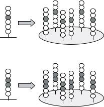
This Section describes two principles for the de novo identification of peptides with a predefined biological activity (mostly binding to another protein): (1) Using combinatorial libraries the aim is to completely cover the potential sequence space (see 7.3.2.1.). Although peptide arrays can be prepared with a high spot density (>40,000/cm2; [151]) there is no technology yet available to synthesize and handle billions of different compounds individually. The solution is to synthesize peptide mixtures with degenerated or randomized positions by statistically incorporating amino acids of a certain set (Figure 7.42). Defined amino acids are only used at a limited number of positions. This results in a manageable number of peptide pools screened on the peptide arrays. The randomized positions of active pools must then be deconvoluted iteratively using deconvolution libraries individually designed for the project, ultimately selecting the active compounds. (2) Since deconvolution is a time-consuming process, arrays of randomly generated peptides (see 7.3.2.2.) have also been applied. Since such libraries only cover a small percentage of the potential sequence space, initially selected peptides often have low affinities to the binding partner and must subsequently be optimized, for example using substitutional analyses.
7.3.2.1. Combinatorial Libraries The five most critical parameters for identifying peptide ligands from combinatorial library arrays are: (1) the number of peptide mixtures tested, (2) the number of defined positions, (3) the ratio between defined and randomized positions, (4) the appropriate spacing of the defined positions within the entire sequence length, and (5) the overall length of the peptides. These parameters determine the ratio between active and inactive compounds in the peptide mixtures and consequently the signal to noise ratio and likelihood of identifying bioactive peptides.
The development of multiple peptide synthesis robots and devices for peptide array preparation have enabled a continuous increase in peptide library complexity, which can be extrapolated to the future based on novel developments in the field of high density microarray production. The first arrays to be developed were hexamer libraries with two defined positions (mostly abbreviated in the literature as “O” or “B”) and four randomized, or mixed positions (X): e.g., XXO1O2XX [269]. Initially, these libraries were used to map

212 |
ULRICH REINEKE, JENS SCHNEIDER-MERGENER AND MIKE SCHUTKOWSKI |
||||||||||||||||||||
|
|
OXXXXX |
|
O = A |
|
|
|
|
|
|
|
|
|
|
ABDFGI |
|
|
|
|||
|
|
|
|
|
|
|
|
|
|
|
|
|
ABDFHI |
|
|
|
|||||
|
A |
XOXXXX |
|
O = B,C |
|
|
|
|
|
|
|
|
|
|
ABEFGI |
|
|
|
|||
|
|
|
|
|
|||||||||||||||||
|
XXOXXX |
|
O = D,E |
|
|
|
|
|
|
|
|
|
|
ABEFHI |
|
|
|
||||
|
|
|
|
|
|
|
|||||||||||||||
|
|
|
|
|
|
|
|
|
|
|
|
|
|
|
|||||||
|
|
XXXOXX |
|
O = F |
|
|
|
|
|
|
|
|
|
|
ACDFGI |
|
|
|
|||
|
|
XXXXOX |
|
O = G,H |
|
|
|
|
|
|
|
|
|
|
ACDFHI |
|
|
|
|||
|
|
XXXXXO |
|
|
O = I |
|
|
|
|
|
|
|
|
|
|
ACEFGI |
|
|
|
||
|
|
|
|
|
|
|
|||||||||||||||
|
|
|
|
|
|
|
|
|
|
|
|
|
|
|
|
ACEFHI |
|
|
|
||
|
|
|
|
|
|
|
|
|
|
|
|
|
|
|
|
|
|
|
|
|
|
|
|
|
|
|
|
|
|
|
|
|
|
|
|
|
|
AB |
|
DF |
|
GH |
|
|
|
|
|
|
|
|
|
|
|
|
|
|
|
|
|
|
|
|
|
|
|
|
|
|
|
|
|
|
|
|
|
|
|
|
|
|
|
|
|
|
|
|
|
|
|
|
|
|
|
|
|
|
|
|
|
|
|
|
|
AB |
|
DF |
|
IJ |
|
|
|
|
|
|
|
|
|
|
|
|
|
|
|
|
|
|
|
|
|
|
|
|
|
|
|
|
|
|
|
|
|
|
|
|
|
|
|
AB |
|
EF |
|
GH |
|
|
|
|
|
|
|
|
|
|
|
|
|
|
|
|
|
|
|
||||
|
B |
OOXXXX |
|
O = AB,AC |
|
|
|
|
|
|
|
AB |
|
EF |
|
IJ |
|
||||
|
XXOOXX |
|
O = DF,EF |
|
|
|
|
|
|
|
|
|
|
|
|
|
|||||
|
|
XXXXOO |
|
O = GH,IJ |
|
|
|
|
|
|
|
AC |
|
DF |
|
GH |
|
||||
|
|
|
|
|
|
|
|
|
|
|
|
|
|
|
|
|
|
|
|
|
|
|
|
|
|
|
|
|
|
|
|
|
|
|
|
|
|
AC |
|
DF |
|
IJ |
|
|
|
|
|
|
|
|
|
|
|
|
|
|
|
|
|
|
|
|
|
|
|
|
|
|
|
|
|
|
|
|
|
|
|
|
|
|
|
AC |
|
EF |
|
GH |
|
|
|
|
|
|
|
|
|
|
|
|
|
|
|
|
|
|
|
|
|
|
|
|
|
|
|
|
|
|
|
|
|
|
|
|
|
|
|
AC |
|
EF |
|
IJ |
|
|
|
|
|
|
|
|
|
|
|
|
|
|
|
|
|||||||
FIGURE 7.43. Deconvolution of active peptide mixtures (see 7.3.2.1). (A) In the positional scanning approach the most active amino acids at each position are identified from the initial library with peptide mixtures (X = randomized position; O = defined position with an individual amino acid). The deconvolution library consists of individual peptides representing all possible combinations of the most active amino acids. (B) In the dual positional scanning approach two positions are defined interdependently in the starting library.
linear antibody epitopes, but several other applications also emerged (see Section 7.5.; Table 7.4).
The randomized positions have to be deconvoluted to obtain single active peptides. Two general procedures have been described: (1) In the positional scanning approach (Figure 7.43A) the entire library is subdivided into a small number of peptide mixtures that have single amino acids at certain positions: O1XXXXX, XO2XXXX, XXO3XXX, XXXO4XX, XXXXO5X and XXXXXO6 (O and X as defined above). If the 20 naturally encoded amino acids are used for the defined positions (O) this library comprises 6 × 20 = 120 separate mixtures that are screened for binding, e.g., to an antibody [414]. Subsequently, individual peptides representing all possible combinations of the most active amino acids at each position are synthesized and screened. Alternatively, two (dual positional scanning approach) ([416]; Frank and Overwin, 1996; Figure 7.43B) or even more positions are defined in the first library. Although two defined positions involve greater synthesis efforts (202 = 400 peptide mixtures) the chance of successful primary screening is significantly better due to interactions with higher affinity and specificity. All randomized positions have to be deconvoluted in a second step based on the results with the starting library. Whereas the initial library is not predefined for a given screening molecule and can be applied universally, the follow-up libraries are tailor-made for specific purposes. The positional scanning approach assumes that the contributions of preferred amino acids at each position are additive or at least not interfering. However, this cannot be taken for granted in every system. (2) In order to circumvent this limitation, the randomized positions can be deconvoluted by an iterative process (Figure 7.44). Here, each deconvolution library is designed based on screening results from the starting or precursor library [269, 270]. Finally, a re-evaluation is recommended since there might be other amino acids at positions