11.7 ● Hydrolysis of Nucleic Acids |
351 |
Type II Restriction Endonucleases
Type II restriction enzymes have received widespread application in the cloning and sequencing of DNA molecules. Their hydrolytic activity is not ATP-depen- dent, and they do not modify DNA by methylation or other means. Most importantly, they cut DNA within or near particular nucleotide sequences that they specifically recognize. These recognition sequences are typically four or six nucleotides in length and have a twofold axis of symmetry. For example, E. coli has a restriction enzyme, EcoRI, that recognizes the hexanucleotide sequence GAATTC:
5 |
|
|
|
N |
|
N |
|
N |
|
N |
|
G |
|
A |
|
A |
|
T |
|
T |
|
C |
|
N |
|
N |
|
N |
|
N |
|
|
|
3 |
|
|
|
|
|
|
|
|
|
|
|
|
|
|
|
|
|
3 |
|
|
|
N |
|
N |
|
N |
|
N |
|
C |
|
T |
|
T |
|
A |
|
A |
|
G |
|
N |
|
N |
|
N |
|
N |
|
|
|
5 |
|
|
|
|
|
|
|
|
|
|
|
|
|
|
|
|
|
|
Note the twofold symmetry: the sequence read 5 n 3 is the same in both strands.
When EcoRI encounters this sequence in dsDNA, it causes a staggered, double-stranded break by hydrolyzing each chain between the G and A residues:
5 |
|
|
|
N |
|
N |
|
N |
|
N |
|
G |
|
A |
|
A |
|
T |
|
T |
|
C |
|
N |
|
N |
|
N |
|
N |
|
|
|
3 |
|
|
|
|
|
|
|
|
|
|
|
|
|
|
|
|
3 |
|
|
|
N |
|
N |
|
N |
|
N |
|
C |
|
T |
|
T |
|
A |
|
A G |
|
N |
|
N |
|
N |
|
N |
|
|
|
5 |
|
|
|
|
|
|
|
|
|
|
|
|
|
|
|
|
|
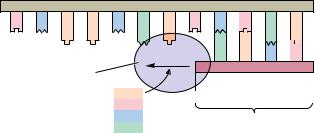
Staggered cleavage results in fragments with protruding single-stranded 5 - ends:
5 |
|
N |
|
N |
|
N |
|
N |
|
G |
5 A |
|
A |
|
T |
|
T |
|
C |
|
N |
|
N |
|
N |
|
N |
|
|
|
3 |
|
|
|
|
|
|
|
|
|
|
|
|
|
|
|
3 |
|
N |
|
N |
|
N |
|
N |
|
C |
|
T |
|
T |
|
A |
|
A 5 |
|
|
|
|
|
|
|
|
G |
|
N |
|
N |
|
N |
|
N |
|
|
|
5 |
|
|
|
|
|
|
|
|
|
|
|
|
|
|
|
|
|
|
|
|
|
|
|
Because the protruding termini of EcoRI fragments have complementary base sequences, they can form base pairs with one another.
N N N N G A A T T C N N N N
N N N N C T T A A G N N N N
Therefore, DNA restriction fragments having such “sticky” ends can be joined together to create new combinations of DNA sequence. If the fragments are derived from DNA molecules of different origin, novel recombinant forms of DNA are created.
EcoRI leaves staggered 5 -termini. Other restriction enzymes, such as Pst I, which recognizes the sequence 5 -CTGCAG-3 and cleaves between A and G, produce cohesive staggered 3 -ends. Still others, such as BalI, act at the center of the twofold symmetry axis of their recognition site and generate blunt ends that are noncohesive. BalI recognizes 5 -TGGCCA-3 and cuts between G and C.
352 Chapter 11 ● Nucleotides and Nucleic Acids
Table 11.5
Restriction Endonucleases
About 1000 restriction enzymes have been characterized. They are named by italicized three-letter codes, the first a capital letter denoting the genus of the organism of origin, while the next two letters are an abbreviation of the particular species. Because prokaryotes often contain more than one restriction enzyme, the various representatives are assigned letter and number codes as they are identified. Thus, EcoRI is the initial restriction endonuclease isolated from Escherichia coli, strain R. With one exception (NciI), all known type II restriction endonucleases generate fragments with 5 -PO4 and 3 -OH ends.
|
Common |
Recognition |
|
|
Enzyme |
Isoschizomers |
Sequence |
|
Compatible Cohesive Ends |
|
|
|
|
|
AluI |
|
AGgCT |
|
Blunt |
ApyI |
AtuI, EcoRII |
CCg(TA)GG |
|
|
AsuII |
|
TTgCGAA |
|
ClaI, HpaII, TaqI |
AvaI |
|
GgPyCGPuG |
SalI, XhoI, XmaI |
AvrII |
|
CgCTAGG |
|
|
BalI |
|
TGGgCCA |
|
Blunt |
BamHI |
|
GgGATCC |
|
BclI, BglII, MboI, Sau3A, XhoII |
BclI |
|
TgGATCA |
|
BamHI, BglII, MboI, Sau3A, XhoII |
BglII |
|
AgGATCT |
|
BamHI, Bcl I, MboI, Sau3A, XhoII |
BstEII |
|
GgGTNACC |
|
BstXI |
|
CCANNNNNgNTGG |
|
ClaI |
|
ATgCGAT |
|
AccI, AcyI, AsyII, HpaII, TaqI |
DdeI |
|
CgTNAG |
|
|
EcoRI |
|
GgAATTC |
|
|
EcoRII |
AtuI, ApyI |
gCC (A)GG |
|
|
|
|
T |
|
|
FnuDII |
ThaI |
CGgCG |
|
Blunt |
HaeI |
|
(TA)GGgCC(AT) |
Blunt |
HaeII |
|
PuGCGCgPy |
|
HaeIII |
|
GGgCC |
|
Blunt |
HincII |
|
GTPygPuAC |
Blunt |
HindIII |
|
AgAGCTT |
|
|
HpaI |
|
GTTgAAC |
|
Blunt |
HpaII |
|
CgCGG |
|
AccI, AcyI, AsuII, ClaI, TaqI |
KpnI |
|
GGTACgC |
|
BamHI, BclI, BglII, XhoII |
MboI |
Sau3A |
gGATC |
|
|
MspI |
|
CgCGG |
|
|
MstI |
|
TGCgGCA |
|
Blunt |
NotI |
|
GCgGGCCGC |
|
PstI |
|
CTGCAgG |
|
|
SacI |
SstI |
GAGCTgC |
|
|
SalI |
|
GgTCGAC |
|
AvaI, XhoI |
Sau3A |
|
gGATC |
|
BamHI, BclI, BglII, MboI, XhoII |
SfiI |
|
GGCCNNNNgNGGCC |
|
SmaI |
XmaI |
CCCgGGG |
|
Blunt |
SphI |
|
GCATGgC |
|
|
SstI |
SacI |
GAGCTgC |
|
|
TaqI |
|
TgCGA |
|
AccI, AcyI, AsuII, ClaI, HpaII |
XbaI |
|
TgCTAGA |
|
|
XhoI |
|
CgTCGAG |
|
AvaI, SalI |
XhoII |
|
(A)gGATC(T) |
BamHI, BclI, BglII, MboI, Sau3A |
|
|
G |
C |
|
XmaI |
SmaI |
CgCCGGG |
|
AvaI |
|
|
|
|
|
FIGURE 11.33
354 Chapter 11 ● Nucleotides and Nucleic Acids
Treatment of a linear 10kb DNA molecule with endonucleases gave the following results:
|
|
A |
B |
A + B |
Treatment with restriction endonuclease A gave 2 fragments, one 7 kb in size and one |
kb |
|
|
|
Longer DNA |
9 |
|
|
|
|
fragments |
3 kb in size, as judged by gel electrophoresis. |
|
|
|
|
|
|
|
Treatment of another sample of the 10 kb DNA with restriction endonuclease B gave |
|
|
|
|
|
|
|
|
|
|
|
|
|
|
|
|
7 |
|
|
|
|
|
|
|
three fragments, 8.5 kb, 1.0 kb, and 0.5 kb. |
|
|
|
|
|
|
|
|
|
|
|
|
|
|
Treatment of a third sample with both restriction endonucleases A and B yielded |
|
|
|
|
|
|
|
|
|
|
|
|
|
|
|
|
5 |
|
|
|
|
|
|
|
fragments 6.5, 2, 1, and 0.5 kb. |
|
|
|
|
|
|
|
3 |
|
|
|
|
|
|
|
|
|
|
|
|
|
|
|
|
|
|
|
|
|
|
|
|
|
|
|
|
|
|
|
|
|
|
|
|
|
|
|
|
|
|
|
|
|
|
|
|
|
|
|
|
|
|
|
|
|
|
1 |
|
|
|
|
Shorter DNA |
|
|
|
|
|
|
|
|
|
|
|
fragments |
|
The observed electrophoretic |
|
|
|
|
1 |
|
|
|
|
|
|
2 |
|
|
|
|
|
|
|
|
|
|
|
|
pattern |
|
|
|
|
|
|
|
|
|
|
|
|
|
|
|
|
|
|
|
|
|
|
|
|
Enzyme A |
|
3 |
7 |
|
|
|
|
|
|
7 |
3 |
|
|
|
|
|
|
|
|
|
|
|
|
|
|
|
|
|
|
|
|
|
|
|
|
|
|
|
|
|
|
|
|
|
|
|
|
|
|
|
|
|
|
|
|
|
|
|
|
|
|
|
|
|
|
|
|
|
|
|
|
|
|
|
|
|
|
|
|
|
|
|
|
|
|
|
|
|
|
|
|
|
|
|
|
|
|
|
|
|
|
|
|
|
|
|
|
|
|
|
|
|
|
|
|
|
|
|
|
|
|
3 |
|
|
|
|
|
|
4 |
|
|
|
|
|
|
|
|
|
5 |
|
|
Restriction mapping: consider |
|
|
|
|
0.5 |
1 |
|
|
|
1 0.5 |
1 |
|
|
0.5 |
|
|
|
|
8.5 |
|
|
|
8.5 |
|
|
8.5 |
the possible arrangements: |
|
|
|
|
|
|
|
|
|
Enzyme B |
|
|
|
|
|
|
|
|
|
|
|
|
|
|
|
|
|
|
|
|
|
|
|
|
|
|
|
|
|
|
|
|
|
|
|
|
|
|
|
|
|
|
|
|
|
|
|
|
|
|
|
|
|
|
|
|
|
|
|
|
|
|
|
|
|
|
|
|
|
|
|
|
0.5 |
1 |
|
8.5 |
|
|
|
|
0.5 |
8.5 |
1 |
|
1 0.5 |
8.5 |
|
|
|
|
|
|
|
|
|
|
|
|
|
|
|
|
|
|
|
|
|
|
|
|
|
|
|
|
|
|
|
|
|
|
|
|
|
|
|
|
|
|
|
|
|
|
|
|
|
|
|
|
|
|
|
|
|
|
|
|
|
|
|
|
|
|
|
|
6 |
|
|
|
|
|
|
7 |
|
|
|
|
|
|
|
|
|
8 |
|
|
|
|
|
|
|
|
|
|
|
|
|
|
|
|
|
|
|
|
|
|
|
|
|
|
|
|
|
|
|
|
|
|
|
|
|
|
|
|
|
|
|
|
|
|
|
|
Which arrangements are correct? |
|
The only combinations giving the observed A + B digests are 1 + 5 and 2 + 7 |
|
|
|
|
|
|
B |
|
|
|
B |
|
|
|
|
|
|
|
|
|
|
|
|
|
|
|
Possible maps of the 10kb fragment:
1 |
|
|
8.5 |
0.5 |
|
+ 5 |
|
|
|
|
|
|
|
Digests |
1 |
|
|
|
|
|
|
|
|
|
|
|
|
|
|
|
|
|
3 A |
|
7 |
|
|
|
|
|
|
|
B |
|
|
|
B |
|
|
0.5 |
|
|
8.5 |
1 |
|
|
+ 7 |
|
|
|
|
|
|
|
|
Digests |
2 |
|
|
|
|
|
|
|
|
7 |
|
A 3 |
|
|
To decide between these alternatives, a fixed point of reference, such as one of the ends of the fragment, must be identified or labeled. The task increases in complexity as DNA size, number of restriction sites, and/or number of restriction enzymes used increases.
● Restriction mapping of a DNA molecule as determined by an analysis of the electrophoretic pattern obtained for different restriction endonuclease digests. (Keep in mind that a dsDNA molecule has a unique nucleotide sequence and therefore a definite polarity; thus, fragments from one end are distinctly different from fragments derived from the other end.)
PROBLEMS
1.Draw the chemical structure of pACG.
2.Chargaff’s results (Table 11.3) yielded a molar ratio of 1.56 for A to G in human DNA, 1.75 for T to C, 1.00 for A to T, and 1.00 for G to C. Given these values, what are the mole fractions of A, C, G, and T in human DNA?
3.Adhering to the convention of writing nucleotide sequences in the 5 n 3 direction, what is the nucleotide sequence of the DNA strand that is complementary to d-ATCGCAACTGTCACTA?
4.Messenger RNAs are synthesized by RNA polymerases that read along a DNA template strand in the 3 n 5 direction, polymerizing ribonucleotides in the 5 n 3 direction (see Figure 11.24). Give the nucleotide sequence (5 n 3 ) of the DNA template strand from which the following mRNA segment was transcribed: 5 -UAGUGACAGUUGCGAU-3 .
5.The DNA strand that is complementary to the template strand copied by RNA polymerase during transcription has a nucleotide
FIGURE 12.1
12.1 ● The Primary Structure of Nucleic Acids |
357 |
12.1 ● The Primary Structure of Nucleic Acids
As recently as 1975, determining the primary structure of nucleic acids (the nucleotide sequence) was a more formidable problem than amino acid sequencing of proteins, simply because nucleic acids contain only four unique monomeric units whereas proteins have twenty. With only four, there are apparently fewer specific sites for selective cleavage, distinctive sequences are more difficult to recognize, and the likelihood of ambiguity is greater. The much greater number of monomeric units in most polynucleotides as compared to polypeptides is a further difficulty. Two important breakthroughs reversed this situation so that now sequencing nucleic acids is substantially easier than sequencing polypeptides. One was the discovery of restriction endonucleases that cleave DNA at specific oligonucleotide sites, generating unique fragments of manageable size (see Chapter 11). The second is the power of polyacrylamide gel electrophoresis separation methods to resolve nucleic acid fragments that differ from one another in length by just one nucleotide.
Sequencing Nucleic Acids
Two basic protocols for nucleic acid sequencing are in widespread use: the chain termination or dideoxy method of F. Sanger and the base-specific chemical cleavage method developed by A. M. Maxam and W. Gilbert. Because both methods are carried out on nanogram amounts of DNA, very sensitive analytical techniques are used to detect the DNA chains following electrophoretic separation on polyacrylamide gels. Typically, the DNA molecules are labeled with radioactive 32P,1 and following electrophoresis, the pattern of their separation is visualized by autoradiography. A piece of X-ray film is placed over the gel and the radioactive disintegrations emanating from 32P decay create a pattern on the film that is an accurate image of the resolved oligonucleotides. Recently, sensitive biochemical and chemiluminescent methods have begun to supersede the use of radioisotopes as tracers in these experiments.
Chain Termination or Dideoxy Method
To appreciate the rationale of the chain termination or dideoxy method, we first must briefly examine the biochemistry of DNA replication. DNA is a dou- ble-helical molecule. In the course of its replication, the sequence of nucleotides in one strand is copied in a complementary fashion to form a new second strand by the enzyme DNA polymerase. Each original strand of the double helix serves as template for the biosynthesis that yields two daughter DNA duplexes from the parental double helix (Figure 12.1). DNA polymerase carries out this reaction in vitro in the presence of the four deoxynucleotide monomers and copies single-stranded DNA, provided a double-stranded region of DNA is artificially generated by adding a primer. This primer is merely an oligonucleotide capable of forming a short stretch of dsDNA by base pairing with the ssDNA (Figure 12.2). The primer must have a free 3 -OH end from which the new polynucleotide chain can grow as the first residue is added in the initial step of the polymerization process. DNA polymerases synthesize new strands by adding successive nucleotides in the 5 n 3 direction.
1Because its longer half-life and lower energy make it more convenient to handle, 35S is replacing 32P as the radioactive tracer of choice in sequencing by the Sanger method. 35S- -labeled deoxynucleotide analogs provide the source for incorporating radioactivity into DNA.
|
Old |
Old |
|
|
|
A |
T |
|
|
|
|
T |
|
A |
|
Parental |
A |
|
DNA |
|
G C |
|
|
|
|
|
|
|
|
G |
C |
|
|
|
T |
A |
|
|
|
|
C G |
|
|
|
|
|
A |
T |
|
|
|
|
G |
C |
|
|
|
C |
G |
|
|
|
|
A |
|
|
|
|
|
|
A |
|
|
|
G |
|
C |
|
|
|
A |
|
|
T |
|
C |
|
|
|
G |
C |
|
New |
|
G |
|
C G |
C |
G |
|
|
|
T |
A |
|
T |
A |
|
|
A |
T |
|
A |
T |
|
C |
G |
|
C |
G |
|
|
T |
|
|
T A |
|
|
G C |
|
|
G C |
|
T |
A |
|
T |
A |
T |
A |
|
|
T A |
|
Old New New Old
● DNA replication yields two daughter DNA duplexes identical to the parental DNA molecule. Each original strand of the double helix serves as a template, and the sequence of nucleotides in each of these strands is copied to form a new complementary strand by the enzyme DNA polymerase. By this process, biosynthesis yields two daughter DNA duplexes from the parental double helix.
FIGURE 12.2
358 Chapter 12 ● Structure of Nucleic Acids
● DNA polymerase copies ssDNA in vitro in the presence of the four deoxynucleotide monomers, provided a doublestranded region of DNA is artificially generated by adding a primer, an oligonucleotide capable of forming a short stretch of dsDNA by base pairing with the ssDNA. The primer must have a free 3 -OH end from which the new polynucleotide chain can grow as the first residue is added in the initial step of the polymerization process.
Single- |
5' |
|
|
3' |
stranded DNA |
T C A A C G A T C |
T |
G |
A |
|
G |
A |
C |
T |
|
DNA polymerase |
|
|
5' |
|
Primer |
|
|
|
|
|
+ dATP |
3'– OH |
|
dTTP |
|
|
dCTP |
Annealing of primer |
|
dGTP |
|
creates a short stretch |
|
|
|
|
of double-stranded DNA |
Chain Termination Pro t o c o l
In the chain termination method of DNA sequencing, a DNA fragment of unknown sequence serves as template in a polymerization reaction using some type of DNA polymerase, usually Sequenase 2®, a genetically engineered version of bacteriophage T7 DNA polymerase that lacks all traces of exonuclease activity that might otherwise degrade the DNA. The primer requirement is met by an appropriate oligonucleotide (this method is also known as the primed synthesis method for this reason). Four parallel reactions are run; all four contain the four deoxynucleoside triphosphates dATP, dGTP, dCTP, and dTTP, which are the substrates for DNA polymerase (Figure 12.3 on the facing page). In each of the four reactions, a different 2 ,3 -dideoxynucleotide is included, and it is these dideoxynucleotides that give the method its name.
Because dideoxynucleotides lack 3 -OH groups, these nucleotides cannot serve as acceptors for 5 -nucleotide addition in the polymerization reaction, and thus the chain is terminated where they become incorporated. The concentrations of the four deoxynucleotides and the single dideoxynucleotide in each reaction mixture are adjusted so that the dideoxynucleotide is incorporated infrequently. Therefore, base-specific premature chain termination is only a random, occasional event, and a population of new strands of varying length is synthesized. Four reactions are run, one for each dideoxynucleotide, so that termination, although random, can occur everywhere in the sequence. In each mixture, each newly synthesized strand has a dideoxynucleotide at its 3 -end, and its presence at that position demonstrates that a base of that particular kind was specified by the template. A radioactively labeled dNTP is included in each reaction mixture to provide a tracer for the products of the polymerization process.
Reading Dideoxy Sequencing Gels
The sequencing products are visualized by autoradiography (or similar means) following their separation according to size by polyacrylamide gel electrophoresis (Figure 12.3). Because the smallest fragments migrate fastest upon electrophoresis and because fragments differing by only single nucleotides in length are readily resolved, the autoradiogram of the gel can be read from bottom to top, noting which lane has the next largest band at each step. Thus, the gel in Figure 12.3 is read AGCGTAGC (5 n 3 ). Because of the way DNA polymerase acts, this observed sequence is complementary to the corresponding unknown template sequence. Knowing this, the template sequence now can be written GCTACGCT (5 n 3 ).
360 Chapter 12 ● Structure of Nucleic Acids
Base-Specific Chemical Cleavage Method
The base-specific chemical cleavage (or Maxam–Gilbert) method starts with a single-stranded DNA that is labeled at one end with radioactive 32P. (Doublestranded DNA can be used if only one strand is labeled at only one of its ends.) The DNA strand is then randomly cleaved by reactions that specifically fragment its sugar–phosphate backbone only where certain bases have been chemically removed. There is no unique reaction for each of the four bases. However,
|
|
O |
|
|
|
|
O |
CH3 |
|
Guanine |
HN |
N |
Dimethyl |
|
HN |
N |
OH– |
|
|
+ |
|
|
G |
|
sulfate |
|
|
G |
|
5' |
H2N |
N |
N |
O |
5' |
H2N |
N |
N |
|
|
|
|
|
|
O |
|
|
+ |
H3COSOCH3 |
|
O |
|
|
|
|
|
|
|
|
|
|
|
O P |
O |
CH2 |
|
O |
O |
P O |
CH2 |
|
|
O– |
|
O |
|
|
O– |
O |
|
|
|
|
|
1 |
|
|
|
|
|
|
|
|
|
|
|
|
|
|
|
O |
|
Methylation |
|
|
O |
|
|
|
|
|
|
|
|
|
|
|
–O |
P |
O |
|
|
–O |
P |
O |
2 |
|
|
|
Ring opening |
|
|
O |
|
|
|
|
O |
|
with alkali |
|
|
|
|
|
|
|
|
|
|
|
|
|
|
|
|
|
3 |
|
|
3' |
|
|
|
|
3' |
|
Piperidine |
5'
O
O P O–
O–
5' PO4
DNA fragment
|
|
|
|
|
|
|
|
|
|
|
|
|
|
|
|
|
|
|
|
|
|
|
|
5' |
|
O |
|
|
|
|
|
|
|
|
|
|
|
|
|
|
|
|
|
|
|
|
|
|
|
|
|
|
|
|
|
|
|
|
|
|
|
|
|
|
|
|
|
|
|
|
|
|
|
|
|
|
|
|
|
|
|
|
|
|
|
|
|
|
|
|
|
|
|
|
|
|
|
|
|
|
|
|
|
|
|
|
|
|
|
|
O |
|
|
|
|
|
|
|
|
|
|
O |
CH3 |
|
|
|
|
|
|
|
|
|
|
|
|
|
|
|
|
|
|
|
|
|
|
|
|
|
|
|
|
|
|
|
|
|
|
|
|
|
|
|
|
|
|
|
|
|
|
|
|
|
|
|
|
|
|
|
|
|
|
|
|
|
|
|
|
|
|
|
|
|
|
|
|
|
|
|
|
|
|
|
|
|
|
|
|
OH |
|
|
|
|
|
|
|
|
|
H |
|
|
|
|
P |
|
O |
|
|
CH2 |
|
|
|
N |
|
|
|
|
|
|
|
|
|
|
|
|
|
|
|
|
|
|
|
HN |
|
|
|
|
|
|
|
|
|
|
|
|
|
|
|
OH– |
|
|
|
|
|
|
|
|
|
|
|
|
|
|
|
+ |
|
|
|
|
|
|
|
|
|
|
|
|
|
|
|
|
|
|
+ |
|
|
|
|
|
|
|
|
|
|
|
|
|
|
|
|
|
|
|
|
+ H2C |
|
|
|
|
|
|
|
|
|
|
|
|
|
|
|
|
|
|
O |
– |
|
|
|
OH HC |
|
N |
+ |
|
|
HC |
|
O |
|
|
|
|
|
|
|
|
|
|
|
|
|
|
|
|
|
|
|
|
|
|
|
|
|
|
|
|
|
|
|
|
|
|
|
|
|
|
|
|
|
|
|
|
|
|
|
|
|
|
|
|
|
|
C |
|
|
CH |
|
|
|
CH |
|
C |
|
N |
|
|
|
|
|
|
|
|
|
|
|
|
|
|
|
|
|
|
|
|
|
|
|
|
|
|
|
|
|
|
|
|
|
|
|
|
|
|
|
|
|
|
|
|
|
|
|
|
|
|
|
|
|
|
|
|
|
|
|
|
|
|
|
|
|
|
|
|
|
|
|
|
|
|
|
|
|
|
|
|
|
|
|
|
|
|
+ |
|
|
|
|
|
|
|
|
|
|
4 |
|
|
|
|
|
|
|
|
|
|
|
|
|
|
|
|
|
H2N N |
NH2 |
|
|
|
|
|
|
|
|
|
|
|
|
|
|
|
|
|
|
β -elimination |
|
|
|
|
|
|
|
|
O |
|
|
|
|
|
|
|
|
|
|
|
|
|
|
|
|
|
|
|
|
|
|
|
|
|
|
|
|
|
|
|
|
|
|
|
|
|
|
|
–O |
|
|
|
|
|
|
|
|
|
|
|
|
|
|
|
|
|
|
|
–O |
|
|
|
O– |
|
|
|
|
|
|
|
|
|
|
|
|
|
|
|
|
P |
|
O |
|
|
|
|
|
|
|
|
|
|
|
|
|
|
|
|
|
|
|
|
|
|
|
|
|
|
|
|
|
|
|
|
|
|
|
|
|
|
|
|
|
|
|
|
|
|
|
|
|
|
|
|
|
|
|
|
|
|
|
|
|
|
|
|
|
|
|
|
|
|
|
|
|
|
|
|
|
|
|
|
|
|
|
|
|
|
|
|
|
|
|
P |
|
O |
|
|
|
|
|
|
|
|
|
|
|
|
|
|
|
|
|
|
O |
|
|
|
|
|
|
|
|
|
|
|
|
|
|
|
|
|
|
|
|
|
|
|
|
|
|
|
|
|
|
|
|
|
|
|
|
|
|
|
|
|
|
|
|
|
|
|
|
|
|
|
|
|
|
|
|
|
|
|
|
|
|
|
|
|
|
|
|
|
|
|
|
|
|
|
|
|
|
|
|
|
|
|
|
|
|
|
|
|
|
|
|
|
|
|
|
|
|
|
|
O |
|
|
|
|
|
|
|
|
|
|
|
|
|
|
|
|
3' |
|
|
|
|
|
|
|
|
|
|
|
|
|
|
|
|
|
|
|
|
|
|
|
|
|
|
|
|
|
|
|
|
|
|
|
|
|
|
|
|
|
|
|
|
|
|
|
|
|
|
|
|
|
|
|
|
|
|
|
|
|
|
|
3' |
|
|
|
|
|
|
|
|
|
|
|
|
|
|
|
|
|
|
|
|
|
|
|
|
|
|
|
|
|
|
|
|
|
|
|
|
3' |
|
|
PO4 |
|
|
|
FIGURE 12.4 ● Maxam–Gilbert sequencing of DNA: cleavage at purines uses dimethyl |
|
|
|
|
|
sulfate, followed by strand scission with piperidine. |
|
|
|
|
|
|
|
|
|
|
DNA fragment |
|
|
|
|
|
|
|
|
|
|
|
|
|
|
|
|
|
|
|
|
|
|
|
|
|
|
|
|
|
|
|
|
|
|
|
|
|
|
|
|
|
|
|
|
|
Cleavage at G using dimethyl sulfate, followed by strand scission with piperidine: Under alkaline conditions, dimethyl sulfate reacts with guanine to methylate it at the 7-position (1). This substitution leads to instability of the N-9 glycosidic bond, so that in the presence of OH and the secondary amine piperidine (2), the purine ring is degraded and released. A - elimination reaction facilitated by piperidine (3) then causes the excision of the naked deoxyribose moiety from the sugar–phosphate backbone, with consequent scission of the DNA strand to yield 5 - and 3 -fragments.
Cleavage at A or G: If the DNA is first treated with acid, dimethyl sulfate methylates adenine at the 3-position as well as guanine at the 7-position (not shown). Subsequent reaction with OH and piperidine triggers degradation and displacement of the methylated A or G purine base and strand scission, essentially as indicated here for reaction of dimethyl sulfate with guanine.


 3' G C T A C G C T C T G A
 5'):
5'): 