plasm itself. Eukaryotic cells, however, contain numerous intracellular organelles that perform specialized tasks. Nucleic acid biosynthesis is handled in the nucleus; mitochondria are the site of electron transport, oxidative phosphorylation, fatty acid oxidation, and the tricarboxylic acid cycle; and secretion of proteins and other substances is handled by the endoplasmic reticulum and the Golgi apparatus. This partitioning of labor is not the only contribution of the membranes in these cells. Many of the processes occurring in these organelles (or in the prokaryotic cell) actively involve membranes. Thus, some of the enzymes involved in nucleic acid metabolism are membrane-associated. The electron transfer chain and its associated system for ATP synthesis are embedded in the mitochondrial membrane. Many enzymes responsible for aspects of lipid biosynthesis are located in the endoplasmic reticulum membrane.
Spontaneously Formed Lipid Structures
Monolayers and Micelles
Amphipathic lipids spontaneously form a variety of structures when added to aqueous solution. All these structures form in ways that minimize contact between the hydrophobic lipid chains and the aqueous milieu. For example, when small amounts of a fatty acid are added to an aqueous solution, a monolayer is formed at the air–water interface, with the polar head groups in contact with the water surface and the hydrophobic tails in contact with the air (Figure 9.2). Few lipid molecules are found as monomers in solution.
Further addition of fatty acid eventually results in the formation of micelles. Micelles formed from an amphipathic lipid in water position the hydrophobic tails in the center of the lipid aggregation with the polar head groups facing outward. Amphipathic molecules that form micelles are characterized by a unique critical micelle concentration, or CMC. Below the CMC, individual lipid molecules predominate. Nearly all the lipid added above the CMC, however, spontaneously forms micelles. Micelles are the preferred form of aggregation in water for detergents and soaps. Some typical CMC values are listed in Figure 9.3.
Layers
Micelles
Air
Water
Monolayer
Inside-out
Water
Bilayer
Normal
FIGURE 9.2 ● Several spontaneously formed lipid structures.
262 Chapter 9 ● Membranes and Cell Surfaces
Structure
Mr
CMC
Micelle Mr
Triton X-100
CH3
CH3
CH3
C
CH2
C
(OCH2CH2) OH
625
0.24 mM
90–95,000
10
Bilayer
CH3
CH3
Octyl glucoside
CH2OH
H
O
O
(CH2)
CH3
292
25 mM
H
7
OH
H
(a)
HO
H
H
OH
Unilamellar vesicle
C12E8
(Dodecyl octaoxyethylene ether)
C12H25
(OCH2CH2)
OH
538 0.071 mM
8
FIGURE 9.3 ● The structures of some common detergents and their physical properties. Micelles formed by detergents can be quite large. Triton X-100, for example, typically forms micelles with a total molecular mass of 90 to 95 kD. This corresponds to approximately 150 molecules of Triton X-100 per micelle.
(b)
Lipid Bilayers
Multilamellar vesicle
(c)
(d)
FIGURE 9.4 ● Drawings of (a) a bilayer,
(b) a unilamellar vesicle, (c) a multilamellar vesicle, and (d) an electron micrograph of a multilamellar Golgi structure ( 94,000).
(d, David Phillips/Visuals Unlimited)
Lipid bilayers consist of back-to-back arrangements of monolayers (Figure 9.2). Phospholipids prefer to form bilayer structures in aqueous solution because their pairs of fatty acyl chains do not pack well in the interior of a micelle. Phospholipid bilayers form rapidly and spontaneously when phospholipids are added to water, and they are stable structures in aqueous solution. As opposed to micelles, which are small, self-limiting structures of a few hundred molecules, bilayers may form spontaneously over large areas (108 nm2 or more). Because exposure of the edges of the bilayer to solvent is highly unfavorable, extensive bilayers normally wrap around themselves and form closed vesicles (Figure 9.4). The nature and integrity of these vesicle structures are very much dependent on the lipid composition. Phospholipids can form either unilamellar vesicles (with a single lipid bilayer) known as liposomes, or multilamellar vesicles. These latter structures are reminiscent of the layered structure of onions. Multilamellar vesicles were discovered by Sir Alex Bangham and are sometimes referred to as “Bangosomes” in his honor.
Liposomes are highly stable structures that can be subjected to manipulations such as gel filtration chromatography and dialysis. With such methods, it is possible to prepare liposomes having different inside and outside solution compositions. Liposomes can be used as drug and enzyme delivery systems in therapeutic applications. For example, liposomes can be used to introduce contrast agents into the body for diagnostic imaging procedures, including computerized tomography (CT) and magnetic resonance imaging (MRI) (Figure 9.5). Liposomes can fuse with cells, mixing their contents with the intracellular medium. If methods can be developed to target liposomes to selected cell populations, it may be possible to deliver drugs, therapeutic enzymes, and contrast agents to particular kinds of cells (such as cancer cells).
That vesicles and liposomes form at all is a consequence of the amphipathic nature of the phospholipid molecule. Ionic interactions between the
(Courtesy of Walter Perkins, The Liposome Co., Inc., Princeton, NJ, and Brigham and Women’s Hospital,
9.1 ● Membranes
263
FIGURE 9.5 ● A computerized tomography (CT) image of the upper abdomen of a dog, following administration of liposome-encapsulated iodine, a contrast agent that improves the light/dark contrast of objects in the image. The spine is the bright white object at the bottom and the other bright objects on the periphery are ribs. The liver (white) occupies most of the abdominal space. The gallbladder (bulbous object at the center top) and blood vessels appear dark in the image. The liposomal iodine contrast agent has been taken up by Kuppfer cells, which are distributed throughout the liver, except in tumors. The dark object in the lower right is a large tumor. None of these anatomical features would be visible in a CT image in the absence of the liposomal iodine contrast agent.
Boston, MA)
polar head groups and water are maximized, whereas hydrophobic interactions (see Chapter 2) facilitate the association of hydrocarbon chains in the interior of the bilayer. The formation of vesicles results in a favorable increase in the entropy of the solution, because the water molecules are not required to order themselves around the lipid chains. It is important to consider for a moment the physical properties of the bilayer membrane, which is the basis of vesicles and also of natural membranes. Bilayers have a polar surface and a nonpolar core. This hydrophobic core provides a substantial barrier to ions and other polar entities. The rates of movement of such species across membranes are thus quite slow. However, this same core also provides a favorable environment for nonpolar molecules and hydrophobic proteins. We will encounter numerous cases of hydrophobic molecules that interact with membranes and regulate biological functions in some way by binding to or embedding themselves in membranes.
Fluid Mosaic Model
In 1972, S. J. Singer and G. L. Nicolson proposed the fluid mosaic model for membrane structure, which suggested that membranes are dynamic structures composed of proteins and phospholipids. In this model, the phospholipid bilayer is a fluid matrix, in essence, a two-dimensional solvent for proteins. Both lipids and proteins are capable of rotational and lateral movement.
Singer and Nicolson also pointed out that proteins can be associated with the surface of this bilayer or embedded in the bilayer to varying degrees (Figure 9.6).They defined two classes of membrane proteins. The first, called peripheral proteins (or extrinsic proteins), includes those that do not penetrate the bilayer to any significant degree and are associated with the membrane by virtue
FIGURE 9.6
264 Chapter 9 ● Membranes and Cell Surfaces
● The fluid mosaic model of membrane structure proposed by S. J. Singer and G. L. Nicolson. In this model, the lipids and proteins are assumed to be mobile, so that they can move rapidly and laterally in the plane of the membrane. Transverse motion may also occur, but it is much slower.
of ionic interactions and hydrogen bonds between the membrane surface and the surface of the protein. Peripheral proteins can be dissociated from the membrane by treatment with salt solutions or by changes in pH (treatments that disrupt hydrogen bonds and ionic interactions). Integral proteins (or intrinsic proteins), in contrast, possess hydrophobic surfaces that can readily penetrate the lipid bilayer itself, as well as surfaces that prefer contact with the aqueous medium. These proteins can either insert into the membrane or extend all the way across the membrane and expose themselves to the aqueous solvent on both sides. Singer and Nicolson also suggested that a portion of the bilayer lipid interacts in specific ways with integral membrane proteins and that these interactions might be important for the function of certain membrane proteins. Because of these intimate associations with membrane lipid, integral proteins can only be removed from the membrane by agents capable of breaking up the hydrophobic interactions within the lipid bilayer itself (such as detergents and organic solvents). The fluid mosaic model has become the paradigm for modern studies of membrane structure and function.
Membrane Bilayer Thickness
The Singer–Nicolson model suggested a value of approximately 5 nm for membrane thickness, the same thickness as a lipid bilayer itself. Low angle X-ray diffraction studies in the early 1970s showed that many natural membranes were approximately 5 nm in thickness and that the interiors of these membranes were low in electron density. This is consistent with the arrangement of bilayers having the hydrocarbon tails (low in electron density) in the interior of the membrane. The outside edges of these same membranes were shown to be of high electron density, which is consistent with the arrangement of the polar lipid head groups on the outside surfaces of the membrane.
Hydrocarbon Chain Orientation in the Bilayer
An important aspect of membrane structure is the orientation or ordering of lipid molecules in the bilayer. In the bilayers sketched in Figures 9.2 and 9.4, the long axes of the lipid molecules are portrayed as being perpendicular (or
FIGURE 9.7
Human cell
9.1 ● Membranes
265
normal) to the plane of the bilayer. In fact, the hydrocarbon tails of phospholipids may tilt and bend and adopt a variety of orientations. Typically, the portions of a lipid chain near the membrane surface lie most nearly perpendicular to the membrane plane, and lipid chain ordering decreases toward the end of the chain (toward the middle of the bilayer).
Membrane Bilayer Mobility
The idea that lipids and proteins could move rapidly in biological membranes was a relatively new one when the fluid mosaic model was proposed. Many of the experiments designed to test this hypothesis involved the use of specially designed probe molecules. The first experiment demonstrating protein lateral movement in the membrane was described by L. Frye and M. Edidin in 1970. In this experiment, human cells and mouse cells were allowed to fuse together. Frye and Edidin used fluorescent antibodies to determine whether integral membrane proteins from the two cell types could move and intermingle in the newly formed, fused cells. The antibodies specific for human cell proteins were labeled with rhodamine, a red fluorescent marker, and the antibodies specific for mouse cell proteins were labeled with fluorescein, a green fluorescent marker. When both types of antibodies were added to newly fused cells, the binding pattern indicated that integral membrane proteins from the two cell types had moved laterally and were dispersed throughout the surface of the fused cell (Figure 9.7). This clearly demonstrated that integral membrane proteins possess significant lateral mobility.
Just how fast can proteins move in a biological membrane? Many membrane proteins can move laterally across a membrane at a rate of a few microns per minute. On the other hand, some integral membrane proteins are much more restricted in their lateral movement, with diffusion rates of about 10 nm/sec or even slower. These latter proteins are often found to be anchored to the cytoskeleton (Chapter 17), a complex latticelike structure that maintains the cell’s shape and assists in the controlled movement of various substances through the cell.
Lipids also undergo rapid lateral motion in membranes. A typical phospholipid can diffuse laterally in a membrane at a linear rate of several microns per second. At that rate, a phospholipid could travel from one end of a bacterial cell to the other in less than a second or traverse a typical animal cell in a few minutes. On the other hand, transverse movement of lipids (or proteins) from one face of the bilayer to the other is much slower (and much less likely). For example, it can take as long as several days for half the phospholipids in a bilayer vesicle to “flip” from one side of the bilayer to the other.
Membranes Are Asymmetric Structures
Biological membranes are asymmetric structures. There are several kinds of asymmetry to consider. Both the lipids and the proteins of membranes exhibit lateral and transverse asymmetries. Lateral asymmetry arises when lipids or proteins of particular types cluster in the plane of the membrane.
Lipids Exhibit Lateral Membrane Asymmetry
Lipids in model systems are often found in asymmetric clusters (see Figure 9.8). Such behavior is referred to as a phase separation, which arises either spontaneously or as the result of some extraneous influence. Phase separations can be induced in model membranes by divalent cations, which interact with negatively charged moieties on the surface of the bilayer. For example, Ca2 induces phase separations in membranes formed from phosphatidylserine (PS)
Mouse cell
● The Frye–Edidin experiment. Human cells with membrane antigens for red fluorescent antibodies were mixed and fused with mouse cells having membrane antigens for green fluorescent antibodies. Treatment of the resulting composite cells with redand green- fluorescent–labeled antibodies revealed a rapid mixing of the membrane antigens in the composite membrane. This experiment demonstrated the lateral mobility of membrane proteins.
FIGURE 9.8
266 Chapter 9 ● Membranes and Cell Surfaces
Addition
of Ca2+
● An illustration of the concept of lateral phase separations in a membrane. Phase separations of phosphatidylserine (green
circles) can be induced by divalent cations such as Ca2 .
and phosphatidylethanolamine (PE) or from PS, PE, and phosphatidylcholine. Ca2 added to these membranes forms complexes with the negatively charged serine carboxyls, causing the PS to cluster and separate from the other lipids. Such metal-induced lipid phase separations have been shown to regulate the activity of membrane-bound enzymes.
There are other ways in which the lateral organization (and asymmetry) of lipids in biological membranes can be altered. For example, cholesterol can intercalate between the phospholipid fatty acid chains, its polar hydroxyl group associated with the polar head groups. In this manner, patches of cholesterol and phospholipids can form in an otherwise homogeneous sea of pure phospholipid. This lateral asymmetry can in turn affect the function of membrane proteins and enzymes. The lateral distribution of lipids in a membrane can also be affected by proteins in the membrane. Certain integral membrane proteins prefer associations with specific lipids. Proteins may select unsaturated lipid chains over saturated chains or may prefer a specific head group over others.
Proteins Exhibit Lateral Membrane Asymmetry
Membrane proteins in many cases are randomly distributed through the plane of the membrane. This was one of the corollaries of the fluid mosaic model of Singer and Nicholson and has been experimentally verified using electron microscopy. Electron micrographs show that integral membrane proteins are often randomly distributed in the membrane, with no apparent long-range order.
However, membrane proteins can also be distributed in nonrandom ways across the surface of a membrane. This can occur for several reasons. Some proteins must interact intimately with certain other proteins, forming multisubunit complexes that perform specific functions in the membrane. A few integral membrane proteins are known to self-associatein the membrane, forming large multimeric clusters. Bacteriorhodopsin, a light-driven proton pump protein, forms such clusters, known as “purple patches,” in the membranes of Halobacterium halobium (Figure 9.9). The bacteriorhodopsin protein in these purple patches forms highly ordered, two-dimensional crystals.
FIGURE 9.9 ● The purple patches of
Halobacterium halobium.
Transverse Membrane Asymmetry
Membrane asymmetries in the transverse direction (from one side of the membrane to the other) can be anticipated when one considers that many properties of a membrane depend upon its two-sided nature. Properties that are a consequence of membrane “sidedness” include membrane transport, which is driven in one direction only, the effects of hormones at the outsides of cells, and the immunological reactions that occur between cells (necessarily involving only the outside surfaces of the cells). One would surmise that the proteins involved in these and other interactions must be arranged asymmetrically in the membrane.
Protein Transverse Asymmetry
Protein transverse asymmetries have been characterized using chemical, enzymatic, and immunological labeling methods. Working with glycophorin, the major glycoprotein in the erythrocyte membrane (discussed in Section 9.2), Mark Bretscher was the first to demonstrate the asymmetric arrangement of an integral membrane protein. Treatment of whole erythrocytes with trypsin released the carbohydrate groups of glycophorin (in the form of several small glycopeptides). Because trypsin is much too large to penetrate the erythrocyte membrane, the N-terminus of glycophorin, which contains the carbohydrate
9.1 ● Membranes
267
moieties, must be exposed to the outside surface of the membrane. Bretscher showed that [35S]-formylmethionylsulfone methyl phosphate could label the C-terminus of glycophorin with 35S in erythrocyte membrane fragments but not in intact erythrocytes. This clearly demonstrated that the C-terminus of glycophorin is uniformly exposed to the interior surface of the erythrocyte membrane. Since that time, many integral membrane proteins have been shown to be oriented uniformly in their respective membranes.
Lipid Transverse Asymmetry
Phospholipids are also distributed asymmetrically across many membranes. In the erythrocyte, phosphatidylcholine (PC) comprises about 30% of the total phospholipid in the membrane. Of this amount, 76% is found in the outer monolayer and 24% is found in the inner monolayer. Since this early observation, the lipids of many membranes have been found to be asymmetrically distributed between the inner and outer monolayers. Figure 9.10 shows the asymmetric distribution of phospholipids observed in the human erythrocyte membrane. Asymmetric lipid distributions are important to cells in several ways. The carbohydrate groups of glycolipids (and of glycoproteins) always face the outside surface of plasma membranes where they participate in cell recognition phenomena. Asymmetric lipid distributions may also be important to various integral membrane proteins, which may prefer particular lipid classes in the inner and outer monolayers. The total charge on the inner and outer surfaces of a membrane depends on the distribution of lipids. The resulting charge differences affect the membrane potential, which in turn is known to modulate the activity of certain ion channels and other membrane proteins.
How are transverse lipid asymmetries created and maintained in cell membranes? From a thermodynamic perspective, these asymmetries could only occur by virtue of asymmetric syntheses of the bilayer itself or by energy-depen- dent asymmetric transport mechanisms. Without at least one of these, lipids of all kinds would eventually distribute equally between the two monolayers of a membrane. In eukaryotic cells, phospholipids, glycolipids, and cholesterol are synthesized by enzymes located in (or on the surface of) the endoplasmic reticulum (ER) and the Golgi system (discussed in Chapter 25). Most if not all of these biosynthetic processes are asymmetrically arranged across the membranes of the ER and Golgi. There is also a separate and continuous flow of phospholipids, glycolipids, and cholesterol from the ER and Golgi to other membranes in the cell, including the plasma membrane. This flow is mediated by specific lipid transfer proteins. Most cells appear to contain such proteins.
Outer leaflet
Inner leaflet
Phosphatidylcholine
Phosphatidylethanolamine
Phosphatidylserine
Sphingomyelin
Total phospholipid
50
40
30
20
10
0
10
20
30
40
50
Percentage
FIGURE 9.10 ● Phospholipids are arranged asymmetrically in most membranes, including the human erythrocyte membrane, as shown here. Values are mole percentages. (After Rothman and Lenard, 1977. Science 194:1744.)
268 Chapter 9 ● Membranes and Cell Surfaces
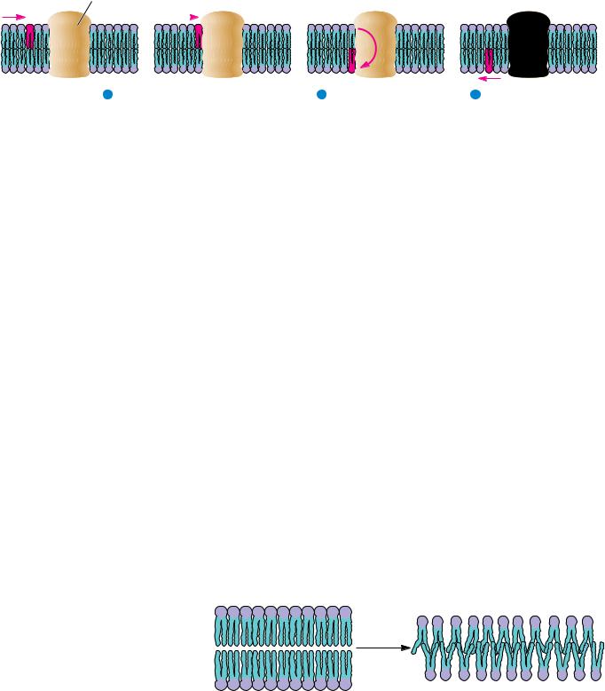
Flippase protein
1 Lipid molecule diffuses
2 Flippase flips lipid to
to flippase protein
opposite side of bilayer
3Lipid diffuses away from flippase
FIGURE 9.11 ● Phospholipids can be “flipped” across a bilayer membrane by the action of flippase proteins. When, by normal diffusion through the bilayer, the lipid encounters a flippase, it can be moved quickly to the other face of the bilayer.
Flippases: Proteins That Flip Lipids Across the Membrane
Proteins that can “flip” phospholipids from one side of a bilayer to the other have also been identified in several tissues (Figure 9.11). Called flippases, these proteins reduce the half-time for phospholipid movement across a membrane from 10 days or more to a few minutes or less. Some of these systems may operate passively, with no required input of energy, but passive transport alone cannot establish or maintain asymmetric transverse lipid distributions. However, rapid phospholipid movement from one monolayer to the other occurs in an ATP-dependentmanner in erythrocytes. Energy-dependent lipid flippase activity may be responsible for the creation and maintenance of transverse lipid asymmetries.
Membrane Phase Transitions
Lipids in bilayers undergo radical changes in physical state over characteristic narrow temperature ranges. These changes are in fact true phase transitions, and the temperatures at which these changes take place are referred to as transition temperatures or melting temperatures (Tm). These phase transitions involve substantial changes in the organization and motion of the fatty acyl chains within the bilayer. The bilayer below the phase transition exists in a closely packed gel state, with the fatty acyl chains relatively immobilized in a tightly packed array (Figure 9.12). In this state, the anti conformation is adopted by all the carbon–carbon bonds in the lipid chains. This leaves the lipid chains in their fully extended conformation. As a result, the surface area per lipid is minimal and the bilayer thickness is maximal. Above the transition temperature, a liquid crystalline state exists in which the mobility of fatty acyl chains is intermediate between solid and liquid alkane. In this more fluid, liquid crystalline state, the carbon–carbon bonds of the lipid chains more readily adopt gauche conformations (Figure 9.13). As a result, the surface area per lipid increases and the bilayer thickness decreases by 10 to 15%.
Heat
Gel
Liquid crystal
FIGURE 9.12 ● An illustration of the gel-to-liquid crystalline phase transition, which occurs when a membrane is warmed through the transition temperature, Tm. Notice that the surface area must increase and the thickness must decrease as the membrane goes through a phase transition. The mobility of the lipid chains increases dramatically.
Heat absorption
9.1 ● Membranes
269
Main transition
Pretransition
Temperature
(a) Before transition
(b) Post transition
Gauche conformations
Anti conformation
The sharpness of the transition in pure lipid preparations shows that the phase change is a cooperative behavior. This is to say that the behavior of one or a few molecules affects the behavior of many other molecules in the vicinity. The sharpness of the transition then reflects the number of molecules that are acting in concert. Sharp transitions involve large numbers of molecules all “melting” together.
Phase transitions have been characterized in a number of different pure and mixed lipid systems. Table 9.1 shows a comparison of the transition temperatures observed for several different phosphatidylcholines with different fatty acyl chain compositions. General characteristics of bilayer phase transitions include the following:
1.The transitions are always endothermic; heat is absorbed as the temperature increases through the transition (Figure 9.13).
2.Particular phospholipids display characteristic transition temperatures (Tm). As shown in Table 9.1, Tm increases with chain length, decreases with unsaturation, and depends on the nature of the polar head group.
3.For pure phospholipid bilayers, the transition occurs over a narrow temperature range. The phase transition for dimyristoyl lecithin has a peak width of about 0.2°C.
4.Native biological membranes also display characteristic phase transitions, but these are broad and strongly dependent on the lipid and protein composition of the membrane.
FIGURE 9.13 ● Membrane lipid phase transitions can be detected and characterized by measuring the rate of absorption of heat by a membrane sample in a calorimeter (see Chapter 3 for a detailed discussion of calorimetry). Pure, homogeneous bilayers (containing only a single lipid component) give sharp calorimetric peaks. Egg PC contains a variety of fatty acid chains and thus yields a broad calorimetric peak. Below the phase transition, lipid chains primarily adopt the anti conformation. Above the phase transition, lipid chains have absorbed a substantial amount of heat. This is reflected in the adoption of higher-energy conformations, including the gauche conformations shown.
270 Chapter 9 ● Membranes and Cell Surfaces
Table 9.1
Phase Transition Temperatures for Phospholipids in Water
Transition Temperature
Phospholipid
(Tm), °C
Dipalmitoyl phosphatidic acid (Di 16:0 PA)
67
Dipalmitoyl phosphatidylethanolamine (Di 16:0 PE)
63.8
Dipalmitoyl phosphatidylcholine (Di 16:0 PC)
41.4
Dipalmitoyl phosphatidylglycerol (Di 16:0 PG)
41.0
Dilauroyl phosphatidylcholine (Di 14:0 PC)
23.6
Distearoyl phosphatidylcholine (Di 18:0 PC)
58
Dioleoyl phosphatidylcholine (Di 18:1 PC)
22
1-Stearoyl-2-oleoyl-phosphatidylcholine
(1-18:0, 2-18:1 PC)
3
Egg phosphatidylcholine (Egg PC)
15
Adapted from Jain, M., and Wagner, R. C., 1980. Introduction to Biological Membranes. New York: John Wiley and Sons; Martonosi, A., ed., 1982. Membranes and Transport, Vol. 1. New York: Plenum Press.
5.With certain lipid bilayers, a change of physical state referred to as a pretransition occurs 5° to 15°C below the phase transition itself. These pretransitions involve a tilting of the hydrocarbon chains.
6.A volume change is usually associated with phase transitions in lipid bilayers.
7.Bilayer phase transitions are sensitive to the presence of solutes that interact with lipids, including multivalent cations, lipid-soluble agents, peptides, and proteins.
Cells adjust the lipid composition of their membranes to maintain proper fluidity as environmental conditions change.
9.2 ● Structure of Membrane Proteins
The lipid bilayer constitutes the fundamental structural unit of all biological membranes. Proteins, in contrast, carry out essentially all of the active functions of membranes, including transport activities, receptor functions, and other related processes. As suggested by Singer and Nicolson, most membrane proteins can be classified as peripheral or integral. The peripheral proteins are globular proteins that interact with the membrane mainly through electrostatic and hydrogen-bonding interactions with integral proteins. Although peripheral proteins are not discussed further here, many proteins of this class are described in the context of other discussions throughout this textbook. Integral proteins are those that are strongly associated with the lipid bilayer, with a portion of the protein embedded in, or extending all the way across, the lipid bilayer. Another class of proteins not anticipated by Singer and Nicolson, the lipid-anchored proteins, are important in a variety of functions in different cells and tissues. These proteins associate with membranes by means of a variety of covalently linked lipid anchors.