

Garrett R.H., Grisham C.M. - Biochemistry (1999)(2nd ed.)(en)
.pdf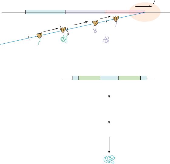


 H
H


348 Chapter 11 ● Nucleotides and Nucleic Acids
Enzymatic Hydrolysis
Enzymes that hydrolyze nucleic acids are called nucleases. Virtually all cells contain various nucleases that serve important housekeeping roles in the normal course of nucleic acid metabolism. Organs that provide digestive fluids, such as the pancreas, are rich in nucleases and secrete substantial amounts to hydrolyze ingested nucleic acids. Fungi and snake venom are often good sources of nucleases. As a class, nucleases are phosphodiesterases because the reaction that they catalyze is the cleavage of phosphodiester bonds by H2O. Because each internal phosphate in a polynucleotide backbone is involved in two phosphoester linkages, cleavage can potentially occur on either side of the phosphorus (Figure 11.30). Convention labels the 3 -side as a and the 5 -side as b. Cleavage on the a side leaves the phosphate attached to the 5 -position of the adjacent nucleotide, while b-side hydrolysis yields 3 -phosphate products. Enzymes or reactions that hydrolyze nucleic acids are characterized as acting at either a or b. A second convention denotes whether the nucleic acid chain was cleaved at some internal location, endo, or whether a terminal nucleotide residue was hydrolytically removed, exo. Note that exo a cleavage characteristically occurs at the 3 -end of the polymer, whereas exo b cleavage involves attack at the 5 -terminus (Figure 11.31).
Nuclease Specificity
Like most enzymes (see Chapter 14), nucleases exhibit selectivity or specificity for the nature of the substance on which they act. That is, some nucleases act only on DNA (DNases), while others are specific for RNA (the RNases). Still
A |
|
G |
C |
T |
|
A |
|
|
|
P |
P |
P |
P |
|
P |
OH |
|
|
|
a |
b |
a |
b a b |
a |
|
b |
|
|
|
Convention: The 3'-side of each phosphodiester |
|
|
|||||||
is termed a ; the 5 '-side is termed b. |
|
|
|
||||||
Hydrolysis of the a bond yields |
5'–PO4 products: |
|
|
||||||
A |
|
|
G |
|
|
C |
T |
|
A |
|
|
|
|
|
|
|
|
|
Mixture of |
OH |
|
OH |
|
|
OH |
|
OH |
OH 5' –nucleoside |
|
P |
|
P |
|
P |
|
|
P |
|
monophosphates (NMPs) |
|
|
|
|
|
P |
||||
Hydrolysis of the b bond yields 3'–PO4 products: |
|
|
|||||||
A |
|
|
G |
|
|
C |
T |
|
A |
|
P |
|
P |
|
|
P |
P |
|
OH |
P |
|
HO |
|
HO |
|
HO |
|
|
HO |
A 3', 5' –diPO4 |
|
|
|
|
|
A nucleoside from |
|||
|
A mixture of |
|
the 3'– OH end |
||||||
nucleotide |
|
|
|
||||||
from the 5'– end |
|
3'– NMPs |
|
|
|
||||
● Cleavage in polynucleotide chains: a cleavage yields 5 -phosphate products, whereas b cleavage gives 3 -phosphate products.

11.7 ● Hydrolysis of Nucleic Acids |
349 |
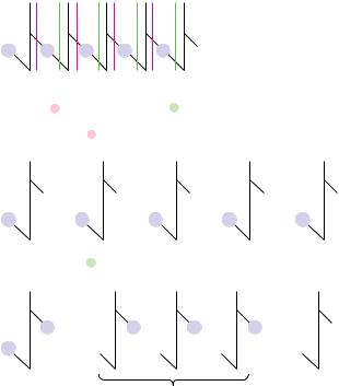
Snake venom phosphodiesterase: an “a” specific exonuclease:
|
C |
G |
A |
C |
G |
A |
|
|
|
|
|
Snake venom |
OH + |
|
|
etc. |
|
|
|
OH phosphodiesterase etc. |
OH |
||
P |
P |
|
P |
P |
P |
P |
|
|
a |
b a |
b |
|
|
5'–AMP |
|
Sequential removal of |
Snake venom |
||||||
|
|||||||
5'–NMP from 3'–end |
phosphodiesterase |
|
|||||
|
|
|
|
attacks here next |
|
||
Spleen phosphodiesterase: a “b” specific exonuclease:
|
C |
G |
A |
C |
G |
|
A |
|
|
|
Spleen |
|
+ |
|
|
|
|
|
phosphodiesterase |
P |
|
|
|
HO |
P |
P |
etc. |
HO |
HO |
P |
etc. |
|
|
|
|
3'–CMP |
Spleen |
|
|
Sequential removal of |
|
phosphodiesterase |
|||||
3'–NMP from 5'–end |
|
attacks here next |
|||||
● Snake venom phosphodiesterase and spleen phosphodiesterase are exonucleases that degrade polynucleotides from opposite ends.
others are nonspecific and are referred to simply as nucleases, as in nuclease S1 (see Table 11.4). Nucleases may also show specificity for only single-stranded nucleic acids or may only act on double helices. Single-stranded nucleic acids are abbreviated by an ss prefix, as in ssRNA; the prefix ds denotes doublestranded. Nucleases may also display a decided preference for acting only at
Table 11.4
Specificity of Various Nucleases
|
DNA, RNA, |
|
|
Enzyme |
or Both |
a or b |
Specificity |
|
|
|
|
Exonucleases |
|
|
|
Snake venom phosphodiesterase |
Both |
a |
Starts at 3 -end, 5 -NMP products |
Spleen phosphodiesterase |
Both |
b |
Starts at 5 -end, 3 -NMP products |
Endonucleases |
|
|
|
RNase A (pancreas) |
RNA |
b |
Where 3 -PO4 is to pyrimidine; oligos with pyrimidine |
|
|
|
3 -PO4 ends |
Bacillus subtilis RNase |
RNA |
b |
Where 3 -PO4 is to purine; oligos with purine 3 -PO4 ends |
RNase T1 |
RNA |
b |
Where 3 -PO4 is to guanine |
RNase T2 |
RNA |
b |
Where 3 -PO4 is to adenine |
DNase I (pancreas) |
DNA |
a |
Preferably between Py and Pu; nicks dsDNA, creating |
|
|
|
3 -OH ends |
DNase II (spleen, thymus, |
DNA |
b |
Oligo products |
Staphylococcus aureus) |
|
|
|
Nuclease S1 |
Both |
a |
Cleaves single-stranded but not double-stranded nucleic |
|
|
|
acids |
|
|
|
|

350 Chapter 11 ● |
Nucleotides and Nucleic Acids |
|
|
|
|
|
|
|
|
|
|
|
|||
Pancreatic RNase is an enzyme specific for b cleavage where a pyrimidine base lies |
|
|
|
|
|||||||||||
to the 3'–side of the phosphodiester; it acts endo. The products are oligonucleotides |
|
|
|
|
|||||||||||
with pyrimidine–3'–PO4 ends: |
|
|
|
|
|
|
|
|
|
|
|
|
|||
|
A |
G |
U |
A |
C |
C |
G |
|
A |
A |
U |
G |
A |
|
|
|
OH |
OH |
OH |
OH |
OH |
OH |
|
OH |
|
OH |
OH |
OH |
OH OH |
|
|
5' |
|
|
|
|
|
|
|
|
|
|
|
|
OH |
|
|
P |
P |
P |
P |
P |
P |
|
P |
|
P |
P |
P |
P |
|
|
|
|
|
|
|
|
|||||||||||
|
|
|
|
|
|
|
|
|
|
|
|
|
3' |
|
|
|
|
|
|
|
|
Pancreatic RNase |
|
|
|
|
|
|
|
||
A |
G |
U |
|
A |
C |
|
C |
|
|
G |
A |
A |
U |
G |
A |
OH |
OH |
OH |
+ |
OH |
OH |
+ |
OH |
|
OH |
OH |
OH |
OH |
OH |
OH |
|
|
|
P |
|
P |
P |
+ |
|
|
|
|
P |
|
OH |
||
P |
P |
|
P |
|
|
|
P |
P |
P |
P |
|||||
|
|
|
|
|
|
|
|
|
|||||||
|
|
|
|
|
|
|
|
|
|
|
|
|
|
|
3' |
Purine–only oligonucleotides arise from the 3'–end
● An example of nuclease specificity: The specificity of RNA hydrolysis by bovine pancreatic RNase. This RNase cleaves b at 3 -pyrimidines, yielding oligonucleotides with pyrimidine 3 -PO4 ends.
certain bases in a polynucleotide (Figure 11.32), or, as we shall see for restriction endonucleases, some nucleases will act only at a particular nucleotide sequence four to eight nucleotides in length. Table 11.4 lists the various permutations in specificity displayed by these nucleases and gives prominent examples of each. To the molecular biologist, nucleases are the surgical tools for the dissection and manipulation of nucleic acids in the laboratory.
Exonucleases degrade nucleic acids by sequentially removing nucleotides from their ends. Two in common use are snake venom phosphodiesterase and bovine spleen phosphodiesterase (Figure 11.31). Because they act on either DNA or RNA, they are referred to by the generic name phosphodiesterase. These two enzymes have complementary specificities. Snake venom phosphodiesterase acts by a cleavage and starts at the free 3 -OH end of a polynucleotide chain, liberating nucleoside 5 -monophosphates. In contrast, the bovine spleen enzyme starts at the 5 -end of a nucleic acid, cleaving b and releasing 3 -NMPs.
Restriction Enzymes
Restriction endonucleases are enzymes, isolated chiefly from bacteria, that have the ability to cleave double-stranded DNA. The term restriction comes from the capacity of prokaryotes to defend against or “restrict” the possibility of takeover by foreign DNA that might gain entry into their cells. Prokaryotes degrade foreign DNA by using their unique restriction enzymes to chop it into relatively large but noninfective fragments. Restriction enzymes are classified into three types, I, II, or III. Types I and III require ATP to hydrolyze DNA and can also catalyze chemical modification of DNA through addition of methyl groups to specific bases. Type I restriction endonucleases cleave DNA randomly, while type III recognize specific nucleotide sequences within dsDNA and cut the DNA at or near these sites.