7.4 ● Polysaccharides |
231 |
|
O |
OH |
|
|
|
OH |
O |
|
OH |
|
|
|
|
|
OH O HO |
|
HO |
|
|
|
O |
|
|
OH |
|
O |
O |
HO |
|
O |
HO |
|
|
|
|
|
OH |
|
O |
|
|
|
|
α -1,4-Linked D-glucose units |
β -1,4-Linked D-glucose units |
(a) |
(b) |
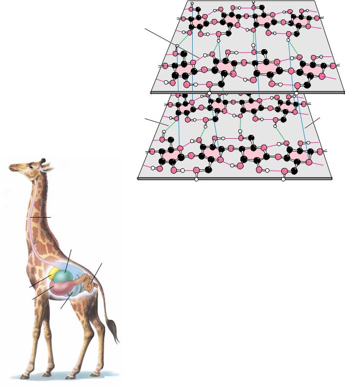
FIGURE 7.26 ● (a) Amylose, composed exclusively of the relatively bent (1n4) linkages, prefers to adopt a helical conformation, whereas (b) cellulose, with (1n4)-glycosidic linkages, can adopt a fully extended conformation with alternating 180° flips of the glucose units. The hydrogen bonding inherent in such extended structures is responsible for the great strength of tree trunks and other cellulose-based materials.
abundant natural polymer found in the world. Found in the cell walls of nearly all plants, cellulose is one of the principal components providing physical structure and strength. The wood and bark of trees are insoluble, highly organized structures formed from cellulose and also from lignin (see Figure 27.35). It is awe-inspiring to look at a large tree and realize the amount of weight supported by polymeric structures derived from sugars and organic alcohols. Cellulose also has its delicate side, however. Cotton, whose woven fibers make some of our most comfortable clothing fabrics, is almost pure cellulose. Derivatives of cellulose have found wide use in our society. Cellulose acetates are produced by the action of acetic anhydride on cellulose in the presence of sulfuric acid and can be spun into a variety of fabrics with particular properties. Referred to simply as acetates, they have a silky appearance, a luxuriously soft feel, and a deep luster and are used in dresses, lingerie, linings, and blouses.
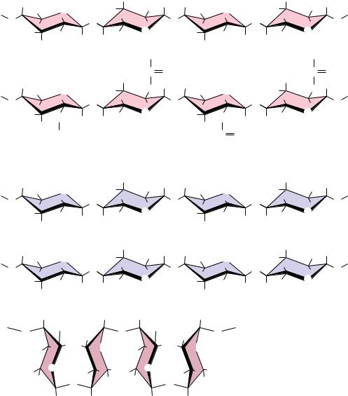
Cellulose is a linear homopolymer of D-glucose units, just as in -amylose. The structural difference, which completely alters the properties of the polymer, is that in cellulose the glucose units are linked by (1n4)-glycosidic bonds, whereas in -amylose the linkage is (1n4). The conformational difference between these two structures is shown in Figure 7.26. The (1n4)-linkage sites of amylose are naturally bent, conferring a gradual turn to the polymer chain, which results in the helical conformation already described (see Figure 7.22). The most stable conformation about the (1n4) linkage involves alternating 180° flips of the glucose units along the chain so that the chain adopts a fully extended conformation, referred to as an extended ribbon. Juxtaposition of several such chains permits efficient interchain hydrogen bonding, the basis of much of the strength of cellulose.
The structure of one form of cellulose, determined by X-ray and electron diffraction data, is shown in Figure 7.27. The flattened sheets of the chains lie side by side and are joined by hydrogen bonds. These sheets are laid on top of one another in a way that staggers the chains, just as bricks are staggered to give strength and stability to a wall. Cellulose is extremely resistant to hydrolysis, whether by acid or by the digestive tract amylases described earlier. As a result, most animals (including humans) cannot digest cellulose to any significant degree. Ruminant animals, such as cattle, deer, giraffes, and camels, are an exception because bacteria that live in the rumen (Figure 7.28) secrete the enzyme cellulase, a -glucosidase effective in the hydrolysis of cellulose. The resulting glucose is then metabolized in a fermentation process to the benefit of the host animal. Termites and shipworms (Teredo navalis) similarly digest cellulose because their digestive tracts also contain bacteria that secrete cellulase.
FIGURE 7.29
O
O
7.4 ● Polysaccharides |
233 |
Cellulose
OH |
|
HO |
OH |
OH |
|
OH |
2 |
|
2 |
|
CH |
O |
|
CH |
O |
|
|
|
|
|
|
O |
|
O |
O |
|
O |
O |
|
HO |
HO |
CH |
O |
HO |
HO |
CH |
O |
|
OH |
OH |
|
|
|
2 |
|
|
2 |
|
|
|
|
|
|
|
|
|
|
CH3 |
|
|
CH3 |
Chitin |
|
|
C O |
|
|
C O |
|
|
|
|
|
|
|
OH |
|
NH |
OH |
|
NH |
|
2 |
|
2 |
|
|
CH |
O |
|
CH |
O |
|
|
|
|
|
|
O |
|
O |
O |
|
O |
O |
|
HO |
HN |
CH |
O |
HO |
HN |
CH |
O |
|
OH |
OH |
|
|
|
2 |
|
|
2 |
|
|
|
|
|
|
|
C |
|
O |
C O |
|
|
|
|
|
|
N–Acetylglucosamine units |
|
|
|
|
|
|
|
CH3 |
CH3 |
Mannan
O |
HO |
O |
|
HO |
O |
HO |
O |
|
HO |
|
|
|
|
|
HO |
|
CH |
O |
HO |
|
|
CH |
O |
|
|
OH |
|
|
OH |
|
|
|
|
|
|
|
|
|
2 |
|
|
|
2 |
|
|
|
|
|
|
|
|
|
|
|
|
|
Mannose units |
|
|
|
|
Poly (D-Mannuronate) |
|
|
|
|
|
|
|
|
Poly (L-Guluronate) |
|
|
|
|
|
COO– |
|
O |
COO– |
|
O |
O |
HO |
|
HO |
|
|
|
|
O |
|
O |
O |
|
O |
H |
|
|
H |
|
|
O H O H
O O
● Like cellulose, chitin, mannan, and poly(D-mannuronate) form extended ribbons and pack together efficiently, taking advantage of multiple hydrogen bonds.
A l g i n a t e s
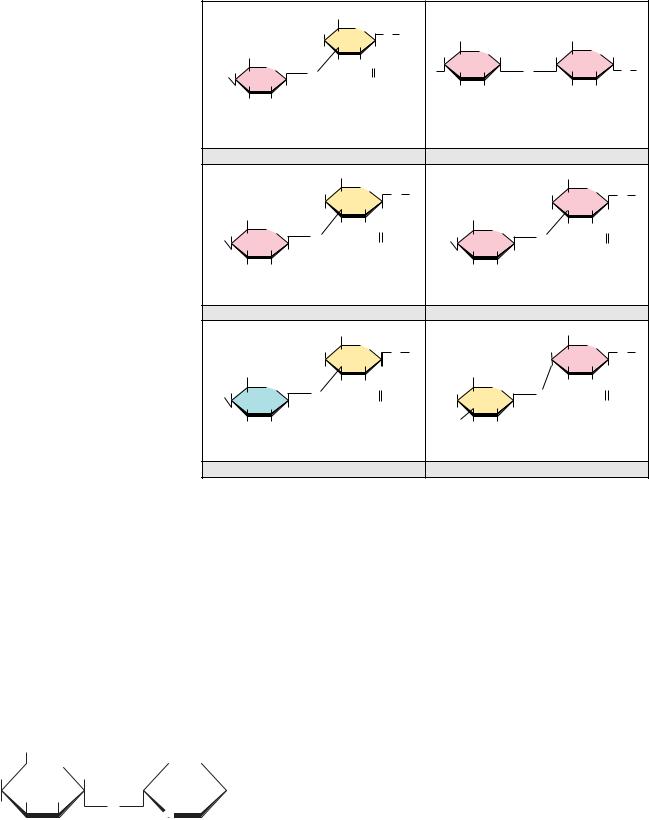
A family of novel extended ribbon structures that bind metal ions, particularly calcium, in their structure are the alginate polysaccharides of marine brown algae
(Phaeophyceae). These include poly( -D-mannuronate) and poly( -L-guluronate), which are (1n4) linked chains formed from -mannuronic acid and -L-guluronic acid, respectively. Both of these homopolymers are found together in most marine alginates, although to widely differing extents, and mixed chains containing both monomer units are also found. As shown in Figure 7.29, the conformation of poly( -D-mannuronate) is similar to that of cellulose. In the solid state, the free form of the polymer exists in cellulose-like form. However, complexes of the polymer with cations (such as lithium, sodium, potassium, and calcium) adopt a threefold helix structure, presumably to accommodate the bound cations. For poly( -L-guluronate) (Figure 7.29), the axial–axial configuration of the glycosidic linkage leads to a distinctly buckled ribbon with limited flexibility. Cooperative interactions between such buckled ribbons can only
234 Chapter 7 ● Carbohydrates
A D E E P E R L O O K
Billiard Balls, Exploding Teeth, and Dynamite— The Colorful History of Cellulose
Although humans cannot digest it and most people’s acquaintance with cellulose is limited to comfortable cotton clothing, cellulose has enjoyed a colorful and varied history of utilization. In 1838, Théophile Pelouze in France found that paper or cotton could be made explosive if dipped in concentrated nitric acid. Christian Schönbein, a professor of chemistry at the University of Basel, prepared “nitrocotton” in 1845 by dipping cotton in a mixture of nitric and sulfuric acids and then washing the material to remove excess acid. In 1860, Major E. Schultze of the Prussian army used the same material, now called guncotton, as a propellant replacement for gunpowder, and its preparation in brass cartridges soon made it popular for this purpose. The only problem was that it was too explosive and could detonate unpredictably in factories where it was produced. The entire town of Faversham, England, was destroyed in such an accident. In 1868, Alfred Nobel mixed guncotton with ether and alcohol, thus preparing nitrocellulose, and in turn mixed this with nitroglycerine and sawdust to produce dynamite. Nobel’s income from dynamite and also
from his profitable development of the Russian oil fields in Baku eventually formed the endowment for the Nobel Prizes.
In 1869, concerned over the precipitous decline (from hunting) of the elephant population in Africa, the billiard ball manufacturers Phelan and Collander offered a prize of $10,000 for production of a substitute for ivory. Brothers Isaiah and John Hyatt in Albany, New York, produced a substitute for ivory by mixing guncotton with camphor, then heating and squeezing it to produce celluloid. This product found immediate uses well beyond billiard balls. It was easy to shape, strong, and resilient, and it exhibited a high tensile strength. Celluloid was used eventually to make dolls, combs, musical instruments, fountain pens, piano keys, and a variety of other products. The Hyatt brothers eventually formed the Albany Dental Company to make false teeth from celluloid. Because camphor was used in their production, the company advertised that their teeth smelled “clean,” but, as reported in the New York Times in 1875, the teeth also occasionally exploded!
Portions adapted from Burke, J., 1996. The Pinball Effect: How Renaissance Water Gardens Made the Carburetor
Possible and Other Journeys Through Knowledge. New York: Little, Brown, & Company.
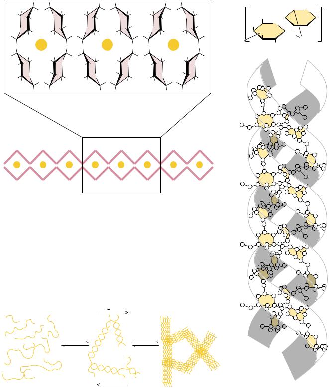
be strong if the interstices are filled effectively with water molecules or metal ions. Figure 7.30 shows a molecular model of a Ca2 -induced dimer of poly( - L-guluronate).
Agaro s e
An important polysaccharide mixture isolated from marine red algae (Rhodophyceae) is agar, which consists of two components, agarose and agaropectin. Agarose (Figure 7.31) is a chain of alternating D-galactose and 3,6- anhydro-L-galactose, with side chains of 6-methyl-D-galactose. Agaropectin is similar, but contains in addition sulfate ester side chains and D-glucuronic acid. The three-dimensional structure of agarose is a double helix with a threefold screw axis, as shown in Figure 7.31. The central cavity is large enough to accommodate water molecules. Agarose and agaropectin readily form gels containing large amounts (up to 99.5%) of water. Agarose can be processed to remove most of the charged groups, yielding a material (trade name Sepharose) useful for purification of macromolecules in gel exclusion chromatography. Pairs of chains form double helices that subsequently aggregate in bundles to form a stable gel, as shown in Figure 7.32.
G l y c o s a m i n o g l y c a n s
A class of polysaccharides known as glycosaminoglycans is involved in a variety of extracellular (and sometimes intracellular) functions. Glycosaminoglycans consist of linear chains of repeating disaccharides in which one of the monosaccharide units is an amino sugar and one (or both) of the monosaccharide units contains at least one negatively charged sulfate or carboxylate group. The repeating disaccharide structures found commonly in glycosaminoglycans are
236 Chapter 7 ● Carbohydrates
FIGURE 7.33 ● Glycosaminoglycans are formed from repeating disaccharide arrays. Glycosaminoglycans are components of the proteoglycans.
|
|
|
|
|
|
CH2OH |
|
|
|
|
|
|
|
|
|
|
|
|
|
|
|
|
–O3SO |
H |
O |
|
β |
|
COO– |
|
|
CH2OSO3– |
|
|
|
|
|
|
4 |
H |
1 |
O |
|
|
|
|
|
COO– |
|
|
H |
3 |
H |
|
|
|
O |
H |
H |
|
O |
H |
|
|
|
|
|
H |
H |
|
H |
|
|
|
|
|
H |
NHCCH3 |
|
|
|
|
|
|
|
|
|
|
|
|
|
|
1 α |
|
H |
H |
O |
|
β |
|
4 |
OH |
H |
1 |
O |
4 OH |
H |
O |
H 1 |
O |
|
|
|
|
|
|
|
2 |
|
α |
|
2 |
|
4 |
OH |
|
|
|
|
O |
|
|
|
OSO3– |
|
|
NHSO3– |
|
|
|
H |
|
|
|
|
|
|
|
H |
|
|
H |
|
H |
OH |
|
|
N-Acetyl- |
|
|
|
|
|
|
N-Sulfo- |
|
|
|
|
|
|
|
|
|
|
|
|
|
|
D-Glucuronate |
D-galactosamine-4-sulfate |
D-Glucuronate- |
D-glucosamine-6-sulfate |
|
|
|
|
|
|
|
|
|
|
|
2-sulfate |
|
|
|
|
|
|
Chondroitin-4-sulfate |
|
|
|
|
|
|
|
|
Heparin |
|
|
|
|
|
|
|
|
|
|
CH2OSO3– |
|
|
|
|
|
|
CH2OH |
|
|
|
|
|
|
HO |
|
O |
β |
|
|
|
|
|
H |
|
O |
β |
O |
|
|
|
|
|
|
H |
|
|
O |
|
|
|
|
|
|
H |
H 1 |
|
|
|
|
|
4 |
H 1 |
|
|
|
|
|
|
|
|
COO– |
|
|
H |
|
|
|
H |
|
|
COO– |
HO |
3 |
|
H |
|
|
|
|
|
|
|
|
|
|
|
|
H |
H |
O |
|
β |
|
H |
NHCCH3 |
|
H |
H |
|
O |
β |
H |
NHCCH3 |
H 1 |
O |
|
|
|
|
|
|
|
O |
|
|
|
|
4 |
OH |
|
|
|
|
|
O |
|
4 |
OH H 1 |
|
|
|
O |
|
|
|
|
H |
|
|
|
|
|
|
|
|
|
|
H |
|
|
|
|
|
|
H |
OH |
|
|
N-Acetyl- |
|
|
H |
|
OH |
|
N-Acetyl- |
|
|
|
|
|
|
|
|
|
|
|
|
|
|
D-Glucuronate |
D-galactosamine-6-sulfate |
D-Glucuronate |
|
D-glucosamine |
|
Chondroitin-6-sulfate |
|
|
|
|
|
|
|
Hyaluronate |
|
|
|
|
|
|
|
|
|
CH2OH |
|
|
|
|
|
|
|
CH2OSO3– |
|
|
|
|
|
–O |
SO |
|
O |
β |
|
|
|
|
|
H |
6 |
O |
β |
|
|
|
|
|
H |
|
|
|
|
|
H |
O |
|
|
|
|
3 |
|
|
|
O |
|
|
|
|
|
4 |
H 1 |
|
|
|
|
|
4 |
|
H 1 |
|
|
|
|
|
OH |
|
|
H |
|
|
|
H |
3 |
|
|
H |
|
|
CH2OH |
|
|
|
H |
|
|
|
|
|
|
H |
NHCCH3 |
|
|
|
H |
NHCCH3 |
H |
|
O |
|
β |
|
HO |
H |
|
O |
β |
COO– |
1 |
O |
|
|
|
|
|
|
O |
|
|
|
|
4 |
OH |
H |
|
|
|
|
|
O |
|
|
|
|
H |
|
|
|
O |
|
|
H |
|
|
|
|
|
|
H |
3 |
|
|
|
|
|
|
|
|
|
|
|
|
|
|
|
|
H |
|
|
|
|
|
|
H |
OH |
|
|
N-Acetyl-D- |
|
|
H |
|
OH |
|
N-Acetyl- |
|
|
|
|
|
|
|
|
D-Galactose |
|
|
L-Iduronate |
galactosamine-4-sulfate |
|
D-glucosamine-6-sulfate |
|
|
Dermatan sulfate |
|
|
|
|
|
|
|
Keratan sulfate |
|
|
|
tant components of the vitreous humor in the eye and of synovial fluid, the lubricant fluid of joints in the body. The chondroitins and keratan sulfate are found in tendons, cartilage, and other connective tissue, whereas dermatan sulfate, as its name implies, is a component of the extracellular matrix of skin. Glycosaminoglycans are fundamental constituents of proteoglycans (discussed later).
PROBLEMS
1.Draw Haworth structures for the two possible isomers of D- altrose (Figure 7.2) and D-psicose (Figure 7.3).
2.Give the systematic name for stachyose (Figure 7.19).
3.Trehalose, a disaccharide produced in fungi, has the following structure:
CH2OH |
|
|
H |
|
OH |
|
|
|
|
|
|
|
|
|
|
|
|
O |
|
|
|
|
|
|
|
|
|
|
|
|
|
|
|
|
|
|
H |
|
|
H |
H |
|
|
|
|
H |
|
|
OH |
H |
OH |
H |
|
|
|
HOCH2 |
|
|
O |
|
|
|
|
|
|
|
|
HO |
|
|
|
O |
|
|
OH |
|
|
|
|
OH |
|
|
|
|
|
|
|
|
|
|
|
|
|
|
|
|
|
|
|
|
|
|
|
|
|
|
|
|
H |
|
|
|
|
|
|
|
|
|
|
|
|
|
H |
|
|
a.What is the systematic name for this disaccharide?
b.Is trehalose a reducing sugar? Explain.
4.Draw a Fischer projection structure for L-sorbose (D-sorbose is shown in Figure 7.3).
5.-D-Glucose has a specific rotation, [ ]D20, of 112.2°, whereas-D-glucose has a specific rotation of 18.7°. What is the composition of a mixture of -D- and -D-glucose, which has a specific
rotation of 83.0°?
6.A 0.2-g sample of amylopectin was analyzed to determine the fraction of the total glucose residues that are branch points in the structure. The sample was exhaustively methylated and then digested, yielding 50 mol of 2,3-dimethylglucose and 0.4 mol of 1, 2, 3, 6-tetramethylglucose.
a. What fraction of the total residues are branch points? b. How many reducing ends does this amylopectin have?
8.1 ● Fatty Acids
A fatty acid is composed of a long hydrocarbon chain (“tail”) and a terminal carboxyl group (or “head”). The carboxyl group is normally ionized under physiological conditions. Fatty acids occur in large amounts in biological systems, but rarely in the free, uncomplexed state. They typically are esterified to glycerol or other backbone structures. Most of the fatty acids found in nature have an even number of carbon atoms (usually 14 to 24). Certain marine organisms, however, contain substantial amounts of fatty acids with odd numbers of carbon atoms. Fatty acids are either saturated (all carbon–carbon bonds are single bonds) or unsaturated (with one or more double bonds in the hydrocarbon chain). If a fatty acid has a single double bond, it is said to be monounsaturated, and if it has more than one, polyunsaturated. Fatty acids can be named or described in at least three ways, as listed in Table 8.1. For example, a fatty acid composed of an 18-carbon chain with no double bonds can be called by its systematic name (octadecanoic acid), its common name (stearic acid), or its shorthand notation, in which the number of carbons is followed by a colon and the number of double bonds in the molecule (18:0 for stearic acid). The structures of several fatty acids are given in Figure 8.1. Stearic acid (18:0) and palmitic acid (16:0) are the most common saturated fatty acids in nature.
Free rotation around each of the carbon–carbon bonds makes saturated fatty acids extremely flexible molecules. Owing to steric constraints, however, the fully extended conformation (Figure 8.1) is the most stable for saturated fatty acids. Nonetheless, the degree of stabilization is slight, and (as will be seen) saturated fatty acid chains adopt a variety of conformations.
Unsaturated fatty acids are slightly more abundant in nature than saturated fatty acids, especially in higher plants. The most common unsaturated fatty acid
Table 8.1
Common Biological Fatty Acids
Number |
|
|
|
|
of Carbons |
Common Name |
Systematic Name |
Symbol |
Structure |
|
|
|
|
Saturated fatty acids |
|
|
|
12 |
Lauric acid |
Dodecanoic acid |
12:0 |
CH3(CH2)10COOH |
14 |
Myristic acid |
Tetradecanoic acid |
14:0 |
CH3(CH2)12COOH |
16 |
Palmitic acid |
Hexadecanoic acid |
16:0 |
CH3(CH2)14COOH |
18 |
Stearic acid |
Octadecanoic acid |
18:0 |
CH3(CH2)16COOH |
20 |
Arachidic acid |
Eicosanoic acid |
20:0 |
CH3(CH2)18COOH |
22 |
Behenic acid |
Docosanoic acid |
22:0 |
CH3(CH2)20COOH |
24 |
Lignoceric acid |
Tetracosanoic acid |
24:0 |
CH3(CH2)22COOH |
Unsaturated fatty acids (all double bonds are cis) |
|
|
16 |
Palmitoleic acid |
9-Hexadecenoic acid |
16:1 |
CH3(CH2)5CHPCH(CH2)7COOH |
18 |
Oleic acid |
9-Octadecenoic acid |
18:1 |
CH3(CH2)7CHPCH(CH2)7COOH |
18 |
Linoleic acid |
9,12-Octadecadienoic acid |
18:2 |
CH3(CH2)4(CHPCHCH2)2(CH2)6COOH |
18 |
-Linolenic acid |
9,12,15-Octadecatrienoic acid |
18:3 |
CH3CH2(CHPCHCH2)3(CH2)6COOH |
18 |
-Linolenic acid |
6,9,12-Octadecatrienoic acid |
18:3 |
CH3(CH2)4(CHPCHCH2)3(CH2)3COOH |
20 |
Arachidonic acid |
5,8,11,14-Eicosatetraenoic acid |
20:4 |
CH3(CH2)4(CHPCHCH2)4(CH2)2COOH |
24 |
Nervonic acid |
15-Tetracosenoic acid |
24:1 |
CH3(CH2)7CHPCH(CH2)13COOH |
|
|
|
|
|
240 |
Chapter 8 |
● Lipids |
|
|
|
|
O |
OH O |
OH |
O |
OH |
O |
OH |
C |
|
C |
|
C |
|
C |
|
CH2 |
|
|
|
|
|
H2C |
|
|
|
|
|
|
|
CH2 |
|
|
|
|
|
H2C |
|
|
|
|
|
|
|
CH2 |
|
|
|
|
|
H2C |
|
|
|
|
|
|
|
CH2 |
|
|
|
|
|
H2C |
|
|
|
|
|
|
|
CH2 |
Palmitic acid |
|
Stearic acid |
|
Oleic acid |
H2C |
|
|
|
|
|
|
|
CH2 |
|
|
|
|
|
H2C |
|
|
|
|
|
|
|
CH2 |
|
|
|
|
|
H2C |
|
|
|
|
|
|
|
CH3 |
|
|
|
|
|
|
O |
OH |
O |
OH |
O |
OH |
|
|
C |
|
C |
|
C |
FIGURE 8.1 ● The structures of some typical fatty acids. Note that most natural fatty acids contain an even number of carbon atoms and that the double bonds are nearly always cis and rarely conjugated.
is oleic acid, or 18:1(9), with the number in parentheses indicating that the double bond is between carbons 9 and 10. The number of double bonds in an unsaturated fatty acid varies typically from one to four, but, in the fatty acids found in most bacteria, this number rarely exceeds one.
The double bonds found in fatty acids are nearly always in the cis configuration. As shown in Figure 8.1, this causes a bend or “kink” in the fatty acid chain. This bend has very important consequences for the structure of biological membranes. Saturated fatty acid chains can pack closely together to form ordered, rigid arrays under certain conditions, but unsaturated fatty acids prevent such close packing and produce flexible, fluid aggregates.
Some fatty acids are not synthesized by mammals and yet are necessary for normal growth and life. These essential fatty acids include linoleic and -linolenic acids. These must be obtained by mammals in their diet (specifically from plant sources). Arachidonic acid, which is not found in plants, can only be synthesized by mammals from linoleic acid. At least one function of the essential fatty acids is to serve as a precursor for the synthesis of eicosanoids, such as