

Flow Cytometry - First Principles (Second Edition)
.pdf150 |
Flow Cytometry |
|
||
255 |
|
|
|
|
|
|
|
2 |
1 |
|
|
4 |
3 |
|
|
|
|
5 |
|
33258 |
|
8 |
6 |
|
Hoechst |
|
X |
7 |
|
|
13 |
9-12 |
|
|
|
|
|
||
|
|
|
|
|
|
|
14 |
|
|
18 |
|
15 |
|
|
|
16 |
|
|
|
Y |
20 |
17 |
|
|
|
|
|
||
21 |
19 |
|
|
|
22 |
|
|
|
|
0 |
|
|
|
255 |
0 |
|
|
|
|
Chromomycin A3
Fig. 8.21. A bivariate ¯ow karyotype for human chromosomes stained with Hoechst 33258 and chromomycin A3. From Gray and Cram (1990).
There has been much discussion about the potential utility of ¯ow cytometry of chromosomes for clinical diagnosis. As regards its sensitivity, this technique appears to stand somewhere between the technique of ¯ow analysis of whole cells for DNA content and that of microscope analysis of banded chromosomes. It may be a useful intellectual exercise for readers to ask themselves which technique or techniques would be most appropriate for detecting the following types of chromosome abnormalities: (1) tetraploidy, where the normal chromosome content of cells is exactly doubled because of failure of cytokinesis after mitosis; (2) an inversion in an arm of one particular chromosome; and (3) trisomy (the existence of cells with three instead of two) of one of the small chromosomes. In addition to these limitations, the use of ¯ow cytometry to look for abnormal chromosomes has been confounded by the fact that several human chromosomes are highly polymorphic, and ¯ow karyotypes, therefore, vary considerably among normal individuals.
APOPTOSIS
We can close this chapter with discussion of two ``end of life'' applicationsÐapoptosis and necrosis. Although a naõÈve observer
DNA in Life and Death |
151 |
might see death as an unfortunate (and scienti®cally boring) endpoint, recent work indicates that cell death is neither boring nor necessarily unfortunate. Cells often die by an active process that is an important part of the maintenance of organismal homeostasis. This process is called apoptosis. Apoptosis, or programmed death, can, for example, prevent the survival of potentially malignant cells with damaged DNA.
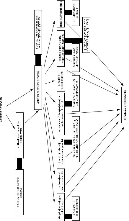
Cells that are undergoing apoptosis progress through a series of events. Many of the events that we measure in association with apoptosis, however, may be overlapping in time and therefore may not be a ``pathway'' so much as symptoms of the underlying process. Some of the events are not obligatory and may di¨er depending on the nature of the apoptotic trigger and the cell type. In any case, several of these apoptotic-associated events can be analyzed by ¯ow cytometry (Fig. 8.22). One of the events is the ¯ipping and stabilization of phosphatidylserine from the inner surface of the cytoplasmic membrane to the outer surface. On the outer surface, phosphatidylserine appears to identify cells as targets for phagocytosis. Because annexin V binds to phosphatidylserine, staining of intact cells with ¯uorochrome-conjugated annexin V will detect cells that are in early stages of apoptosis.
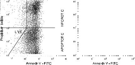
A two-color dot plot (Fig. 8.23) of cells stained with propidium iodide and annexin V±FITC will indicate cells in three of the four quadrants. Unstained cells are alive and well and are the double negatives; they neither express phosphatidylserine on their surface nor take up propidium iodide through leaky membranes. Cells that stain just with annexin V are apoptotic; they have begun to express phosphatidylserine on their surface, but have not yet gone through the process that leads to permeabilization of their cytoplasmic membrane. Cells that stain both with propidium iodide and annexin V are necrotic (that is, dead); they take up propidium iodide and also stain with annexin V. With a permeable cell, the ¯ow cytometer cannot tell us whether the annexin V is on the outside of the membrane (because the cells have gone through apoptosis before membrane permeabilization) or on the inside of the membrane (because the cells have died by the necrotic pathway without apoptosis).
Another event associated with apoptosis is the fragmentation of DNA due to endonuclease activation. This fragmentation results in the appearance of increased numbers of ``free ends'' on the DNA molecules in the cell. By incubating ®xed and permeabilized cells with
|
|
|
|
DNA in Life and Death |
153 |
|||||||||
|
|
|
|
|
|
|
|
|
|
|
|
|
|
|
|
|
|
|
|
|
|
|
|
|
|
|
|
|
|
|
|
|
|
|
|
|
|
|
|
|
|
|
|
|
|
|
|
|
|
|
|
|
|
|
|
|
|
|
|
|
|
|
|
|
|
|
|
|
|
|
|
|
|
|
|
|
|
|
|
|
|
|
|
|
|
|
|
|
|
|
|
|
|
|
|
|
|
|
|
|
|
|
|
|
|
|
|
|
|
|
|
|
|
|
|
|
|
|
|
|
|
|
|
|
|
|
|
|
|
|
|
|
|
|
|
|
|
|
|
|
|
|
|
|
|
|
|
|
|
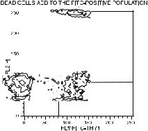
Fig. 8.23. Cells stained with propidium iodide for permeable membranes and with annexin V for phosphatidylserine can distinguish live (double negative), apoptotic (annexin V-positive, propidium iodide±negative), and necrotic (double positive) cells. This ®gure (with data from Robert Wagner) shows cultured bovine aortic endothelial cells; the left plot displays data from adherent cells, and the right plot displays data from the ``¯oaters.''
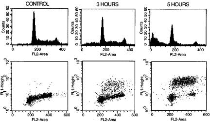
the enzyme terminal deoxynucleotidyl transferase (TdT) and with ¯uorochrome-labeled nucleotides, cells with greater numbers of DNA fragments incorporate more of the ¯uorescent nucleotides onto their increased number of termini. The resulting increased cellular ¯uorescence is indicative of an apoptotic cell (Fig. 8.24, lower dot plots). Appearance of positive cells in this TdT ¯ow assay correlates well with the classic gel electrophoresis assay for apoptosis, where ``ladders'' of small DNA fragments are indicators of endonuclease activation. In a desperate attempt to ®nd an acronym, this ¯ow assay for apoptosis has been called the TUNEL assay (for Terminal deoxynucleotidyl transferase±mediated dUTP N ick End-Labeling).
Propidium iodide staining alone can be used to detect later stages of apoptosis (Fig. 8.24, upper histograms). When the DNA becomes extensively fragmented, the small fragments begin to be ejected from
gРРРРРРРРРРРРРРРРРРРРРРРРРРРРРРРРРРРРРРРР
Fig. 8.22. A diagram of some of the events associated with apoptosis, showing related ¯ow cytometric assays (outlined in bold boxes). Although it is generally agreed that calcium ¯ux and caspase activation are early events following the trigger signal, the events following on from there may be essential steps in an ordered pathway or may be independent and merely symptomatic of apoptosis.
154 |
Flow Cytometry |
Fig. 8.24. Cells in late apoptosis have fragmented DNA. They can be visualized as cells with increased incorporation of ¯uorochrome-labeled nucleotides to the ends of the fragments in the ¯ow cytometric TUNEL assay (lower dot plots, with FL1 as FITC-dUTP and FL2 as propidium iodide bound to DNA). Alternatively, they can be visualized as cells with less-than-normal (sub-G0/G1) DNA content (upper histograms of propidium iodide ¯uorescence). Data from etoposide-treated ML-1 cells courtesy of Mary Kay Brown and Alan Eastman.
the cells and/or can be washed from the cells during the staining protocol. In either case, the apoptotic cell at this stage will have signi®cantly less DNA than a healthy, viable cell. This lowered DNA content can be detected as a ``sub-G0/G1'' peak in a simple, onecolor DNA histogram. It needs to be mentioned that staining for the cells with sub-G0/G1 DNA content requires a protocol that encourages apoptotic cells to release their fragmented DNA. In contrast, the TUNEL assay uses ®xation to ensure that the fragmented DNA is retained.
NECROSIS
Unlike apoptosis, which involves the scheduled and active coordination of metabolic processes, necrosis is a passive response to a toxic or injurious environment. Whereas in apoptosis the cell membrane remains intact until very late in the game, permeabilization of the cell membrane is an early event in necrosis. Because propidium iodide
|
|
|
|
DNA in Life and Death |
155 |
||||||||||||||
|
|
|
|
|
|
|
|
|
|
|
|
|
|
|
|
|
|
|
|
|
|
|
|
|
|
|
|
|
|
|
|
|
|
|
|
|
|
|
|
|
|
|
|
|
|
|
|
|
|
|
|
|
|
|
|
|
|
|
|
|
|
|
|
|
|
|
|
|
|
|
|
|
|
|
|
|
|
|
|
|
|
|
|
|
|
|
|
|
|
|
|
|
|
|
|
|
|
|
|
|
|
|
|
|
|
|
|
|
|
|
|
|
|
|
|
|
|
|
|
|
|
|
|
|
|
|
|
|
|
|
|
|
|
|
|
|
|
|
|
|
|
|
|
|
|
|
|
|
|
|
|
|
|
|
|
|
|
|
|
|
|
|
|
|
|
|
|
|
|
|
|
|
|
|
|
|
|
|
|
|
|
|
|
|
|
|
|
|
|
|
|
|
|
|
|
|
|
|
|
|
|
|
|
|
|
|
|
|
|
|
|
|
|
|
|
|
|
|
|
|
|
|
|
|
|
|
|
|
|
|
|
|
|
|
|
|
|
|
|
|
|
|
|
|
|
|
|
|
|
|
|
|
|
|
|
|
|
|
|
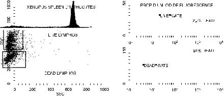
Fig. 8.25. The use of propidium iodide to monitor cell death. Dead lymphocytes have a less intense forward scatter signal than do live lymphocytes. Cultured Xenopus spleen lymphocytes courtesy of Jocelyn Ho and John Horton.
is excluded from entering cells by an intact plasma membrane and because it only ¯uoresces when intercalated between the bases of double-stranded nucleic acid, it will not ¯uoresce if it is added to a suspension of intact cells. The intact plasma membrane forms a barrier, keeping propidium iodide and nucleic acids apart. It is only when the outer membrane has been breached that the cells will emit red ¯uorescence. Propidium iodide (or other membrane-impermeant DNA ¯uorochromes) is therefore a stain (like trypan blue) that can be used to mark necrotic cells (on the reasonable but not necessarily valid assumption that cells with holes in their membranes large enough to allow the penetration of propidium iodide are actually dead according to other viability criteria and vice versa). By this method, cell viability can be monitored in the presence of various cytotoxic conditions (Fig. 8.25).
The only di½culty in using ¯ow cytometry to monitor cell death is that, as mentioned in Chapter 3, dead cells have di¨erent scatter properties than living cells. In particular, because of their perforated outer membrane, they have a lower refractive index than living cells and therefore have forward scatter signals of lower intensity. For this reason, it is important not to use a gate or forward scatter threshold when analyzing a population for the proportion of dead and live cells. Any forward versus side scatter gate drawn around normal lymphocytes, for example, will always show most if not all of the cells
156 |
Flow Cytometry |
within that gate to have excluded propidium iodide no matter how many cells in the preparation are dead; this is simply because the dead cells drop out of the gate (Fig. 8.25). The lesson here (and one that needs repeating in reference to most ¯ow analysis) is that gating is an analytic procedure that needs to be performed carefully and with explicit purpose. If we do not de®ne our population of interest (by gating) with care, our results may be accurate answers to inappropriate questions (e.g., ``what percentage of living lymphocytes are alive?'').
The use of membrane-impermeant DNA ¯uorochromes to mark dead cells has a more routine application in simply allowing the exclusion of dead cells from ¯ow analysis. This is important because dead cells have the habit of staining nonspeci®cally when their broken membranes tend to trap monoclonal antibodies directed against surface antigens. Therefore a cell suspension including many dead cells may show high levels of nonspeci®c stain. By adding propidium iodide to cells just before analysis, the cells ¯uorescing red can be excluded from further analysis by gating on red negativity and only the living cells then examined for surface marker staining. Figure 8.26 shows the way that this technique can be used when looking at ¯uorescein staining for surface antigens on cultured mouse thymocytes.
Fig. 8.26. The use of propidium iodide to exclude dead cells from analysis of a mouse spleen cell population for the expression of the Thy-1 surface antigen. The cells were stained with ¯uorescein for Thy-1 and then with propidium iodide to mark the dead cells. It can be seen that the dead cells (upper right quadrant) contribute to the ¯uorescein-positive population. Stained cells courtesy of Maxwell Holscher.
DNA in Life and Death |
157 |
The use of propidium iodide or equivalent dyes to exclude dead cells from analysis is a procedure that is recommended as routine for any system staining for surface markers. It is particularly important in analysis of mixed populations where a high percentage of the cells may be dead.
It should, however, be remembered that ®xed cells cannot be stained with propidium iodide for live/dead discrimination. Fixed cells are, in fact, all dead and will therefore all take up propidium iodide even if some were alive and some dead before ®xation. Ethidium monoazide o¨ers an alternative to propidium iodide if cells will be ®xed before ¯ow analysis. It is a dye that, like propidium iodide, only enters dead cells. It has, however, the added advantage of forming permanent cross-links with DNA when photoactivated. Therefore, cells can be stained for surface proteins, incubated with ethidium monoazide under a desk lamp, washed, and then ®xed. At the time of acquisition of data on the ¯ow cytometer, red ¯uorescence will mark the cells that were dead before ®xation.
FURTHER READING
Chapters 13±16 and 24 in Melamed et al. and Chapters 11±13 in Watson are detailed reviews of the applications of ¯ow cytometry to various aspects of cell cycle research.
Several chapters in Ormerod and several sections in the Purdue Handbook, in Diamond and DeMaggio, and in Current Protocols in Cytometry give good practical protocols. Darzynkiewicz (both the 1994 and 2001 editions) include many chapters with protocols and advice on both the simpler and the more complex methods in nucleic acid, apoptosis, and cell cycle analysis.
Good discussions of the mathematical algorithms for cell cycle analysis can be found in Chapter 6 of Van Dilla et al., Chapter 23 of Melamed et al., and in the multiauthor book Techniques in Cell Cycle Analysis, edited by Gray JW and Darzynkiewicz Z (1987), Humana Press, Clifton, NJ.
The ``Cytometry of Cyclin Proteins'' is reviewed by Darzynkiewicz Z, et al. (1996) in Cytometry 25:1±13.
Molecular biology and chromosome analysis are discussed in Chapters 25± 28 of Melamed et al. A special issue (Volume 11, Number 1, 1990) of the journal Cytometry, although somewhat aged, is devoted to the subject of
158 |
Flow Cytometry |
analytical cytogenetics; it contains articles about work at the interface between theory and clinical practice (as well as some beautiful pictures).
A review article by Darzynkiewicz Z, et al. (1997) on ``Cytometry in Cell Necrobiology: Analysis of Apoptosis and Accidental Cell Death (Necrosis)'' appeared in Cytometry 27:1±20. Boehringer Mannheim produces a free, but substantial manual (now in its second edition) on methods related to the study of cell death.

Flow Cytometry: First Principles, Second Edition. Alice Longobardi Givan
Copyright 2001 by Wiley-Liss, Inc.
ISBNs 0-471-38224-8 (Paper); 0-471-22394-8 (Electronic)
9
The Sorting of Cells
Although early ¯ow cytometers were developed as instruments that could separate or sort particles based on analysis of the signals coming from those particles, it is now true that most ¯ow cytometry involves analyzing particles but not actually sorting them. This is just one of many examples of the way in which ¯ow technology has moved in unpredictable directions. Many cytometers now do not even possess a sorting capability, and the instruments that can sort particles may be used only infrequently for that purpose. Nevertheless, sorting cells or chromosomes with a ¯ow cytometer is an elegant technology; it may be the only method available for obtaining pure preparations of particles with certain kinds of characteristics of interest. On both of those counts it is certainly worth knowing about sorting even if you are not interested in using the technique at the moment. Now that you are comfortable with ¯ow cytometry and its basic applications, this chapter will present a bit more technical information about the way a sorting ¯ow cytometer (Fig. 9.1) can be used to sort cells.
SORTING THEORY
If a stream of liquid is vibrated along its axis, the stream will break up into drops (pick a nice summer day and try it with a garden hose). The characteristics of this drop formation are governed by an equation that will be familiar to anyone who has studied light waves: v ˆ fl, where v is the velocity of the stream, f is the frequency of the vibration applied, and l is the ``wave length'' or distance between the drops. One other fact that needs to be known before we can under-
159