

Flow Cytometry - First Principles (Second Edition)
.pdf170 |
Flow Cytometry |
Whether at high or low speed, every sort needs to be optimized according to the particular requirements of the experiment in question. Speed, purity, and sorting e½ciency interact and are not, all three, optimizable under the same conditions. For example, a sort for a minor subpopulation of cells from a large, easily obtainable cell suspension may require optimization of speed, without concern about low sorting e½ciency. On the other hand, a requirement for as many speci®c cells as possible from among a scarce mixed suspension will need to maximize recovery without much concern for speed.
ALTERNATIVE METHODS FOR SORTING
There are, of course, other methods for separating cells. Traditional methods have been based on physical properties (e.g., density gradient centrifugation) or on biochemical properties (e.g., the adherence of T cells to sheep erythrocytes or of monocytes to plastic or the resistance of white cells [as opposed to erythrocytes] to lysis by ammonium chloride). Additional separation methods involve the use of comple- ment-®xing antibodies, which can be used to stain unwanted cells; complement lysis will enrich the suspension for the unstained cells. More recent methods involve the staining of cells with antibodies that have been bound with magnetic beads; the cells of choice, after being stained with the antibodies, become magnetic and can be removed from a mixed suspension by passing the suspension through a magnetic ®eld.
All these methods are ``bulk'' or ``batch'' methods; that is, the amount of time that it takes to perform a separation is not related to the number of cells that are being separated. For example, if blood needs to be centrifuged for 20 minutes over a density gradient in order to separate monocytes and lymphocytes from neutrophils and erythrocytes, the separation will take 20 minutes whether the original volume of blood is 1 or 100 ml (assuming enough buckets in the centrifuge). Batch procedures are therefore ideally combined with ¯ow sorting; they provide an initial rapid pre-separation that can greatly decrease the amount of time required for the ®nal ¯ow separation (refer back to Table 9.1).
By way of a practical example, consider CD5-positive B lymphocytes. Because CD5-positive B lymphocytes may be 10% of all B
Cell Sorting |
171 |
lymphocytes, B lymphocytes are perhaps 10% of all lymphocytes, and lymphocytes could be 50% of all mononuclear white cells, if we want to sort out CD5-positive B cells (0.5%) from a mononuclear cell preparation, they will be ¯owing through our system at a rate of 36,000 per hour if our total particle ¯ow rate is limited to about 2000 per second. It would therefore take 28 h to collect 1 million CD5-positive B cells. However, if we remove all nonlymphocytes by adherence to plastic and then remove all T lymphocytes by rosetting with sheep erythrocytes (both techniques are rapid batch processes that might take an immunologist about an hour to perform), we can then send pure B lymphocytes through the cytometer. The desired CD5-positive particles (now 10% of the total) will therefore be ¯owing at a rate of 720,000 per hour. As a result, the time it will take to get 1 million desired cells will go from 28 hours to 1.4 hours. This has obvious bene®ts both in terms of cost, if you are paying for cytometer time, and the general health and viability of the sorted cells at the end of the procedure.
Batch procedures can be useful as pre-¯ow enrichment of the cells of interest. They can also be used as alternatives to ¯ow sorting if the cells of interest can be appropriately selected. Flow sorting, although slow, is generally the only alternative if multiple parameters are required to de®ne the cells of interest, if antigen density on the cell surface is low, or if forward or side scattered light is part of the de®- nition of the desired cells.
In recent years, alternative ¯ow cytometric methods for sorting have been developed. Instead of sorting cells by applying a charge to the drops generated by a vibrating stream, one of these methods involves sorting cells according to their ¯ow signals by ``grabbing'' the cells of interest into a catcher tube that moves into the center of the stream when a desired cell ¯ows by. Another ¯ow sorting technology involves the use of a piston that applies pressure to one arm of a Y-shaped channel downstream from the laser intercept. Cells normally ¯ow down the waste arm of the ``Y.'' By application of pressure after a desired cell ¯ows through the analysis point, selected cells are diverted from the waste arm to the sorting arm of the ``Y.'' While these are cheaper alternatives than traditional, droplet-based, electronic sorting, speed limitations and the large volume of sheath ¯uid that dilutes the sorted cells make these methods primarily suitable for collection of small numbers of cells.
172 |
Flow Cytometry |
Another method for ¯ow sorting is currently under development. It permits extremely rapid selection of cells at speeds beyond those possible even under very high-speed traditional (drop de¯ection) conditions. In drop de¯ection sorting, the sort rate is restricted by the rate of drop formation. This alternative method could be called reverse sorting or optical zapping and does not involve drop formation. Cells or chromosomes are stained with a phototoxic dye as well as with antibodies or a DNA stain. The particles are sent through a more or less conventional cytometer. Downstream from the analysis point, a cell or chromosome that does not possess the light scatter or ¯uorescent properties of interest is zapped by a second (killer) laser that activates the phototoxic dye. All particles end up in the collection vessel, but only those that have not been zapped survive. The rate of selection/destruction is limited, therefore, only by the rate at which the zapping laser can be de¯ected away from the stream. In proof-of-principle experiments, it appears that sorting rates may be increased 5-fold over those of the high-speed sorters and 25-fold over conventional sorters.
THE CONDITION OF CELLS AFTER SORTING
This brings us to a brief discussion of the condition of cells after they have been sorted. Depending on the purpose for which the sorted cells will be used, there may be di¨erent requirements for sterility and viability. If you are sorting cells for subsequent polymerase chain reaction ampli®cation, for example, then neither sterility nor viability is important. On the other hand, if you are using a ¯ow sorter because you want to clone cells expressing a certain characteristic, then both sterility and viability are necessary. Sterile sorting is possible (with care and a fair amount of 70% ethanol used to sterilize the ¯ow lines). Sorted cells can remain sterile through the sort procedure and can subsequently be cultured for functional analysis or cloning. Given that cells in a sorting cytometer have been subjected to acceleration, decompression, illumination, vibration, enclosure, charging, and de- ¯ection toward high voltage plates, it seems reasonable to expect some trauma and, therefore, to coddle the cells as quickly as possible after the sort if viability is required. If you remember that the cells move down the stream in a core that is just a small percentage of the total volume of the sheath stream, you will realize that each drop is
Cell Sorting |
173 |
mainly sheath ¯uid and that, following drop de¯ection, the cells are deposited in medium that is partly sample medium, but mainly sheath ¯uid. Therefore, it is recommended to sort the cells into tubes that already contain a volume of appropriate culture medium. Some cells lose viability after sorting; many cells seem to be remarkably oblivious to the process.
It is also important to remember that most sorting procedures involve staining of the cells with antibodies conjugated to ¯uorochromes so that the cells can be sorted according to whether or not they are ¯uorescent. There is always the possibility that the binding of antibodies to the cell surface might by itself a¨ect the function of the selected cells. In other words, the sorted cells that are low in ¯uorescence intensity may function di¨erently from the cells that are bright not because they are essentially di¨erent but simply because the staining itself has blocked or tweaked surface receptors. By now you should be able to design a control for this potentially confusing phenomenon. If the staining is functionally neutral, stained cells (without any sorting at all) should function identically to unstained cells with regard to the assay of interest. In addition, it is important to be sure that the sorting procedure does not a¨ect cell function. The control for this is to compare unsorted cells with cells that have gone through the sorter with large sort gates that do not actually exclude any cells. The sorting procedure should not change the functional ability of the cells if the proportion of di¨erent kinds of cells is not changed in this control sort. If the functional ability of the cells is changed either by sorting or by staining, then this needs to be considered in the interpretation of the subsequent functional analysis of the sorted populations.
FURTHER READING
Practical issues in sorting are discussed well in Chapter 4 of Ormerod and in Section 7 of Diamond and DeMaggio.
Theoretical principles of droplet generation sorting are discussed in Chapter 8 in Melamed, in Chapter 6 of Watson, and in Chapter 3 of Van Dilla.
High speed sorting is described in Section 1.7 of Current Protocols in Cytometry.
174 |
Flow Cytometry |
For discussion of chromosome sorting by optical zapping, see Roslaniec MC, Reynolds RJ, Martin JC, et al. (1996). Advances in ¯ow cytogenetics: Progress in the development of a high speed optical chromosome sorter based on photochemical adduct formation between psoralens and chromosomal DNA. NATO Advanced Studies Series, Flow and Image Cytometry 95:104±114.

Flow Cytometry: First Principles, Second Edition. Alice Longobardi Givan
Copyright 2001 by Wiley-Liss, Inc.
ISBNs 0-471-38224-8 (Paper); 0-471-22394-8 (Electronic)
10
Disease and Diagnosis:
The Clinical Laboratory
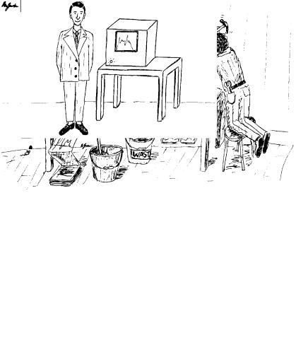
Early in this book I stated that one of the areas in which cytometry has had great impact is in clinical diagnosis. Research work, beginning with Kamentsky's foray into cytological screening, but particularly during the last 20 years, has made it clear that the information that can be obtained by ¯ow cytometry could be of clear use to clinicians. However, as long as ¯ow cytometers maintained their image of cumbersome and ®ddly instruments, di½cult to standardize and requiring constant attention from a team of unconventional but devoted hackers (see Figs. 1.3 and 1.4 for an understanding of the origins of this image), there was little chance that ¯ow cytometry would become integrated into routine hospital laboratories.
The direct cause of the rapid introduction of ¯ow technology into many hospitals was the commercial development and marketing of ``black box'' or ``benchtop'' ¯ow cytometers (in imitation of the pattern set by blood counters, which were, by the 1980s, standard equipment in hematology laboratories). Installation of these benchtop ¯ow instruments involves placing them on a laboratory bench and plugging them in. They are optically stable and therefore can, in principle, be maintained with relatively little human intervention; they can produce clinical printouts that can be read without any requirement for knowledge of the intricacies of ¯ow data analysis; and they can, we are told, be run by anyone with the ability to push a key on a computer keyboard (Fig. 10.1). It was only with the development and marketing of these friendly machines (starting in the mid to late 1980s) that provision of ¯ow cytometric information for routine diagnosis became a practical proposition.
175
176 |
Flow Cytometry |
|
|
|
|
|
|
|
|
|
|
|
|
|
|
|
|
|
|
|
|
|
|
|
|
|
|
|
|
|
|
|
|
|
Fig. 10.1. Two opposing fantasies of what ¯ow cytometry is all about. Drawings by Ben Givan.
As a result of the introduction of cytometers into the hospital setting, three aspects of clinical practice have led to some general reassessment of the nature of ¯ow analysis. First, clinical laboratories are, because of the import of their results, overwhelmingly concerned with so-called quality control. This concern has forced all cytometrists to become more aware of the standardization and calibration
Disease and Diagnosis |
177 |
of their instruments. Neither standardization nor calibration comes naturally to a ¯ow cytometer. In response to this clinical requirement, beads, ®xed cells, and mock cells have been developed to help in assessing the stability of conditions from day to day. Instruments can be set up from stored computer information so that machine parameters are constant from run to run. Furthermore, the analysis of data can be automated so that clinical information can be derived and reported quickly and in standard format. In addition, quality control schemes of various sorts have resulted in samples ¯ying around the world; reports appear in the literature documenting the factors that lead to variation in results obtained from di¨erent laboratories and with di¨erent operators. Quality control in ¯ow cytometry is still far from secure, but progress is being made toward producing technical recommendations (both for DNA and for leukocyte surface marker analysis), toward providing schemes for accrediting personnel and laboratories, and toward monitoring the performance of laboratories Ðwith comparisons between laboratories and within any one laboratory over the course of time.
The second aspect of clinical practice that has led to a reassessment of the nature of ¯ow cytometry is the occasional clinical requirement for ``rare-event analysis.'' Methods have been developed, particularly with the use of multiparameter gating, to lower background noise in order to provide increased sensitivity for detection of rare cells. In the clinic, this increased sensitivity translates, for example, into earlier diagnosis of relapse in leukemia, more sensitive detection of fetal± maternal hemorrhage, and better ability to screen leukocyte-reduced blood transfusion products for residual white blood cells. Outside the clinic, these methods for rare-event detection have begun to stretch the limits of research applications as well.
A third aspect of clinical practice that has led to modi®cations in ¯ow technology has been the requirement for safety in the handling of potentially infectious specimens. The ¯uidic speci®cations of instruments have been modi®ed with attention to the control of aerosols that might occur around the stream, the prevention of leaks around the sample manifold, and the collection of the waste ¯uid after it has been analyzed. However, a more e¨ective means of minimizing biological hazards has been the development of techniques for killing and ®xing specimens in such a way that cells, viruses, and bacteria are no longer viable, but the scatter and ¯uorescent proper-
178 |
Flow Cytometry |
ties of the cells of interest are relatively unchanged. Formaldehyde (1.0%) is the ®xative of choice in most ¯ow laboratories. It is used after cells have been stained. As well as reducing the infectivity of the hepatitis B and AIDS (HIV) viruses, it ®xes leukocytes in such a way that their scatter characteristics are virtually una¨ected. Moreover, the ¯uorescence intensity of their surface stain remains essentially stable (albeit with slightly raised control auto¯uorescence) for several days or more. Therefore, routine ®xation of biological specimens has not only increased the safety of ¯ow procedures but has also made it possible to ship specimens around the world and to store specimens within a laboratory for convenient structuring of access to the cytometer (a euphemism for not working in the middle of the night). It should be said, however, that anyone working with material of known biological hazard needs to check ®xation procedures to con- ®rm the reduction of infectivity. Even after ®xation, anyone working with any biological material at all should use standard precautions for control because any particular ®xation procedure might not be e¨ective against unknown or undocumented hazards. Furthermore, whatever ®xation procedure is used should be checked with individual cell preparations and staining protocols to con®rm the stability of cytometric parameters over the required period of time.
THE HEMATOLOGY LABORATORY
The ¯ow cytometer has, for several reasons, found a very natural home in the hospital hematology laboratory. In the ®rst place, because of the virtually universal use of automated instrumentation for enumerating erythrocytes and leukocytes, people working in hematology laboratories are quite relaxed about the idea of cells ¯owing in one end of an instrument and numbers coming out the other end. As I have said, both historically and technologically there is a close relationship between ¯ow cytometers and automated hematology counters. The second reason is the obvious fact that blood exists as a suspension of individual cellsÐso that red and white cells do not need to be disaggregated before ¯ow analysis. The third reason for the hematologist's ease with ¯ow technology is that hematology/ immunology laboratories were among the ®rst to make routine use
Disease and Diagnosis |
179 |
of ¯uorescently tagged monoclonal antibodies. For many years, microscopists have been using panels of monoclonal antibodies for identifying various subpopulations of lymphocytes that are suggestive or diagnostic of various disease conditions. In particular, because the leukemias and lymphomas represent a group of diseases that involve the uncontrolled clonal proliferation of particular groups of leukocytes, various forms of these malignancies can be identi®ed and classi- ®ed according to the phenotype of the increased number of white cells found in biopsy material or in the patient's peripheral circulation.
Leukemia/Lymphoma Phenotyping
Studies of the staining of surface proteins (CD antigens) to determine the phenotypes of the abnormal cells in patients with various leukemias or lymphomas have been useful, revealing much about ways to classify the diseases but also increasing our knowledge of normal immune cell development. In general, immune cells gain and lose various surface proteins in the course of their normal development in the bone marrow until they become the cells (with mature phenotype and function) that are released from the marrow into the peripheral circulation (Fig. 10.2). Leukemias and lymphomas often involve a block in this development so that cells with immature phenotypes appear in great numbers in the periphery. Alternatively, certain forms of disease may involve rapid expansion of a clone of mature normal cells so that one type of cell predominates in the circulation over all other normal subpopulations. In other cases, cells seem to express abnormal combinations of CD antigens, a condition referred to as lineage in®delity or as lineage promiscuity depending on one's interpretation of the data. These variations in surface protein expression make classi®cation of leukemias and lymphomas a suitable application for ¯ow cytometry, albeit a very complex task. Correlation between classi®cation and predicted clinical course is therefore correspondingly di½cult.
Once the phenotype of a blood cell malignancy in a particular patient is known, the cytometrist can use the multiparameter ¯ow description of that phenotype to de®ne the malignant clone and to look for the absence of these cells in order to diagnose remission after