

structures
.pdfChapter 5 NMR Spectroscopy
(1) Splitting Diagrams
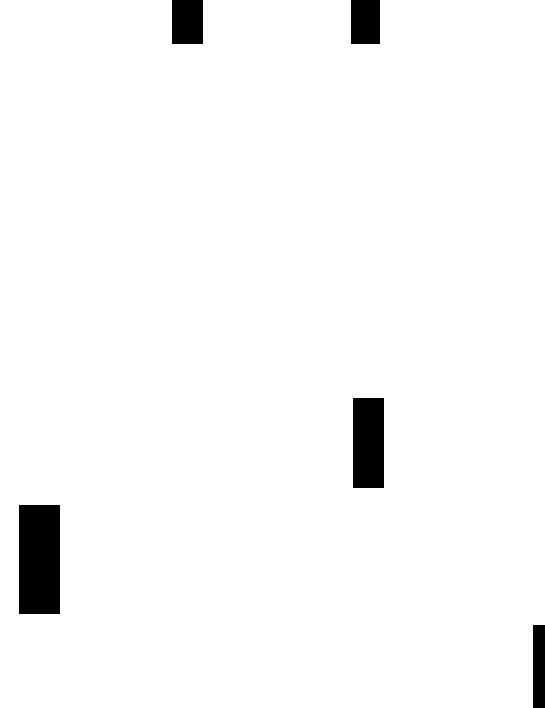
The knowledge of the rules listed above, pennits the development of a simple procedure for the analysis of any spectrum which is suspected of being first order. The first step consists of drawing a splitting diagram, from which the line spacings can be measured and identical (hence related) splittings can be identified (Figure 5.5).
)
3.5 |
3.0 |
2.5 |
|
8 (ppm from TMS) |
|
Figure 5.5 A Portion of the IH NMR Spectrum of Styrene Epoxide
(100 MHz as a 5% solution in CCI4)
The section of the spectrum of styrene epoxide (Figure 5.5) clearly contains the signals from 3 separate protons (identified as HI, Hz and H3) with HI at 8 2.95, Hz at 8 2.58 and H3 at 8 3.67 ppm. Each signal appears as a doublet of doublets and the chemical shift of each proton is simply obtained by locating the centre of the multiplet. The pair of nuclei giving rise to each splitting is clearly indicated by the splitting diagram above each multiplet with 2JH I_HZ =: 5.9 Hz, 3JHI_H3 =: 4.0 Hz and
=: 2.5 Hz.
The validity of a first order analysis can be verified by calculating the ratio !1vlJ for each pair of nuclei and establishing that it is greater than 3.
57
Chapter 5 NMR Spectroscopy
From Figure 5.5
= |
37 |
= 6.3 |
= |
72 |
= 18.0 |
= 109 = 43.6 |
|
5.9 |
|
|
4.0 |
|
2.5 |
Each ratio is greater than 3 so a first order analysis is justified and the 100 MHz spectrum of the aliphatic protons of styrene oxide is indeed a first order spectrum and could be labelled as an AMX spin system.
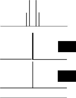
The 60 MHz IH spectrum of a 4 spin AMX2 system is given in Figure 5.6. This system contains 3 separate proton signals (in the intensity ratios 1:1:2, identified as HA> HM and Hx)' The multiplicity of H; is a triplet of doublets, the multiplicity ofHM is a triplet of doublets and the multiplicity ofHx is a doublet of doublets. Again, the nuclei giving rise to each splitting are clearly indicated by the splitting diagram above each multiplet and the chemical shifts of each multiplet are simply obtained by measuring the centres of each multiplet.
HA |
|
HM |
|
HX |
|
|||||||
/) =7.0 ppm |
0= 6.0 ppm |
0= 4.95 ppm |
||||||||||
v = 420 Hz |
v = 360 Hz |
v = 297 Hz |
||||||||||
|
|
|
t>VAM = 60 Hz |
|
|
|
|
t>VMX =63 Hz |
|
|
|
|
|
|
|
|
|
|
|
|
|
||||
|
__------_____ |
|
........E --------- _ |
J AX = 6.0 Hz |
||||||||
|
|
|
|
|
|
|
|
|
/ |
|||
|
|
|
|
|
|
|
|
|
/ |
J MX = 3.5 Hz |
||
|
|
/ JAX =6.0 Hz |
|
|
|
/ |
JMX = 3.5 Hz |
|
|
|
|
|
|
|
|
|
|
|
|
|
|
||||
|
|
|
|
|
|
|
|
|
||||
|
|
|
|
|
|
|
|
|
|
|
||
|
|
/JAM=1.5HZ |
|
|
/ |
JAM = 1.5 Hz |
|
|
|
|
||
|
|
|
|
|
|
|
|
|
|
|
||
|
|
'IH |
|
|
|
|
'IH |
|
|
|
2H |
|
|
|
|
|
|
|
|
|
|
||||
|
|
|
|
|
|
|
|
|
|
|
|
|
400 |
|
|
|
|
350 |
300 |
|
(Hz from TMS) |
||||
|
|
|
I |
|
|
|
|
|
I |
|
||
7.0 |
|
|
|
|
|
|
|
o (ppm from TMS) |
||||
|
|
6.0 |
|
|
5.0 |
|
||||||
Figure 5.6 The 60 MHz IH NMR Spectrum of a 4-Spin AMX2 Spin System
58
Chapter 5 NMR Spectroscopy
A spin system comprising just two protons (i.e. an AX or an AB system) is always exceptionally easy to analyse because, independent ofthe value of the ratio of !1v/J, the spectrum always consists ofjust four lines with each pair of lines separated by the coupling constant J. The only distortion from the first-order pattern consists of the gradual reduction of intensities of the outer lines in favour of the inner lines, a characteristic "sloping" or "tenting" towards the coupling partner. A series of simulated spectra of two-spin systems are shown in Figure 5.7.
|
V1 |
v 2 |
|
|
|||
J12 |
-, |
|
|
|
6v |
12 = 10.0 |
|
|
|
" |
- |
||||
|
|
||||||
|
|
|
|
J12 |
|||
|
|
|
|
|
|
|
|
|
|
|
|
|
|
|
|
-
100 |
50 |
oHz -50 |
-100 |
Figure 5.7 Simulated IH NMR Spectra of a 2-Spin System as the Ratio
txvlJ, is Varied from 10.0 to 0.0
59
Chapter 5 NMR Spectroscopy
(2) Spin Decoupling
In the signal of a proton that is a multiplet due to spin-spin coupling, it is possible to remove the splitting effects by irradiating the sample with an additional Rf source at the exact resonance frequency of the proton giving rise to the splitting. The additional radiofrequency causes rapid flipping of the irradiated nuclei and as a consequence nuclei coupled to them cannot sense them as being in either an a or ~ state for long enough to cause splitting. The irradiated nuclei are said to be decoupled from other nuclei in the spin system. Decoupling simplifies the appearance of complex multiplets by removing some of the splittings. In addition, decoupling is a powerful tool for assigning spectra because the skilled spectroscopist can use a series of decoupling experiments to sequentially identify which nuclei are coupled.
In a 4-spin AM2X spin system, the signal for proton HA would appear as a doublet of triplets (with the triplet splitting due to coupling to the 2 M protons and the doublet splitting due to coupling to the X proton). Irradiation at the frequency ofHx reduces the multiplicity of the A signal to a triplet (with the remaining splitting due to JAM) and irradiation at the frequency ofHM reduces the multiplicity of the A signal to a doublet (with the remaining splitting due to JAX) (Figure 5.8).
HM |
HA |
|
I |
Hx |
|
I |
|
I |
X-C-C-C-y
HI I I
M
Basic spectrum of HA |
with irradiation |
with irradiation |
without irradiation of |
of Hx |
ofH M |
of HM or Hx |
|
|
Figure 5.8 Selective Decoupling in a Simple 4-Spin System
60
Chapter 5 NMR Spectroscopy
(3)Correlation of1H _lH Coupling Constants with Structure
Interproton spin-spin coupling constants are of obvious, value in obtaining structural data about a molecule, in particular information about the connectivity of structural elements and the relative disposition of various protons.
Non-aromatic Spin Systems.
In saturated systems, the magnitude of the geminal coupling constant 2JH_ (two
C_H
protons attached to the same carbon atom) is typically between 10 and 16 Hz but values between 0 and 22 Hz have been recorded in some unusual structures.
2 =10 -16 Hz.
JAB
The vicinal coupling (protons on adjacent carbon atoms) 3JH_C_C_H can have values o- 16 Hz depending mainly on the dihedral angle </>.
H H
A B
I I
- C - C -
I I
3
'JAB= 0 -16 Hz.
The so-called Karplus relationship expresses approximately, the angular dependence of the vicinal coupling constant as:
3JH_C_C_H = 10 cos? </> |
for 0 < </> < 90° and |
3JH_C_C_H = 15 cos- </> |
for 90 < </> < 180° |
It follows from these equations that if the dihedral angle </> between two vicinal protons is near 90° then the coupling constant will be very small and conversely, if the dihedral angle </> between two vicinal protons is near 0° or 180° then the coupling constant will be relatively large. The Karplus relationship is of great value in determining the stereochemistry of organic molecules but must be treated with caution because vicinal coupling constants also depend markedly on the nature of substituents. In systems that assume an average conformation, such as a flexible hydrocarbon chain, 3J H_H generally lies between 6 and 8 Hz.
61
Chapter 5 NMR Spectroscopy
The coupling constants in unsaturated (olefmic) systems depend on the nature of the substituents attached to the C=C but for the vast majority of substituents, the ranges
for 3J _ =C_H(ciS) and 3JH_ =C_H(lr ans) do not overlap. This means that the stereochemistry
H C C
of the double bond can be determined by measuring the coupling constant between vinylic protons. Where the C=C bond is in a ring, the 3JH_C=C_H coupling reflects the ring size.
3
JAB(CiS)
3
JAC(trans)
2
JBC(gem)
= 6 - 11 Hz |
= 5 -7 Hz |
|
|
= 12 - 19 Hz |
|
=a -3 Hz |
= 9 - 11 Hz |
|
The magnitude of the long-range allylic coupling, (41AB) is controlled by the dihedral angle between the C-HA bond and the plane of the double bond in a relationship reminiscent of the Karplus relation.
'" |
/ HA |
|
/ |
CxC=C / |
Hs |
/"-
He
/Aromatic Spin Systems
In aromatic systems, the coupling constant between protons attached to an aromatic ring is characteristic of the relative position ofthe coupled protons i.e. whether they
are ortho, meta or para.
3 |
=6 - 10Hz |
4 |
5 |
=a -1.5 Hz |
'JAB(orthO) |
JAB(meta) = 1 - 3 Hz |
'JAB(para) |
Similarly in condensed polynuclear aromatic compounds and heterocyclic compounds, the magnitude of the coupling constants between protons in the aromatic rings reflects the relative position of the coupled protons.
62
|
|
|
|
|
|
Chapter 5 |
NMR Spectroscopy |
~ |
~ |
|
4 |
|
|
|
|
2 |
50 |
|
|
|
|||
|
,-,:::: |
|
|
3 |
|
|
|
00I |
3 |
6:-.... N I 2 |
|
|
|||
5 |
4 |
|
|
|
|
|
|
3J1,2 =8.3 - |
9.1 Hz |
3J2.3 =4.0 - |
5.7 Hz |
3J2,3 =4.7 Hz |
3J2,3 =1.8 Hz |
||
3J2 ,3 = 6.1 - |
6.9 Hz |
3J 3,4 =6.8 - |
9.1 Hz |
3J3,4 =3.4 Hz |
3J3 ,4 =3.5 Hz |
||
4J1,3 = 1.2 -1.6 Hz |
4J2,4 =0.0 - |
2.5 Hz |
4J2,4 =1.0 Hz |
4J2,4 =0.8 Hz |
|||
SJ1•4 = 0 -1.0 Hz |
4J 3,S = 0.5 - |
1.8 Hz |
4J2.S =2.9 Hz |
4 J2,S = 1.6 Hz |
|||
sJ1,s=0 - 1 . 5Hz |
4J2.6 = 0.0 - |
0.6 Hz |
|
|
|||
|
|
|
sJ2 S =0.0 - |
2.3 Hz |
|
|
|
The splitting patterns of the protons in the aromatic region of the IH spectrum are frequently used to establish the substitution pattern of an aromatic ring. For example, a trisubstituted aromatic ring has 3 remaining protons. There are 3 possible arrangements for the 3 protons - they can have relative positions 1,2,3-; 1,2,4-; or 1,3,5- and each has a characteristic splitting pattern.
|
|
|
H |
|
H |
|
|
H |
|
|
|
|
A |
|
B |
|
|
c |
|
"':q"'y |
3 |
|
3 |
|
3 |
||||
J AB(Of1hO) |
|
JAB(orthO) |
|
JBC(orthO) |
|||||
X |
~I |
Z |
|
4 |
3 |
|
4 |
||
n |
M |
J BC(Of1hO) |
n |
JAC(meta) |
|||||
|
|
|
|
JAC(meta) |
ill |
|
|||
|
|
|
JAJ1L |
|
|
j}JJL |
|||
|
|
|
H |
|
H |
|
|
H |
|
|
|
|
A |
|
B |
|
|
c |
|
|
HB |
|
JuL |
JAJJL |
~ |
||||
"'*zy |
|||||||||
|
|
|
|
3 |
|
3 |
|
4 |
|
X |
~I |
Hc |
|
JAB(OrlhO) |
n |
JAB(orthO) |
|
JBC(meta) |
|
n |
|
~ |
5 |
||||||
|
|
|
|
5 |
|
4 |
|||
|
|
|
|
JAC(para) |
|
|
JBC(meta) |
JAC(para) |
|
|
|
|
H |
|
H |
|
|
H |
|
|
|
|
|
B |
|
|
C |
|
|
|
|
|
A |
|
|
|
|
|
|
|
HA |
|
|
4 |
4 |
|
|
4 |
|
Hcz*x~ I HB |
~ |
JAB(meta) |
JAB(meta) |
|
JAC(meta) |
||||
4 |
4 |
|
|
|
4 |
||||
JAC(meta) |
~JBC(meta) |
MJBC(meta) |
|||||||
|
y |
|
JJIl |
JJl |
|
|
~ |
||
|
|
|
|
|
|||||
63

Chapter 5 NMR Spectroscopy
para-Disubstituted benzenes
para-Disubstituted benzenes have characteristically "simple" and symmetrical lH
NMR spectra in the aromatic region. Superficially, the spectra of p-disubstituted benzenes always appear as two strong doublets with the line positions symmetrically disposed about a central frequency. The spectra are in fact far more complex (many lines make up the pattern for the NMR spectrum when it is analysed in detail) but the symmetry of the pattern of lines makes 1,4-disubstituted benzenes very easy to recognise from their lH NMR spectra. The lH NMR spectrum of p-nitrophenylacetylene is given in Figure 5.9. The expanded section shows the 4 strong prominent signals in the aromatic region, characteristic of 1,4-substitution on a benzene ring.
H-C=C-Q-N02
expansion
I |
i |
i |
8.25 |
7.75 |
ppm |
8 |
7 |
6 |
5 |
ppm from TMS |
Figure 5.9 IH NMR Spectrum of p-Nitrophenylacetylene (200 MHz as
a 10% solution in CDCh)
>";
64

Chapter 6 13C NMR Spectroscopy
6
13C NMR SPECTROSCOPY
The most abundant isotope of carbon (l2C) cannot be observed by NMR. 13C is a rare nucleus (1.1 % natural abundance) and its low concentration coupled with the fact that 13C has a relatively low resonance frequency, leads to its relative insensitivity as an NMR-active nucleus (about 116000 as sensitive as 'H). However, with the increasing availability of routine pulsed FT NMR spectrometers, it is now common to acquire many spectra and add them together (Section 5.3), so l3C NMR spectra of good quality can be obtained readily.
6.1COUPLING AND DECOUPLING IN 13C NMR SPECTRA
Because the 13C nucleus is isotopically rare, it is extremely unlikely that any two adjacent carbon atoms in a molecule will both be 13c. As a consequence, BC_BC coupling is not observed in 13C NMR spectra i.e. there is no signal multiplicity or splitting in a 13C NMR spectrum due 13C_13C coupling. 13C couples strongly to any protons that may be attached (IJCH is typically about 125 Hz for saturated carbon atoms in organic molecules). It is the usual practice to irradiate the 'H nuclei during l3C acquisition so that all ll-l are fully decoupled from the 13C nuclei (usually termed broad band decoupling or noise decoupling). BC NMR spectra usually appear as a series of singlets (when 'H is fully decoupled) and each distinct l3e environment in
the molecule gives rise to a separate signal.
If IH is not decoupled from the 13C nuclei during acquisition, the signals in the 13C spectrum appear as multiplets where the major splittings are due to the lJC_H couplings (about 125 Hz for Sp3 hybridised carbon atoms, about 160 Hz for Sp2 hybridised carbon atoms, about 250 Hz for sp hybridised carbon atoms). CH3- signals appear as quartets, -CH z- signals appear as triplets, -CH- groups appear as doublets and quaternary C (no attached H) appear as singlets. The multiplicity information, taken together with chemical shift data, is useful in identifying and assigning the 13C resonances.
65
Chapter 6 13C NMR Spectroscopy
In BC spectra acquired without proton decoupling, there is usually much more "long range" coupling information visible in the fine structure of each multiplet. The fine structure arises from coupling between the carbon and protons that are not directly
bonded to it (e.g. from 2JC_C_H' The magnitude oflong range C-H coupling
is typically < 10 Hz and this is much less than IJ H• Sometimes a more detailed
C_
analysis of the long-range C-H couplings can be used to provide additional information about the structure of the molecule.
In most BC spectra, BC nuclei which have directly attached protons receive a significant (but not easily predictable) signal enhancement when the protons are decoupled as a result of the Nuclear Overhauser Effect (see Section 7.3) and as a consequence, peak intensity does not necessarily reflect the number of BC nuclei giving rise to the signal.
It is not usually possible to integrate routine 13C spectra directly unless specific precautions have been taken. However with proper controls, BC NMR spectroscopy can be used quantitatively and it is a valuable technique for the analysis of mixtures. To record 13C NMR spectra where the relative signal intensity can be reliably determined, the spectra must be recorded with techniques to suppress the Nuclear Overhauser Effect and with a long delay between the acquisition of successive spectra to ensure that all of the carbons in the molecule are completely relaxed between spectral acquisitions.
,SFORD : Offresonance decoupling. Another method for obtaining BC NMR spectra (still retaining the multiplicity information) involves the application of a strong decoupling signal at a single frequency just outside the range of proton resonances. This has the effect of incompletely or partially decoupling protons from the 13C nuclei. The technique is usually referred to as offresonance decoupling or SFORD (Single Frequency Off Resonance Decoupling). When a SFORD spectrum is acquired, the effect on the BC spectrum is to reduce the values of splittings due to all carbon-proton coupling (Figure 6.1d). The multiplicity due to the larger one-bond C- H couplings remains, making it possible to distinguish by inspection whether a carbon atom is a part of a methyl group (quartet), methylene group (triplet), a methine (CH) group (doublet) or a quaternary carbon (a singlet) just as in the fully proton-coupled spectrum. It should be noted that because the protons are partially decoupled, the magnitude of the splittings observed in the signals in a SFORD BC NMR spectrum is not the C-H coupling constant (the splittings are always less than the real coupling constants). SFORD spectra are useful only to establish signal multiplicity.
66