

• • • • •
Structure and function
Introduction
Insects are segmented animals with an external skeleton (cuticle) in which the segments are grouped in three sections: a head, formed from the protocephalon and seven post-oral segments; a thorax of three segments; and an abdomen of eleven segments plus the telson. The external signs of segmentation are largely lost in the head, except for the segments bearing the mouthparts. In the abdomen the number of visible segments often is reduced, because segments have fused together. The head is the sensory/neural and feeding center of the insect. The thorax is the locomotor center, with three pairs of legs and, in adults, two pairs of wings. The abdomen holds the structures concerned with food processing and reproduction and, externally, the genitalia.
Cuticle
The cuticle is secreted by the epidermis and covers the whole of the outside of the body as well as lining the foregut and hindgut and the tracheal system, which are formed as invaginations of the epidermis. Most of the cuticle is composed of a mixture of proteins and the polysaccharide chitin. Outside this chitinous cuticle is a chemically complex epicuticle that does not contain chitin. It is only a few microns thick.
Chitinous cuticle
Chitin occurs as long molecules that are bound together to form microfibrils. These microfibrils lie parallel to the plane of the surface and, at any depth below the surface, to each other. In successive layers the orientation changes, usually giving rise to a helicoid (spiral) arrangement through the thickness of the cuticle. This gives strength to the cuticle in all directions. Sometimes layers of helicoidally arranged microfibrils alternate with layers in which the microfibrils have a consistent orientation. These layers differ in their refractive indexes, and the metallic colors of insects typically are the result of differences in the optical properties of the successive layers, so that only specific wavelengths of light are reflected.
The helicoid arrangement of microfibrils provides strength to the cuticle, but it does not impart hardness or rigidity. Hardness in insect cuticle derives from the linking together of proteins. The process of linking the proteins is called sclerotization, and the hardened cuticle that results is said to be sclerotized or
tanned. Hardening is restricted to the outer parts of chitinous cuticle, so that the cuticle becomes differentiated into the outer sclerotized exocuticle and an inner endocuticle that remains unsclerotized. Sclerotization does not take place until the cuticle is expanded fully after a molt and depends on the transport of chemicals from the epidermis. This is achieved via a series of slender processes of the epidermal cells that extend through the chitinous cuticle, creating canals in the cuticle that run at right angles to the surface. These are called pore canals.
Sclerotization affords some rigidity in addition to hardness, but in many areas of the cuticle this rigidity is enhanced by shallow folds in the cuticle. Their effect is comparable to that of a T-girder. The folds are seen as grooves, called sulci (singular: “sulcus”), on the outside of the cuticle. Sulci are most common on the head and thorax, where they define areas of cuticle that are given specific names. Additional rigidity is achieved where fingerlike inpushings of the cuticle, called apodemes, meet internally, forming an endophragmal skeleton. This occurs in the head of all insects, where two pairs of apodemes, originating anteriorly and posteriorly on the head, join beneath the brain to form the tentorium, which provides the head with great rigidity in the horizontal plane. In winged insects lateral and ventral apodemes in the thorax may join or be held together by muscles forming a strut that holds the sides (pleura) of the thorax rigid with respect to the ventral surface (sternum). This is essential for the movement of the wings in flight. The tubular form of the legs and other appendages makes them rigid.
Flexibility in the cuticle, which allows different parts of the body to move with respect to each other, depends on regions of movable cuticle between the hardened plates (sclerites). Sclerotization does not occur in this flexible cuticle, which is referred to as “membranous.” It is most extensive in the region of the neck, between the abdominal segments, and between segments of the appendages. Membranous cuticle also is found where the wings join the thorax and at the bases of the antennae, mouthparts, and other appendages, giving them freedom to move. Precision of movement is achieved by points of articulation at which there is only a very small region of membrane between adjacent sclerites.
A rubberlike protein, called resilin, also is known to be present in some insects and may occur more widely. When it is
Grzimek’s Animal Life Encyclopedia |
17 |

Structure and function |
Vol. 3: Insects |
2 |
3 |
4 |
1
5
7 |
8 |
|
6
9
12
11
10
13
Insect antennae function as sensory organs, and have shapes and sizes. 1. Filiform; 2 Serrate; 3. Moniliform; 4. Clavate; 5. Capilate; 6. Setaceous; 7. Flabellate; 8. Lamellate; 9. Geniculate; 10. Stylate; 11. Pectinate; 12. Plumose; 13. Aristate. (Illustration by Marguette Dongvillo)
distorted, it retains the energy imparted to it and, like a rubber ball, returns to its original shape when the tension is released. There is a pad of resilin in the hind wing hinge of the locust and also in the side of the thorax of the flea, where the release of stored energy gives rise to the jump. Small amounts also are present in the hinge of the labrum in the locust and in the abdomen of some beetles.
The strength, rigidity and articulations of the cuticle provide the insect with support, protection, and precision of movement. In larval forms, such as caterpillars and fly larvae, most of the cuticle remains unsclerotized. In these cases, the hemolymph (insects’ blood) functions as a hydrostatic (held by water pressure) skeleton, and movements are much less precise.
Epicuticle
Three or, in some species, four chemically distinct layers are present in the epicuticle. The innermost layer (inner epicuticle) contains lipoproteins but is chemically complex. Its functions are unknown. The next layer, the outer epicuticle, is made of polymerized lipid, though it probably also contains some protein. It is believed to be inextensible, such that it can unfold but not stretch. It defines the details of patterns on the surface of the cuticle. Outside the outer epicuticle is a layer of wax. This comprises a mixture of chemical compounds whose composition varies considerably between insect taxa. The wax limits water loss through the cuticle and so is a major feature contributing to the success of insects as terrestrial organisms, for whom water is at a premium. Because this layer becomes abraded (worn away) during normal activities, it has
18 |
Grzimek’s Animal Life Encyclopedia |

Vol. 3: Insects |
Structure and function |
Transverse view
Dorso-ventral muscles contracted
Dorsal longitudinal muscles relaxed
Dorso-ventral muscles relaxed
Dorsal longitudinal muscles contracted
Muscles involved in insect flight. (Illustration by Wendy Baker)
to be renewed continually. New compounds are synthesized in the epidermis and are thought to be transported to the surface via wax canal filaments that run through the pore canals and the inner and outer epicuticles. A fourth layer sometimes occurs outside the wax, but its functions are unknown.
Epidermis
The epidermis is a single layer of cells. In addition to producing the cuticle, it contains many glands that secrete chemicals to the outside of the insect. These chemicals include many pheromones, involved in communication with other members of the same species, and defensive compounds that often are repellent to potential enemies. In the latter case, the glands frequently include a reservoir in which the noxious substances are accumulated until they are needed.
Feeding and digestion
Mouthparts
The appendages of four segments of the head form the insect’s mouthparts, the structures involved in manipulating food
and passing it back to the alimentary canal. Although the mouthparts functionally resemble the jaws of vertebrates, they differ fundamentally in being outside the mouth. They retain their greatest resemblance to the leglike structures from which they are derived in the more basal groups of insects, the Microcoryphia, Thysanura, Blattodea, Mantodea, and Orthoptera, although they also occur throughout the Coleoptera, in many Hymenoptera, and in larval Lepidoptera. These insects are said to possess “biting and chewing” mouthparts.
Suspended immediately in front of the mouth is the labrum. It is unpaired, and, unlike the remaining mouthparts, its origin from appendages is not obvious. It forms a lip that prevents food from falling out from the mandibles as it is moved toward the mouth. Upwardly pointing cuticular spines on its inner face help keep the food moving in the right direction. On the inside of the labrum, just outside the mouth, are taste receptors that presumably make the final decision concerning the acceptability of food before it is ingested.
The mandibles are the most anterior of the post-oral appendages. They consist of a single, unsegmented unit, which, in all but the Microcoryphia, has two points of articulation
Grzimek’s Animal Life Encyclopedia |
19 |

Structure and function Vol. 3: Insects
Crop |
Proventriculus |
|
Pharynx |
Stomodael |
|
|
|
|
|
Valve |
Ventriculus |
|
|
Ileum |
|
|
Rectum |
Mouth
Anus
Hypopharynx
Salivary |
Salivary |
Gastric |
Malpighian |
|
Tubule |
||||
Duct |
||||
Gland |
Cecum |
|
Basic structure of the alimentary canal. (Illustration by Jarrod Erdody)
with the head capsule. This restricts their movement to the transverse plane and, because of the rigidity of the head capsule, gives them the ability to cut through hard objects. Their power is provided by large adductor muscles that occupy much of the space within the head. The cutting surface of the mandibles bears a series of cusps, whose form and arrangement vary according to the nature of the food. The cusps are hardened with heavy metals, commonly zinc and manganese, in addition to being heavily sclerotized.
Behind the mandibles are the maxillae and labium. They often retain a jointed appendage in the form of a palp that has large numbers of contact chemoreceptors at its tip. Each maxilla has a single articulation with the head capsule, giving it great mobility. The primary function of the maxillae is to manipulate food toward the mouth, although their sensory structures also are involved in food selection. The third pair of appendages forms the labium. The labium resembles the maxillae, but the structures on either side are fused together so that it forms a lower lip behind the mouth. The duct from the salivary glands opens immediately in front of the base of the labium. Consequently, saliva reaches the food before the food enters the mouth, and in some species pre-oral digestion by the salivary enzymes is more important than digestion within the alimentary canal.
Many insects are fluid feeders, and in these insects the mouthparts form tubular structures through which liquid is drawn into the alimentary canal. The Lepidoptera, bees, and many flies feed on nectar that is freely available in floral nectaries. Other fluid-feeding insects, such as the Hemiptera, fleas, and blood-sucking flies, feed on fluids that are contained within their food plants or animals. Consequently, in these groups some components of the mouthparts are modified for
piercing the host tissues, whereas others form the tubes through which food is taken in and saliva is injected into the wound. The tubular and piercing components of the mouthparts of different taxa are derived from different components of the basic biting and chewing mouthparts.
Alimentary canal
Developmentally, the alimentary canal is formed as three units: foregut, midgut, and hindgut. The foregut and hindgut develop as invaginations (in-foldings) of the epidermis and so are lined with cuticle; the midgut has a separate origin and has no cuticular lining. The most anterior part of the alimentary canal (pharynx) has extrinsic muscles that draw food into the mouth and pass it backward. These muscles form a powerful sucking pump in fluid-feeding insects. From the pharynx, the food passes along the esophagus, which often is expanded posteriorly to form a temporary storage chamber, the crop. The cuticle lining the crop is impermeable, so food can be stored without affecting hemolymph composition.
The midgut is involved with enzyme synthesis and secretion and with digestion and absorption of nutrients. The principal cells of which it is formed are large and metabolically active, requiring replacement at relatively frequent intervals. New principal cells are produced from groups of undifferentiated cells at the base of the epithelium. There are also endocrine cells in the midgut wall. They probably regulate enzyme synthesis. The surface area of the midgut often is increased by a number of diverticula (sacs), called “midgut caeca.” Where this occurs, the central tubular part of the midgut is called the ventriculus. Posteriorly the ventriculus connects with the hindgut, and at this point the Malpighian tubules of the excretory system also connect with the hindgut.
20 |
Grzimek’s Animal Life Encyclopedia |

Vol. 3: Insects
The hindgut is differentiated into a tubular ileum and a bar- rel-shaped rectum. A major function of the latter is the removal of water from the urine and feces so that water loss from the body is kept to a minimum. The rectum is lined by a very delicate, freely permeable cuticle.
Excretion
Malpighian tubules are the main excretory organs of most insects. They are long, slender, blindly ending tubes that arise from the hindgut close to its junction with the midgut. The number of tubules varies in different species, ranging from two in scale insects to more than 200 in some grasshoppers. They extend through the hemocoel (body cavity) and are in continual writhing motion.
Ammonia is the primary end product of nitrogen metabolism. It is highly toxic and must be removed from the body, but its safe elimination requires large amounts of water. Because terrestrial insects must conserve water, they eliminate much of their waste nitrogen as uric acid, which has low toxicity. This compound is synthesized in the fat body and transported to the Malpighian tubules, where it is pumped into the primary urine, which also contains inorganic ions that are essential for urine production. Urine flows down the tubules and into the hindgut, joining undigested food as it passes from the midgut. In the rectum, salts and water are removed from the fluid, because it is important for the insect to conserve them, and the uric acid passes out in the feces. Fluid urine, without fecal material, is produced only when insects have too much water.
Thoracic Abdominal spiracles spiracles
spiracle
Insect respiratory system. Oxygen and carbon dioxide move through a system of tubes (trachea) that branch to all parts of the body. Air enters via the spiracles on the insects’ bodies. (Illustration by Wendy Baker)
Structure and function
This bumblebee is equipped with a long tongue for collecting nectar. (Photo by Dwight Kuhn. Bruce Coleman, Inc. Reproduced by permission.)
Gas exchange
Gas exchange in insects takes place via a system of tubes, the tracheae, that carry air directly to the tissues; there is no respiratory pigment in the blood, as there is in most other animals. The tubes arise as invaginations of the epidermis, one on either side of each body segment. The invaginations from adjacent segments join to form longitudinal trunks running the length of the body; from these trunks, and from transverse connections, finer branches extend to all the tissues. At their innermost ends, the tracheae continue as fine intracellular tubes—tracheoles—less than a micron in diameter; it is here that gas exchange with the tissues occurs. In flight muscles, which have huge demands for oxygen when the insect flies, the tracheoles indent the muscle membrane so that they become functionally intracellular, ending adjacent to the muscle mitochondria, where oxidation occurs. In this way, the tissue diffusion path is reduced to only a few microns. This is important, because the rate of diffusion of oxygen is 100,000 times greater in air than in the tissues.
The segmental openings of the tracheal system are called spiracles. Dragonflies, cockroaches, grasshoppers, and the lar-
Grzimek’s Animal Life Encyclopedia |
21 |

Structure and function
A leaf-footed bug (Diactor bilineatus, family Coreidae) from Brazil showing the three pairs of legs, one pair of antennae, and three body parts typical of insects. (Photo by Rosser W. Garrison. Reproduced by permission.)
vae of some Diptera and Hymenoptera have 10 pairs of spiracles, two thoracic and eight abdominal. Most other terrestrial insects have eight or nine pairs. In the immature stages of aquatic insects the number of spiracles is greatly reduced, and they may be absent altogether in insects that obtain oxygen directly from the water, such as dragonfly and mayfly nymphs. These insects are said to be “apneustic,” but even in them the tracheal system is retained. This allows for much more rapid diffusion of oxygen around the body than if oxygen were dissolved in the hemolymph.
The spiracles of most terrestrial insects have valves that close. Closure minimizes water loss from the tracheal system, and insects keep the spiracles closed as long as is consistent with efficient respiration. With the spiracles closed, the removal of oxygen from the tracheae causes a reduction in pressure. This is not offset by the production of carbon dioxide, because this gas is much more soluble and much goes into solution in the hemolymph. The tracheae do not collapse as the pressure decreases. Because they are formed from epidermis, they are lined with cuticle, and this is made into thickened spiral ridges, called taenidia, running along all the tracheae. This spiral thickening resists collapse, just as the spiral construction of the wall of a vacuum hose does. Consequently, when the spiracles are opened, air flows into the tracheae.
Diffusion alone is sufficient to account for the oxygen requirements of the tissues of small insects at rest. Larger insects and active insects, however, require some form of forced ventilation of the tracheal system. This is made possible by sections of the tracheae that are expanded into balloon-like air sacs. Unlike the tracheae themselves, the air sacs are sub-
Vol. 3: Insects
ject to expansion and collapse. During expansion, air is drawn into the tracheal system through the spiracles; when the air sacs collapse, the air is forced out again. The changes in volume of the air sacs result from changes in the pressure of the hemolymph in which they lie. In active insects the pressure changes result from changes in body volume, often involving changes in the length of the abdomen. Ventilation in some resting insects also may take place without changes in body volume, by movement of hemolymph between the thorax and the abdomen so that the air sacs in the thorax expand while those in the abdomen collapse and vice versa.
Wings and flight
Most adult insects have two pairs of wings, one pair on each of the second and third thoracic segments, or the mesothoracic and metathoracic segments. A wing consists of a double layer of cuticle that is continuous with the cuticle of the thorax. In most insects the cuticle of the wing is unsclerotized, although in Orthoptera, Blattodea, and Mantodea the forewings are weakly sclerotized, and in Coleoptera they are heavily sclerotized. These harder forewings provide protection for the more extensive hindwings, which furnish most of the power for flight in these groups. The flexibility of the membranous wings allows them to be folded at rest and also permits changes in shape during flight, which are important aerodynamically. The production of power, however, requires the wings to be rigid to some extent, and rigidity is conferred by the wing veins. These veins are tubular, and their cuticle is sclerotized, so that they provide girders to support the wing membrane. Differences in cross-sectional shape and the degree of sclerotization, as well as small breaks in the veins, allow the wing to bend in certain directions during parts of the wing stroke. These details are critical in generating the forces that keep the insect airborne. There are
The two sets of wings on this brown-spotted yellow wing dragonfly (Celithemis eponina) are clearly visible. (Photo by Larry West. Bruce Coleman, Inc. Reproduced by permission.)
22 |
Grzimek’s Animal Life Encyclopedia |

Vol. 3: Insects |
Structure and function |
antennae
THORAX ABDOMEN
femur
forewing
tympanum hindwing ovipositor
metathorax
HEAD
mesothorax prothorax
eye |
|
|
|
|
|
gena |
|
tergites |
|
|
scape of |
coxae |
|
|
|
|
cercus |
||
|
antenna |
sternites |
||
ocelli |
maxilla |
|
|
|
|
|
|
||
frons |
labium |
|
|
tibia |
clypeus |
femur |
trochanters |
spiracles |
|
labrum |
|
|
|
|
|
|
tarsus |
claws |
|
|
|
tibiae |
||
maxillary palp |
labial palp |
|
|
|
|
|
tarsomeres |
||
mandible |
tarsomeres |
|
||
|
|
|||
|
claws |
pulvilli |
claws |
pulvillus |
|
|
|||
|
|
|
|
|
A lateral view showing the major features of an insect. (Illustration by Bruce Worden)
broad similarities in the arrangement of the wing veins in the different orders of insects, but there are also many differences that reflect the ways in which the wings are used. At the base of the wing the veins articulate with the cuticle of the thorax via several axillary sclerites. These give a degree of mobility somewhat analogous to the carpal bones in the human wrist, so that the wing can be folded and unfolded and its camber changed during flight.
The movements of wings that produce aerodynamic forces result, in most insects, from changes in the shape of the thorax and not, primarily, from the action of muscles attached directly to the wings. Downward movement of the wings (depression) is produced when the upper surface of the thoracic segment (notum) is raised relative to the sides (pleura). Upward movement (levation) occurs when the notum is lowered. These changes in shape are produced by the indirect flight muscles in the mesothoracic and metathoracic segments. Dorsal longitudinal muscles extend from the front of one segment to the front of the next. When they contract, they raise the notum and cause wing depression. Dorsoven-
tral longitudinal muscles, running from the notum to the sternum in the wing-bearing segment, pull the notum down and cause wing levation. Because the power needed for flight is so great, these muscles are very large and occupy the greater part of the thorax. Direct flight muscles, which are attached to the underside of the wing at its base, produce changes in the shape of the wing during the downstroke. In Odonata and Blattodea, however, these muscles are the main wing depressors, and the indirect dorsoventral muscles are only weakly developed.
The wings move up and down at a high frequency in flight, to provide sufficient power to support the insect in the air. In general, smaller insects have a higher wing-beat frequency than larger ones. Odonata, Orthoptera, and most Lepidoptera have relatively low wing-beat frequencies, usually less than 40 cycles per second. Many Diptera and Hymenoptera, and some Hemiptera, on the other hand, have wing-beat frequencies greater than 200 cycles per second. These very high frequencies require anatomical and physiological specializations of the indirect flight muscles. Because the muscles use so
Grzimek’s Animal Life Encyclopedia |
23 |
Structure and function |
Vol. 3: Insects |
1 |
2 |
3 |
4 |
6 |
|
5 |
Insects have different mouth parts for feeding. 1. Cricket (chewing); 2. House fly (mopping); 3. Horse fly (piercing and sucking); 4. Mosquito (piercing and sucking); 5. Moth (sucking); 6. Froghopper (piercing and sucking). (Illustration by Ryan Burkhalter)
much energy, they also require a great deal of oxygen, and the tracheal system of the thorax is modified to ensure good airflow to the muscles while not depriving other parts of the body, and especially the nervous system, of oxygen.
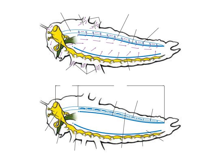
two pulsatile organs in the thorax pull hemolymph from the trailing edges of the wings, causing new hemolymph to be drawn into the veins anteriorly. Other structures at the bases of the antennae pump hemolymph through a vessel that opens close to the tip of the antenna. Hemolymph returns to the body cavity in the antennal lumen outside the vessel.
Circulatory system
The insect’s body cavity is called a hemocoel because of its mode of formation, and the liquid it contains is called hemolymph rather than blood. The hemolymph bathes the organs of the body directly and is not, in general, enclosed in vessels.
Circulation
Circulation of the hemolymph within the body cavity results from the action of a vessel lying dorsally in the hemocoel and extending the length of the body. The posterior part of this vessel pulsates and is known as the heart. As it expands, in diastole, hemolymph is drawn in through a series of valved openings, ostia. When it contracts, in systole, the valves close, and hemolymph is driven forward through the noncontractile anterior section of the vessel, the aorta, that opens into the hemocoel in the head. This creates a sluggish circulation, forcing hemolymph back from the head to the abdomen, where it moves up and into the heart to be pumped forward again. In adult Coleoptera, Diptera, and Lepidoptera, however, the hemocoel is partially occluded between the thorax and the abdomen, and hemolymph is shunted back and forth between the two as the heart undergoes reversals in its direction of pumping.
Accessory pulsatile organs maintain the circulation in the wings and antennae and sometimes also in the legs. One or
Hemolymph
Hormones and nutrients are circulated in the hemolymph, but in most insects the hemolymph is not involved in gas exchange. The water in the hemolymph provides a reservoir for the tissues, and in desiccated insects its volume is greatly reduced. In insects with a soft cuticle, such as caterpillars, and in newly molted insects before the cuticle has hardened, the hemolymph serves as a hydrostatic skeleton.
Amino acids are stored in the hemolymph as proteins. These storage proteins are synthesized in the fat body as the insect accumulates nitrogenous nutrients. The proteins are broken down at molts and, especially, at metamorphosis, when the amino acids are re-synthesized into tissue proteins. The hemolymph also contains cells known as hemocytes. There are several different types of hemocytes, all of which are nucleated. A principal function of the hemocytes is defense against potential pathogens. Bacteria are engulfed by single cells; larger organisms are encapsulated by large numbers of hemocytes.
Fat body
The fat body is an ill-defined structure in the hemolymph between the alimentary canal and the body wall. It is made
24 |
Grzimek’s Animal Life Encyclopedia |
Vol. 3: Insects |
|
|
Structure and function |
|
Accessory |
|
|
Aorta |
Pulsatile |
|
|
Organ |
Heart |
|
|
|
|
Wing Bases |
|
|
|
Dorsal |
Ostia |
|
|
Diaphragm |
Septum |
|
Ventral Diaphragm |
|
Leg Bases |
|
|
|
|
Aorta |
Heart |
|
|
Dorsal |
Alary Muscle |
|
Diaphragm |
|
|
|
Ostia |
Brain |
|
|
|
Esophagus |
|
|
Perivisceral |
|
Perineural |
Sinus |
|
|
Pericardial |
||
|
|
||
Ventral |
Sinus |
|
|
Sinus |
|
||
Nerve Cord |
|
|
|
|
|
|
Insect circulatory system. (Illustration by Jarrod Erdody)
up of sheets of cells, and its development varies. In a newly molted insect, the fat body may be barely visible, but as the insect feeds and accumulates nutrients, the fat body comes to occupy a large part of the hemocoel. The cells become filled with lipids and carbohydrates in the form of glycogen, a polysaccharide comparable to starch in plants. The fat body is also a major site for the synthesis of storage and yolk proteins. In addition, it is involved in detoxification of toxic compounds that may be derived from food or, like insecticides, may penetrate the cuticle.
Nervous system and sense organs
Central nervous system
The insect central nervous stem consists of a brain, lying dorsally within the head above the esophagus, and a chain of ventral ganglia joined together by a pair of connectives. Fundamentally, each body segment has a ganglion, but some degree of fusion always occurs, so that the number of visible
ganglia invariably is less than the number of body segments. The ganglia of the mandibular, maxillary, and labial segments are fused into a single sub-esophageal ganglion, and the last four abdominal ganglia also fuse into one. In the most extreme situation, all the thoracic and abdominal ganglia combine into a single ganglion. This occurs in the adult house fly, for example. Even where fusion takes place, the segmental components usually remain discrete within the compound ganglion.
The basic unit of the insect nervous system is the nerve cell, or neuron. The neuron is produced into a (usually) short process, the dendrite, that receives information from other neurons or from the environment, and a much longer process, the axon, that conveys information to other nerve cells and effector organs. There are three types of neurons: sensory neurons that receive information from the outside environment; motor neurons that convey instructions to effector organs, muscles, or glands; and interneurons that connect sensory input with motor output and provide for flexibility of response. The cell bodies of sensory neurons are
Grzimek’s Animal Life Encyclopedia |
25 |

Structure and function |
Vol. 3: Insects |
Hypocerebral
Ganglion
Brain
Circumesophageal
Connective
Thoracic Ganglia |
Abdominal Ganglia |
|
|
||||
1 |
2 |
3 |
|
|
|
|
6 |
|
|
|
4 |
5 |
|||
|
|
2 |
3 |
||||
|
|
1 |
|
||||
Frontal
Ganglion
Caudal
Ganglion
Subesophageal |
Longitudinal |
Ganglion |
Connectives |
Insect central nervous system, including the brain and segmental ganglia. (Illustration by Jarrod Erdody)
within or immediately beneath the epidermis. The cuticle above them is modified to allow environmental stimuli— light, touch, or a chemical—to reach the dendrite, and the information is conveyed to the central nervous system along the axon. The cell bodies of interneurons and motor neurons are grouped together in the ganglia, where all the synapses between neurons occur. From each ganglion, peripheral nerves radiate outward to the various structures of the appropriate body segment. The nerves typically include the axons of sensory cells carrying information to the central nervous system and the axons of motor neurons carrying information to the effector organs.
Insect neurons transmit information in the same way as neurons in other animals, by the passage of changes in the electrical potential across the cell membrane. Because the movement of ions into and out of the neuron produces these changes, it is critical that the ionic environment of the neurons should remain constant. The ionic composition of the insect’s blood is subject to change, however. To ensure that such changes do not affect the functioning of the neurons, the central nervous system is separated by a barrier of cells called the perineurium, known more generally as the “bloodbrain barrier.” These cells regulate the ionic composition of the fluid immediately surrounding the neurons.
Mechanoreception
Insects have many different kinds of mechanoreceptors concerned either with the perception of mechanical signals from the environment (exteroception) or with providing positional information about the parts of the body (proprioception). Most of the larger hairs on the cuticle are stimulated by contact, giving the insect its sense of touch. They are
present on all parts of the body. Sometimes the hairs are very long and slender, and their articulation with the cuticle is so sensitive that they move with airborne vibrations. Such hairs on the prothorax of some caterpillars are stimulated by the sound of the wing beat of a wasp that might be a predator. Similar hairs are present on the cerci of crickets. Essentially similar but very small hairs occur in groups called hair plates at some joints in the cuticle, where they function as proprioceptors. The hairs are bent over and stimulated when one segment of cuticle moves toward another. Hair plates are present on the basal segments of the antenna and on the small sclerites in the neck membrane. They give information on the position of the antenna relative to the head and the head relative to the thorax, respectively.
Many other types of mechanoreceptors are present in the cuticle, detecting stress on the cuticle and within the body. They have a variety of functions. Some may be involved in the detection of airborne or substrate-borne vibrations; others are stretch receptors that give the insect information about the degree of distension of its foregut, for instance.
Taste
Taste in insects is more appropriately called contact chemoreception or gustation, because similar receptors are involved not only in detecting food but also in the perception of oviposition substrates and sometimes pheromones. Contact chemoreceptors are short, hairlike or conical projections of the cuticle with a terminal pore through which chemicals in the environment reach the dendrites of the sensory neurons. These receptors occur in groups on the mouthparts and also commonly on the tarsi. Small numbers are present on the ovipositors of some insects.
26 |
Grzimek’s Animal Life Encyclopedia |

Vol. 3: Insects
There often are four sensory neurons in a single contact chemoreceptor. These are functionally of two kinds: they may initiate behavior, such as feeding or oviposition, or they may inhibit it. Each cell responds to a different range of compounds with varying degrees of sensitivity. Nearly all insects examined have neurons that respond to some sugars and amino acids, but not all the essential nutrients are tasted. Similarly, cells that inhibit behavior respond differentially to compounds that inhibit feeding or oviposition. Whether an insect feeds or oviposits on a particular substrate depends on the balance between the sensory input from neurons signaling acceptability and those signaling unacceptability.
Olfaction
The antennae are the principal olfactory organs of insects, and their length or extent of branching is an indication of the numbers of olfactory receptors and the importance of olfaction in the insects’ lives. The number of receptors typically is large. The antenna of a large grasshopper may have about 6,000 receptors; that of a cockroach may have as many as 250,000. These very large numbers increase the likelihood that odor molecules, often in low concentration, will reach a receptor and so be detected by the insect. Often there is sexual dimorphism in antennal form, again reflecting the relative importance of olfaction to the two sexes. Some male moths that are attracted to females by a sex pheromone have plumose antennae, whereas those of the female are filiform.
Olfactory receptors have large numbers of small pores in the cuticle through which odor molecules can reach the neurons inside. The form of the receptors varies. Often they are hairlike; sometimes they are flat plates in the cuticle, and sometimes they are short pegs in a depression of the cuticle. Each receptor has numerous associated sensory neurons that convey olfactory information directly to antennal lobes, which are connected to the centers for learning and memory. The antennal lobe is similar to the olfactory lobe in vertebrates, and the processing of olfactory information probably is broadly similar. Insects have the ability to respond to a wide range of odors in addition to those that are of particular importance to the species, such as pheromones and specific hostassociated odors.
Vision
Adult insects and the larvae of apterygote and endopterygote insects have a pair of compound eyes on the head. A compound eye comprises a number of similar units called ommatidia, each of which has a light-gathering system (lens), made partly from the transparent cuticle covering the eye, and a light receptor consisting of light-sensitive neurons called retinula cells. In dragonflies each eye contains about 10,000 ommatidia, the eye of a worker honey bee about 5,500 and that of a fruit fly about 800. The ommatidia of day-flying insects are separated from each other by cells containing a screening pigment that prevents light from passing from one ommatidium to another. Consequently, the retinula cells of one ommatidium receive light only from the lens system of the same ommatidium. The image formed by the eye as a whole consists of a series of spots of light, one from each ommatidium, varying in intensity and giving a fairly sharp rep-
Structure and function
Structure of the Cuticle
A
B
C
D
E
F
G
H
I
The structure of the cuticle forms a rigid casing that supports the soft inner body parts. A. Cement; B. Wax; C. Outer cuticle; D. Inner epicuticle; E. Epicuticular filament; F. Exocuticle; G. Endocuticle; H. Pore canal; I. Epidermis. (Illustration by Marguette Dongvillo)
resentation of the object. The image has a superficial resemblance to an old newsprint photograph. This type of eye is called an apposition eye.
Night-flying insects experience low light intensities, and their compound eyes have a structure that permits maximal use of the available light. The screening pigment between ommatidia is withdrawn to the extreme outer and inner parts of the eye, where it does not interfere with the movement of light between ommatidia. As a consequence, the retinula cells of one ommatidium are illuminated by light from several lens systems. As a result, the image formed by the eye is much less sharp than that produced by an apposition eye. This type of eye is called a superposition eye.
Some insects are known to have color vision. In most species that have been studied, the visual spectrum is slightly different from that of humans. Insects can detect ultraviolet light, whereas humans cannot, but many insects cannot distinguish red. The fact that insects typically do not pollinate red flowers is a reflection of this; birds usually pollinate red flowers. Some insects, notably bees and ants, have the ability to distinguish the plane of polarization of light. Light from the sky is partially polarized, and the dominant plane of polarization is determined by the position of the sun. Bees and ants can learn the pattern of polarization of light from the sky and can use this information to navigate, usually back to the nest, even if the sun is not visible.
Grzimek’s Animal Life Encyclopedia |
27 |

Structure and function |
Vol. 3: Insects |
Dorsal view of a bee head
lens
crystalline compound cone
eye
photoreceptor cell
ocelli
compound eye
ocellus
ommatidia
one ommatidium
Compound insect eye. (Illustration by Jacqueline Mahannah.)
In addition to compound eyes, adult insects and exopterygote larvae typically have three ocelli arranged in a triangle on the front of the head between the compound eyes. Ocelli have single lenses and are sensitive to rapid changes in light intensity. Larval endopterygote insects have no compound eyes but often have stemmata on the side of the head. Sawfly larvae have only one stemma on each side, but caterpillars and tiger beetle larvae have six. Like ocelli, each stemma has only a single lens, and although the arrangement of sensory cells varies, the connections within the insect’s brain are quite different from those of ocelli. Even the single stemma of sawfly larvae is capable of image formation, but the six stemmata of caterpillars seem capable of only crude form perception.
Endocrine system and hormones
Normal metabolism and major events, such as molting and metamorphosis, are controlled by hormones. Insects have three endocrine glands: the prothoracic gland, a diffuse structure in the prothorax; the corpora allata, small round bodies, one on either side of the foregut just behind the brain; and the corpora cardiaca adjacent to the heart and also just behind the brain. In addition, there are numerous isolated endocrine cells in the midgut epithelium. Many hormones also
are produced by neurosecretory cells in the central nervous system. These cells release their products (neurohormones) into the hemolymph at numerous neurohemal organs, where the cell axons are exposed to the hemolymph.
The prothoracic gland produces the molting hormone, ecdysone. Because insects do not molt after becoming adult, these glands generally are absent from adult insects. The corpora allata produce juvenile hormone, which plays a major role in metamorphosis and also regulates yolk production in many insect species. The corpora cardiaca produce adipokinetic hormone, which controls the mobilization of lipids from the fat body. This is especially important in insects, such as locusts, that use lipids as the major fuel in flight. The corpora cardiaca also act as neurohemal organs for many neurohormones produced by cells in the brain.
Reproductive system
The paired gonads of both sexes lie in the dorsal part of the abdomen above the alimentary canal. From the gonads, a duct on either side passes around the gut, and the two ducts join beneath the gut to form a single median duct from which the gametes are discharged. In addition, both sexes have storage organs and accessory glands associated with the reproductive system.
28 |
Grzimek’s Animal Life Encyclopedia |

Vol. 3: Insects
Close-up of horsefly head showing its compound eyes. (Photo by Animals Animals © J. A. L. Cooke, OSF. Reproduced by permission.)
Structure and function
diploid, having a set of chromosome from each parent, but their gametes have only one set (haploid). The reduction in number (meiosis) occurs at two divisions of the spermatocytes, so that each spermatocyte gives rise to four spermatozoa. These events occur in sequence along the length of each sperm tube. From the testes, the sperm move to seminal vesicles, where they are stored until they are transferred to a female. At the time of transfer, in many insects, the sperm are packaged in a structure called a spermatophore, which protects them from desiccation during transfer. The spermatophore is produced by material from the male accessory glands, which usually open separately into the median ejaculatory duct. Other glands, at least in some species, produce chemicals that affect the female’s physiology or behavior. These chemicals may cause her to reject subsequent mating attempts by males (fruit flies), they may provide protein that is added to the protein put into eggs by the female (many insects), or they may confer protection against predators to the female and her eggs (some moths).
Male
Each testis is made up of tubes varying in number from only one in some beetles to more than 100 in grasshoppers. Germ cells at the tip of each testis tube divide, giving rise after several divisions to spermatocytes. Most insects are
Female
Each ovary is made up of ovarioles. The number of ovarioles varies in different taxa. Lepidoptera have four in each ovary, fruit flies have 10–30; and large grasshoppers have more than 100. Stem cells at the tip of each ovariole divide
A mayfly larva with its cast skin (top). (Photo by P. A. Hinchliffe. Bruce Coleman, Inc. Reproduced by permission.)
Grzimek’s Animal Life Encyclopedia |
29 |

Structure and function
Terminal
filament
Ovariole
Calyx
Lateral oviduct
Common
oviduct
Accessory
gland
Vagina
Female
|
|
Vol. 3: Insects |
|
Spermatogonia |
Apical cell |
|
Epithelial |
Sperm |
|
sheath |
|
|
cyst |
|
|
|
|
Spermatids |
|
Germarium |
|
|
|
Spermatozoa |
Zone of growth |
|
|
||
Vas efferens |
Zone of maturation & reduction |
|
|
Zone of transformation |
|
Ovary |
|
Sperm |
|
Development |
|
|
|
|
|
Vas |
Testes |
|
deferens |
|
|
|
Spermathecal
gland
Accessory
gland
Seminal vesicle
Spermatheca |
Ejaculatory |
|
duct |
Penis
Gonopore
Male
Male and female insect reproductive systems and sperm development. (Illustration Jarrod Erdody)
to produce oocytes (eggs). This parallels the process in males, but in females the cell divisions at which chromosome number is reduced do not occur until the oocyte is laid and penetrated by a sperm. In the ovariole each oocyte is provisioned with yolk, the proteins of which are synthesized in the fat body and then carried to the ovaries in the hemolymph. Finally, a layer of cells surrounding the oocyte produces the chorion (eggshell). As in the testis tubes, the developmental stages occur successively along the ovarioles, with the most advanced stages at the greatest distance from the tip.
The unfertilized eggs move from the ovarioles and are stored in the oviducts. The female accessory glands make the glue that is used by many insects to stick eggs to the substratum as they are laid. In a few cases, such as grasshoppers, cockroaches, mantids, and tortoise beetles, they provide material for the production of an ootheca, in which the eggs are concealed and protected after laying. The female reproductive organs include a spermatheca, in which sperm are stored after the female is inseminated. Fertilization of the eggs does not take place until they are laid, which may be months after insemination, especially in social insects.
Resources
Books |
Reginald F. Chapman, DSc |
Chapman, R. F. The Insects: Structure and Function. 4th edition. |
|
Cambridge: Cambridge University Press, 1998. |
|
30 |
Grzimek’s Animal Life Encyclopedia |