

Учебники / Atlas_anatomii_cheloveka_Frenk_Netter_6-oe_izdanie_Chast3
.pdfWrist and Hand: Deeper palmar Dissections
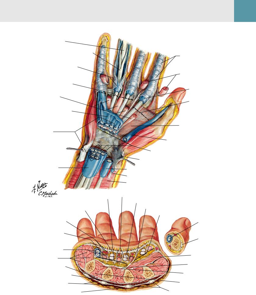
Insertion of flexor digitorum profundus tendon
Insertion of flexor digitorum
superficialis tendon
Midpalmar space (deep to flexor tendons and lumbrical muscles)
Probe in midpalmar space
5th finger (synovial) tendinous sheath
Common flexor sheath (ulnar bursa)
Hypothenar muscles
Common palmar digital branches of median nerve (cut)
Ulnar artery and nerve
Superficial palmar branch of radial artery and recurrent branch of median nerve to thenar muscles
 (Synovial) flexor tendon sheaths of fingers
2nd, 3rd, and 4th lumbrical muscles (in fascial sheaths)
Superficial palmar arterial and venous arches
Common flexor sheath (ulnar bursa)
Median nerve
Palmar digital nerves to 5th finger and medial half of 4th finger
Superficial branch of ulnar nerve
Deep palmar branch of ulnar artery and deep branch of ulnar nerve
Pisiform
Common flexor sheath (ulnar bursa) containing superficialis and profundus flexor tendons
Flexor carpi ulnaris tendon
Ulnar artery with venae comitantes and ulnar nerve
Anular and cruciform parts of fibrous sheath over (synovial) flexor tendon sheaths
Proper palmar digital arteries and nerves
Common palmar digital artery
Septum separating thenar from midpalmar space
Thenar space (deep to flexor tendons and 1st lumbrical muscle)
Probe in dorsal extension of thenar space deep to adductor pollicis muscle
1st dorsal interosseous muscle
Fascia over adductor pollicis muscle
Proper palmar digital nerves of thumb
Anterior (palmar) views
Palmar aponeurosis (reflected)
Septa from palmar aponeurosis forming canals
Proper palmar digital arteries
Common palmar digital artery
Probe in 1st lumbrical fascial sheath
(Synovial) tendinous sheath of flexor pollicis longus (radial bursa)
Proper palmar digital nerves of thumb
Thenar muscles
Flexor retinaculum (transverse carpal ligament)
Palmaris longus tendon and palmar carpal ligament
Median nerve
Tendinous sheath of flexor pollicis longus (radial bursa)
Flexor carpi radialis tendon
Radial artery and venae comitantes
Plate 447 |
Wrist and Hand |

Lumbrical Muscles and Bursae, Spaces, and Sheaths: Schema 6
|
See also Plate 445 |
Common variation |
Usual arrangement |
|
(Synovial) tendon |
|
sheaths of fingers |
|
Lumbrical muscles |
|
(in fascial sheaths) |
Intermediate bursa |
Midpalmar space |
|
|
||
(communication |
Thenar space |
|
between common |
||
flexor sheath [ulnar |
|
|
bursa] and tendinous |
Common flexor sheath |
|
sheath of flexor pollicis |
||
(ulnar bursa) |
||
longus [radial bursa]) |
||
|
||
|
Tendinous sheath of flexor |
|
|
pollicis longus (radial bursa) |
Lumbrical muscles: schema
Flexor digitorum |
|
|
|
|
|
|
|
|
superficialis tendons (cut) |
|
|
|
|
|
|
|
Camper chiasm |
|
|
|
|
|
|
|
|
Distal lumbrical tendons |
|
|
|
|
|
|
|
|
insert into extensor |
|
|
|
|
|
|
|
|
expansion system |
3rd and 4th lumbrical muscles (bipennate) |
|
|
|
|
|
|
1st and 2nd lumbrical muscles |
|
|
|
|
|
|
|
|
||
|
|
|
|
|
|
|
(unipennate) |
|
Proximal lumbrical tendons arise from |
|
|
|
|
|
|
|
|
flexor digitorum profundus tendons |
|
|
|
|
|
|
|
|
Note: Flexor digitorum superficialis and |
|
|
|
|
|
|
|
|
profundus tendons encased in synovial |
|
|
|
|
|
Flexor digitorum |
||
sheaths are bound to phalanges by fibrous |
|
|
|
|
|
profundus tendons |
||
digital sheaths made up of alternating |
|
|
|
|
|
|
|
|
strong anular (A) and weaker cruciform (C) |
|
|
|
|
|
|
|
|
parts (pulleys). |
|
|
|
|
|
|
|
|
A1 |
C1 |
A2 |
C2 |
A3 |
C3 |
A4 |
C4 |
A5 |
Tendons of flexor digitorum superficialis and profundus muscles
(Synovial) tendon sheath
Palmar ligaments (volar plates)
|
|
|
Wrist and Hand |
Plate 448 |
|

Flexor Tendons, Arteries, and Nerves at Wrist
See also Plates 459, 460
Palmar view
Lumbrical muscles
Superficial palmar (arterial) arch
Opponens digiti minimi muscle
Flexor digiti minimi brevis muscle
Abductor digiti minimi muscle
Pisiform
Flexor carpi ulnaris tendon
Ulnar nerve
Ulnar artery
Common flexor sheath (ulnar bursa)
Flexor digitorum superficialis tendons
Flexor pollicis
Adductor pollicis muscle brevis muscle (reflected)
Abductor pollicis
brevis muscle (reflected)
Opponens pollicis muscle (cut)
1st metacarpal bone
Trapezium
Flexor retinaculum (transverse carpal ligament)
(Synovial) tendon sheath
Palmar carpal ligament (reflected)
Flexor pollicis longus tendon in tendon sheath (radial bursa)
Flexor carpi radialis tendon
Radial artery
Median nerve
Palmaris longus tendon
Cross section of wrist demonstrating carpal tunnel
|
|
Flexor retinaculum (transverse |
Palmaris longus |
|
|
|
tendon |
||
|
|
carpal ligament) |
||
|
|
Median nerve* |
||
|
|
|
||
|
|
Ulnar artery |
|
|
|
|
and nerve |
|
|
|
|
Flexor carpi |
|
|
|
|
ulnaris tendon |
|
|
|
|
Flexor |
|
|
|
|
digitorum |
|
|
|
|
superficialis |
|
|
|
|
tendons* |
|
|
|
|
(3, 4 superficial; |
Flexor carpi |
|
|
|
2, 5 deep) |
||
|
|
radialis tendon |
||
|
|
|
||
|
4 |
Flexor |
Flexor pollicis |
|
3 |
digitorum |
|||
|
longus tendon |
|||
|
|
profundus |
||
|
|
in tendon sheath* |
||
|
|
tendons* |
||
2 5 |
|
|
||
|
(2, 3, 4, 5) |
Radial artery |
||
|
|
Hamate |
Trapezium |
|
Simple method of demonstrating |
Trapezoid |
|||
|
|
Capitate |
||
arrangement of flexor |
|
digitorum superficialis |
|
tendons within carpal tunnel |
*Contents of carpal tunnel |
|
Plate 449 Wrist and Hand
Bursae, Spaces, and Tendon Sheaths of Hand 6
Flexor digitorum profundus tendon
Flexor digitorum superficialis tendon
Fibrous and synovial (tendon) sheaths of finger (opened)
Midpalmar space
(deep to flexor tendons and lumbrical muscles)
Lumbrical muscles in fascial sheaths
Common flexor sheath (ulnar bursa) (opened)
Flexor digitorum superficialis tendons (3, 4 superficial;
2, 5 deep)
Common flexor sheath (ulnar bursa)
Midpalmar space
Palmar aponeurosis
Common palmar digital artery and nerve
Lumbrical muscle in its fascial sheath
Flexor tendons to 5th digit in common flexor sheath (ulnar bursa)
Hypothenar muscles
Dorsal interosseous fascia
Dorsal subaponeurotic space
Dorsal fascia of hand
Dorsal subcutaneous space
Anular and cruciform parts (pulleys) of fibrous sheath (over synovial sheath of finger)
Lumbrical muscles in fascial sheaths (cut and reflected)
(Synovial) tendon sheath of finger
Thenar space
(deep to flexor tendon and 1st lumbrical muscle)
Fascia of adductor pollicis muscle
Tendinous sheath of flexor pollicis longus (radial bursa)
Flexor digitorum profundus tendons (2, 3, 4, 5)
Flexor retinaculum (transverse carpal ligament) (reflected)
Tendinous sheath of flexor pollicis longus (radial bursa)
Flexor carpi radialis tendon
Pronator quadratus muscle
Septa forming canals
Profundus and superficialis flexor tendons to 3rd digit
Septum between midpalmar and thenar spaces
Thenar space
Flexor pollicis longus tendon in tendon sheath (radial bursa)
Extensor pollicis longus tendon
Adductor pollicis muscle
Palmar interosseous fascia
Palmar interosseous muscles
Dorsal interosseous muscles
Extensor tendons
|
|
|
Wrist and Hand |
Plate 450 |
|

Flexor and Extensor Tendons in Fingers
See also Plate 445
Insertion of central band of extensor tendon to base of middle phalanx
Triangular aponeurosis
|
Extensor |
Long extensor |
Interosseous muscles |
|
expansion |
tendon |
|
Slips of long |
(hood) |
|
|
extensor tendon |
|
|
|
to lateral bands |
|
|
|
Posterior (dorsal) view
Insertion on extensor tendon |
|
Interosseous |
to base of distal phalanx |
|
|
|
tendon slip |
|
|
|
|
|
Lateral bands |
to lateral band |
|
|
|
|
|
Lumbrical muscle |
Metacarpal bone
Part of interosseous tendon passes to base of proximal phalanx and joint capsule
Insertion of extensor tendon |
Lateral band |
Extensor expansion (hood) |
|
|
Long extensor tendon |
||
to base of middle phalanx |
Central band |
||
|
|||
Insertion of extensor |
|
|
|
tendon to base |
|
|
|
of distal phalanx |
|
Metacarpal bone |
|
|
|
Finger in extension: lateral (radial) view
Collateral |
Vinculum |
Vincula |
Flexor digitorum |
Interosseous muscles |
|
profundus tendon |
|||||
ligaments |
breve |
longa |
|
||
Flexor digitorum |
|
||||
|
|
|
Lumbrical muscle |
||
|
|
|
superficialis tendon |
||
|
|
|
|
Insertion of small deep slip of extensor tendon to proximal phalanx and joint capsule
Attachment of interosseous muscle to base of proximal phalanx and joint capsule
Collateral ligament
Extensor tendon
|
Palmar ligament |
|
Insertion of lumbrical |
(volar plate) |
|
|
||
muscle to extensor tendon |
Flexor digitorum |
|
|
superficialis tendon (cut) |
|
|
Collateral ligaments |
|
Finger in flexion: |
Flexor digitorum |
|
profundus tendon (cut) |
||
lateral (radial) view |
||
|
||
|
Palmar ligament (volar plate) |
Interosseous muscles
Lumbrical muscle
Note: Black arrows indicate pull of long extensor tendon; red arrows indicate pull
of interosseous and lumbrical muscles; dots indicate axis of rotation of joints.
Plate 451 |
Wrist and Hand |

Deep transverse metacarpal ligaments Common palmar digital arteries Palmar metacarpal arteries Deep palmar (arterial) arch Opponens digiti minimi muscle
Flexor digiti minimi brevis muscle (cut)
Deep palmar branch of ulnar artery and deep branch of ulnar nerve
Abductor digiti minimi muscle (cut) Median nerve Pisiform
Palmar carpal arterial arch Flexor carpi ulnaris tendon Ulnar artery and palmar carpal branch Ulnar nerve Pronator quadratus muscle
Intrinsic Muscles of Hand 6
Lumbrical muscles (reflected)
Branches from deep branch of ulnar nerve to 3rd and 4th lumbrical muscles and to all interosseous muscles
1st dorsal interosseous muscle
Adductor pollicis muscle
Flexor pollicis brevis muscle
Abductor pollicis brevis muscle (cut)
Branches of median nerve to thenar muscles and to 1st and 2nd lumbrical muscles
Opponens pollicis muscle
Flexor retinaculum (transverse carpal ligament) (reflected)
Superficial palmar branch of radial artery
Radius
Radial artery and palmar carpal branch
Anterior (palmar) view
Tendinous slips to extensor expansions (hoods)
1 |
2 |
3 |
3 |
2 |
1 |
Deep transverse |
Dorsal |
|
|
4 |
|
|
metacarpal |
interosseous |
|
|
|
|
|
|
|
|
|
|
|
ligaments |
|
muscles |
|
|
|
|
|
|
|
|
|
|
|
|
|
(bipennate) |
|
|
|
|
|
Palmar interosseous |
|
|
|
|
|
|
|
|
|
|
Abductor digiti |
|
|
muscles (unipennate) |
|
|
|
minimi muscle |
|
|
|
Abductor pollicis |
|
|
|
|
|
|
brevis muscle |
|
|
|
|
|
|
Radial artery |
|
|
|
|
|
|
|
|
|
|
|
|
Radius |
Radius |
|
|
Ulna |
|
|
Anterior |
|
|
Ulna |
|
|
||
|
|
|
|
|
|
(palmar) view |
Posterior |
|
|
Note: Arrows indicate action of muscles. |
|||
(dorsal) view |
|
|
||||
|
|
|
Wrist and Hand |
Plate 452 |
|

Arteries and Nerves of Hand: Palmar Views
See also Plate 420
Branches of palmar digital nerves and arteries to dorsum of middle and distal phalanges
Proper palmar digital nerves and arteries
Communicating branch of median nerve with ulnar nerve
Common palmar digital nerves and arteries
Superficial palmar (arterial) arch
Common flexor sheath (ulnar bursa)
Superficial branch of ulnar nerve
Deep palmar branch of ulnar artery and deep branch of ulnar nerve
Flexor retinaculum (transverse carpal ligament)
Palmar carpal ligament (continuous with extensor retinaculum)
Ulnar artery and nerve
Proper palmar digital nerves of ulnar nerve
Communicating branch of median nerve with ulnar nerve
Deep palmar branch of ulnar nerve to 3rd and 4th lumbrical, all interosseous, adductor pollicis, and deep head of flexor pollicis brevis muscles
Hook of hamate
Superficial branch of ulnar nerve
Branches to hypothenar muscles
Deep palmar branch of ulnar artery and deep branch of ulnar nerve
Pisiform
Palmar carpal branches of radial and ulnar arteries
Ulnar artery and nerve
Flexor tendons, synovial and fibrous sheaths
Branches of median nerve to 1st and 2nd lumbrical muscles
Adductor pollicis muscle
Proper palmar digital nerves and arteries to thumb
Flexor pollicis brevis muscle
Recurrent (motor) branch of median nerve to thenar muscles
Opponens pollicis muscle
Abductor pollicis brevis muscle (cut)
Superficial palmar branch of radial artery
Median nerve and palmar branch
Radial artery
Proper palmar digital nerves of median nerve
Proper palmar digital arteries
Common palmar digital arteries
Proper palmar metacarpal arteries
Radialis indicis artery
Proper digital arteries and nerves of thumb
Princeps pollicis artery
Deep palmar (arterial) arch and deep branch of ulnar nerve
Superficial palmar branch of radial artery
Median nerve
Radial artery
Plate 453 |
Wrist and Hand |

Wrist and Hand: Superficial Radial Dissection |
6 |
|
|
See also Plate 466 |
|
Lateral (radial) view
Insertion of extensor pollicis longus tendon
Insertion of extensor pollicis brevis tendon
1st metacarpal bone
Insertion of abductor pollicis longus tendon
Trapezium
Radial artery in anatomical snuffbox*
Scaphoid*
Dorsal digital branches of radial nerve*
Lateral branch
*Snuffbox contents (superficial to deep)
Radial nerve (dorsal digital branch) Cephalic vein branches (cut away) Radial artery and branches Scaphoid bone
Deep fascia (cut)
1st dorsal interosseous muscle
Radial artery
Extensor carpi radialis longus tendon
Extensor carpi radialis brevis tendon
Dorsal carpal branch of radial artery
Extensor retinaculum
Medial branch
Superficial branch of radial nerve
|
|
|
Wrist and Hand |
Plate 454 |
|

Wrist and Hand: Superficial Dorsal Dissection
See also Plates 402, 403
Posterior (dorsal) view
Dorsal branches of proper palmar digital nerves
Dorsal digital nerves and veins
Intercapitular veins
Probe in dorsal subaponeurotic space (between opened dorsal fascia of hand and dorsal interosseous fascia)
Dorsal metacarpal veins
Dorsal venous network of hand
Dorsal branch of ulnar nerve
Basilic vein
Communicating branches of radial and ulnar nerves
Extensor retinaculum (thickening of posterior antebrachial fascia)
Superficial branch of radial nerve
Cephalic vein
Posterior antebrachial cutaneous nerve (from radial nerve)
Note: Lymphatic pathways shown in black; arrows indicate direction of drainage.
Plate 455 |
Wrist and Hand |

Wrist and Hand: Deep Dorsal Dissection 6
See also Plate 445
Dorsal branches of proper palmar digital branches of median nerve and of proper palmar digital arteries to
dorsum of middle and distal phalanges of 2nd, 3rd, and radial half of 5th fingers
Dorsal digital branches of superficial branch of radial nerve to 1st, 2nd, 3rd, and radial half of 4th fingers
Dorsal digital arteries
Extensor carpi radialis brevis tendon
Extensor carpi radialis longus tendon
Extensor pollicis longus tendon Extensor pollicis brevis tendon
Abductor pollicis longus tendon
Radial artery in anatomical snuffbox
Extensor digitorum, extensor digiti minimi, and extensor indicis tendons (cut)
Superficial branch of radial nerve
Lateral antebrachial cutaneous nerve (terminal part of musculocutaneous nerve)
Posterior (dorsal) view
Dorsal branches of proper palmar digital branches of ulnar nerve and of proper palmar digital arteries to dorsum of middle and distal phalanges of 5th and ulnar half of 4th fingers
Dorsal digital branches of dorsal branch
of ulnar nerve
Dorsal metacarpal arteries
Dorsal carpal (arterial) arch
Extensor carpi ulnaris tendon
Dorsal carpal branch of ulnar artery
Dorsal branch of ulnar nerve
Extensor retinaculum
Posterior antebrachial cutaneous nerve (branch of radial nerve)
Medial antebrachial cutaneous nerve
|
|
|
Wrist and Hand |
Plate 456 |
|