

Учебники / Atlas_anatomii_cheloveka_Frenk_Netter_6-oe_izdanie_Chast3
.pdfArteries of Femoral Head and Neck
See also Plates 499, 530
|
Anterior view |
|
|
Posterior view |
|
Retinacular |
Superior |
|
Acetabular |
Superior |
Retinacular |
|
branch of |
||||
arteries |
Anterior |
|
obturator artery |
Posterior |
arteries |
(subsynovial) |
Inferior |
|
(often minute) |
Inferior |
(subsynovial) |
|
|
|
|
|
|
Anastomosis |
|
|
|
|
Anastomosis |
between medial |
|
|
|
|
|
|
|
|
|
|
|
and lateral |
|
|
|
|
|
circumflex |
|
|
|
|
Ischiofemoral |
femoral |
|
|
|
|
|
|
|
|
|
ligament and |
|
arteries |
|
|
|
|
|
|
|
|
|
joint capsule |
|
|
|
|
|
|
|
lliofemoral (Y) |
|
lliopsoas |
Medial |
|
|
|
circumflex |
|
|
||
|
tendon |
|
|
||
ligament and |
|
|
|
||
|
femoral artery |
|
|||
|
|
|
|||
joint capsule |
|
|
|
||
|
Medial circumflex |
|
|
||
|
|
|
|
||
|
|
femoral artery |
Lateral |
|
|
Ascending, |
|
|
circumflex |
|
|
|
|
femoral artery |
|
||
Transverse, |
|
Profunda femoris |
|
||
|
|
|
|
||
Descending |
|
|
|
|
|
|
(deep femoral) |
|
|
|
|
branches of |
|
|
Nutrient artery |
||
|
artery |
|
|||
Lateral circumflex |
|
|
of femur |
||
|
|
|
|||
femoral artery |
|
|
|
|
|
|
|
Nutrient artery |
|
|
|
|
|
of femur |
|
|
|
|
|
|
|
Anterior view in situ |
|
|
Coronal section |
|
|
|
Iliacus |
|
|
Medial circumflex |
|
muscle |
|
|
|
|
|
||
|
|
|
femoral artery |
|
Psoas |
Acetabular labrum |
|
|
|
||
|
Anastomosis |
|
muscle |
||
|
|
|
|
||
|
|
|
|
|
|
Ligaments and |
Lateral circumflex |
|
Femoral |
||
joint capsule |
|
artery |
|||
femoral artery |
|
||||
|
|
|
|
||
Synovial |
|
Ascending, |
|
Pectineus |
|
|
Transverse, |
|
muscle |
||
membrane |
|
|
|||
|
Descending |
|
|
||
|
|
|
|
||
Retinacular |
|
branches |
|
|
|
|
|
|
|
|
|
arteries |
|
|
|
|
|
|
|
Acetabular |
|
|
|
|
|
branch |
|
|
|
|
|
Obturator |
|
|
|
|
|
artery |
|
|
|
|
|
|
|
|
Medial |
|
|
|
|
|
circumflex |
|
|
|
|
|
femoral |
|
|
|
|
|
artery |
|
|
|
|
|
Profunda femoris |
|
|
|
|
|
(deep femoral) |
|
Epiphyseal |
|
|
artery |
|
|
|
|
|
||
|
plate |
|
|
Medial circumflex femoral artery |
|
|
|
|
|
||
Medial circumflex |
|
lliopsoas tendon |
|
||
|
|
|
|
||
femoral artery |
|
Lateral circumflex femoral artery |
|
||
Femur of child: anterior view
Plate 491 Hip and Thigh

Sartorius muscle Profunda femoris (deep femoral) artery and vein
Pectineus muscle lliopsoas muscle
Rectus femoris muscle
Vastus medialis muscle
Lateral femoral cutaneous nerve
Vastus intermedius muscle
Femur
Vastus lateralis muscle
Tensor fasciae latae muscle
lliotibial tract Gluteus maximus muscle Sciatic nerve
Vastus medialis muscle
Rectus femoris muscle
Vastus intermedius muscle
Vastus lateralis muscle
Iliotibial tract
Lateral intermuscular septum of thigh
Biceps |
Short head |
femoris |
|
muscle |
Long head |
Semitendinosus muscle
Semimembranosus muscle
Rectus femoris tendon Vastus intermedius muscle lliotibial tract Vastus lateralis muscle
Articularis genus muscle
Lateral intermuscular septum of thigh
Femur
Biceps femoris muscle
Common fibular (peroneal) nerve
Tibial nerve
Thigh: Serial Cross Sections |
7 |
Fascia lata
Branches of femoral nerve
Femoral artery and vein
Adductor longus muscle
Great saphenous vein
Obturator nerve (anterior branch)
Adductor brevis muscle
Obturator nerve (posterior branch)
Gracilis muscle
Adductor magnus muscle
Posterior femoral cutaneous nerve
Semimembranosus muscle
Semitendinosus muscle
Biceps femoris muscle (long head)
Medial intermuscular septum of thigh
Sartorius muscle
Nerve to vastus medialis muscle
Saphenous nerve
Femoral artery and vein
Great saphenous vein
Adductor longus muscle
Gracilis muscle
Adductor brevis muscle
Profunda femoris (deep femoral) artery and vein
Adductor magnus muscle
Posterior intermuscular septum of thigh
Sciatic nerve
Vastus medialis muscle
Sartorius muscle
Saphenous nerve and descending genicular artery
Great saphenous vein
Gracilis muscle
Adductor magnus tendon
Popliteal vein and artery
Semimembranosus muscle
Semitendinosus muscle
|
|
|
Hip and Thigh |
Plate 492 |
|

Knee: Medial and Lateral Views
Medial view
Vastus medialis muscle
Quadriceps femoris tendon
Medial epicondyle of femur
Patella
Medial patellar retinaculum
Joint capsule
Patellar ligament
Tibial tuberosity
Lateral view
lliotibial tract (cut)
Biceps femoris |
Long head |
muscle (cut) |
Short head |
Bursa deep to iliotibial tract
Fibular collateral ligament and bursa deep to it
Plantaris muscle
Biceps femoris tendon and its inferior subtendinous bursa
Common fibular (peroneal) nerve
Head of fibula
Gastrocnemius muscle
Soleus muscle
Fibularis (peroneus) longus muscle
Sartorius muscle (cut)
Gracilis muscle (cut)
Tendon of semitendinosus muscle (cut)
Semimembranosus muscle and tendon
Adductor magnus tendon
Parallel fibers |
Tibial |
||
collateral |
|||
Oblique fibers |
|||
ligament |
|||
|
|||
Semimembranosus |
|
||
bursa |
|
|
|
Anserine bursa |
|
|
|
deep to |
|
|
|
Semitendinosus, |
|
Pes |
|
Gracilis, and |
|
||
|
anserinus |
||
Sartorius tendons |
|||
|
|||
Gastrocnemius muscle |
|||
Soleus muscle |
|
|
|
Vastus lateralis muscle
Quadriceps femoris tendon
Patella
Lateral patellar retinaculum
Joint capsule of knee
Patellar ligament
Tibial tuberosity
Tibialis anterior muscle
Plate 493 |
Knee |

Right knee in extension
Vastus intermedius muscle
Vastus lateralis muscle
lliotibial tract
Lateral patellar retinaculum
Lateral epicondyle of femur
Fibular collateral ligament and bursa
Biceps femoris tendon and its inferior subtendinous bursa
Dashed oval indicates bursa deep to iliotibial tract
Insertion of iliotibial tract to Gerdy’s tubercle and oblique line of tibia
Common fibular (peroneal) nerve
Head of fibula
Fibularis (peroneus) longus muscle
Extensor digitorum longus muscle
Tibialis anterior muscle
Joint opened,
knee slightly in flexion
Femur
Articularis genus muscle
Synovial membrane (cut edge)
Lateral condyle of femur
Origin of popliteus tendon (covered by synovial membrane)
Subpopliteal recess
Lateral meniscus
Fibular collateral ligament
Head of fibula
Patella (articular surface on posterior aspect)
Vastus lateralis muscle (reflected inferiorly)
Knee: Anterior Views 7
See also Plate 497
Femur
Articularis genus muscle
Vastus medialis muscle
Rectus femoris tendon |
||
becoming |
|
|
Quadriceps femoris tendon |
||
Patella |
|
|
Medial epicondyle of femur |
||
Medial patellar retinaculum |
||
Tibial collateral ligament |
||
Semitendinosus, |
Pes |
|
Gracilis, and |
||
anserinus |
||
Sartorius tendons |
||
|
||
Anserine bursa |
|
|
Medial condyle of tibia |
||
Patellar ligament |
|
|
Tibial tuberosity |
|
|
Gastrocnemius muscle |
||
Suprapatellar (synovial) bursa
Cruciate ligaments (covered by synovial membrane)
Medial condyle of femur
Infrapatellar synovial fold
Medial meniscus
Alar folds (cut)
Infrapatellar fat pads (lined by synovial membrane)
Suprapatellar (synovial) bursa (roof reflected)
Vastus medialis muscle (reflected inferiorly)
|
|
|
Knee |
Plate 494 |
|

Knee: Interior
See also Plate 501
Inferior view
lliotibial tract blended into lateral patellar retinaculum and capsule
Bursa
Subpopliteal recess
Popliteus tendon
Fibular collateral ligament
Bursa
Lateral condyle of femur
Anterior cruciate ligament
Arcuate popliteal ligament
Patellar ligament
Medial patellar retinaculum blended into joint capsule
Suprapatellar synovial bursa
Synovial membrane (cut edge)
Infrapatellar synovial fold
Posterior cruciate ligament
Tibial collateral ligament (superficial and deep fibers)
Medial condyle of femur
Oblique popliteal ligament
Semimembranosus tendon
Posterior aspect
Superior view
Posterior meniscofemoral ligament
Arcuate popliteal ligament
Fibular collateral ligament
Bursa
Popliteus tendon
Subpopliteal recess
Lateral meniscus
Superior articular surface of tibia (lateral facet)
lliotibial tract blended into capsule
Infrapatellar fat pad
Semimembranosus tendon
Oblique popliteal ligament
Posterior cruciate ligament
Tibial collateral ligament (deep fibers bound to medial meniscus)
Medial meniscus
Synovial membrane
Superior articular surface of tibia (medial facet)
Joint capsule
Anterior cruciate ligament
Patellar ligament
Anterior aspect 
Superior view: ligaments and cartilage removed
Attachment of posterior cruciate ligament |
Intercondylar eminence |
|
|
Attachment of synovial membrane |
Attachment of synovial membrane |
|
Superior articular surface Superior articular surface of tibia (medial facet)
of tibia (lateral facet)
Attachment of anterior Attachments of horns cruciate ligament
of lateral meniscus
Attachments of horns of medial meniscus
|
|
|
Tibial tuberosity |
|
Anterior |
|
aspect |
|
|
||
|
|
|
|
Plate 495 |
|
|
Knee |

Knee: Cruciate and Collateral Ligaments 7
Right knee in flexion: anterior view
Anterior cruciate ligament
Lateral condyle of femur (articular surface)
Popliteus tendon
Fibular collateral ligament
Lateral meniscus
Transverse
ligament of knee
Head of fibula
Gerdy’s tubercle
Adductor tubercle on medial epicondyle of femur
Medial condyle of femur (articular surface)
Tibial collateral ligament (superficial and deep fibers)
Medial meniscus
Medial condyle of tibia
Posterior cruciate ligament
Medial condyle of femur (articular surface)
Medial meniscus
Tibial collateral ligament (superficial and deep fibers)
Medial condyle of tibia
Tibial tuberosity
Right knee in extension: posterior view
Posterior cruciate ligament
Anterior cruciate ligament
Posterior meniscofemoral ligament
Lateral condyle of femur (articular surface)
Popliteus tendon
Fibular collateral ligament
Lateral meniscus
Head of fibula
|
|
|
Knee |
Plate 496 |
|

Knee: Anteroposterior Radiograph and Posterior View
See also Plate 494
Femur
Patella
Lateral epicondyle
Lateral condyle of femur Intercondylar eminence Lateral condyle of tibia
Head of fibula
Neck of fibula
Fibula
Semitendinosus muscle
Sartorius muscle
Semimembranosus muscle
Gracilis muscle
Medial head of gastrocnemius muscle (cut)
Medial subtendinous bursa of gastrocnemius muscle (open)
Oblique popliteal ligament
Medial epicondyle
Medial condyle of femur
Medial condyle of tibia
|
Tibia |
Plantaris muscle (cut) |
Iliotibial tract (band) |
|
Biceps femoris muscle |
|
Lateral head of gastrocnemius |
|
muscle (cut and reflected superiorly |
|
and laterally) |
|
Lateral subtendinous |
|
bursa of gastrocnemius muscle |
|
Lateral collateral ligament |
Arcuate popliteal ligament
Tibial collateral ligament
Biceps femoris tendon
Bursa of semimembranosus muscle
Common fibular (peroneal) nerve
Popliteus muscle
Semimembranosus tendon
Fibula
Interosseous membrane
|
Tibia |
Plate 497 |
Knee |

Knee: Posterior and Sagittal Views 7
Right knee: posterior view
|
Femur (popliteal surface) |
|
Adductor magnus tendon |
Attachment of joint capsule |
|
Medial head of gastrocnemius |
|
|
muscle and |
Plantaris muscle |
|
Subtendinous bursa |
|
|
Tibial collateral ligament |
Lateral head of gastrocnemius muscle and |
|
Subtendinous bursa |
||
|
Semimembranosus tendon
Fibular collateral ligament
and its inferior Oblique popliteal ligament Subtendinous bursa (tendinous expansion of
semimembranosus muscle)
Biceps femoris tendon and
Semimembranosus bursa Bursa beneath it deep to tendon (broken line)
Arcuate popliteal ligament
Popliteus muscle (edge of capsule that arches over popliteus muscle)
|
Head of fibula |
|
Posterior ligament of |
|
fibular head |
Femur |
Attachment of joint capsule |
|
|
Articularis |
Interosseous membrane |
genus muscle
|
|
Tibia |
|
|
Quadriceps |
|
|
femoris tendon |
|
|
Suprapatellar fat body |
Lateral |
|
Suprapatellar (synovial) bursa |
subtendinous |
|
|
|
|
|
bursa of |
|
Patella |
gastrocnemius |
|
|
|
|
|
muscle |
|
Subcutaneous prepatellar bursa |
|
|
|
|
|
Articular cavity |
|
|
Synovial membrane |
|
|
Patellar ligament |
Synovial |
|
Infrapatellar fat pad |
membrane |
|
|
|
|
|
|
|
Subcutaneous infrapatellar bursa |
|
|
Deep (subtendinous) infrapatellar bursa |
|
|
Lateral meniscus |
Articular cartilages |
|
Tibial tuberosity |
|
|
|
|
Tibia |
Sagittal section |
|
|
|
|
|
(lateral to midline of knee) |
|
|
|
Knee |
Plate 498 |
|
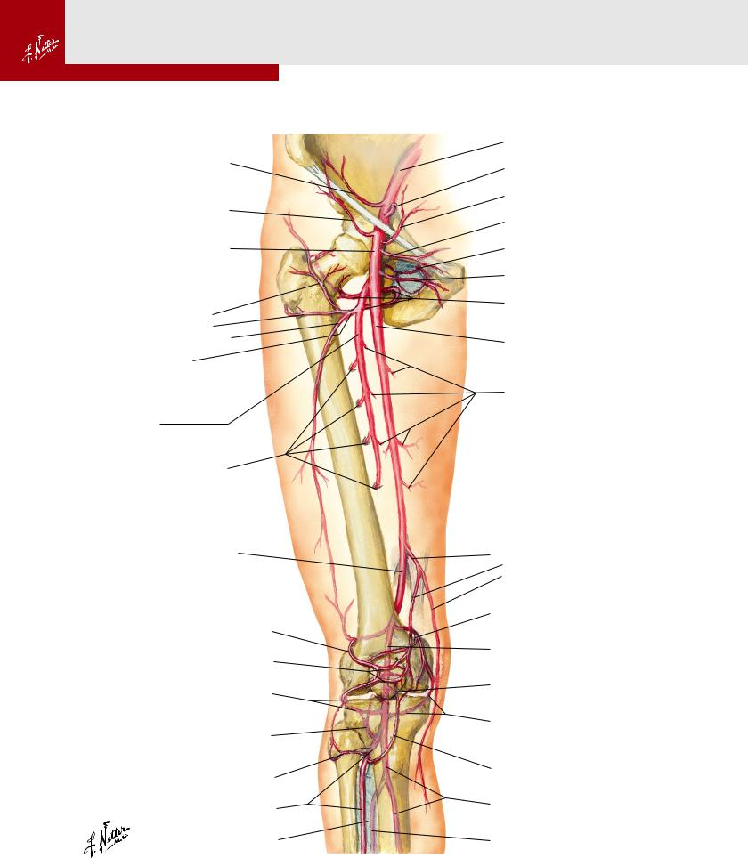
Arteries of Thigh and Knee: Schema
See also Plates 491, 508, 509, 518
Deep circumflex iliac artery
Superficial circumflex iliac artery
Femoral artery
Ascending branch, Transverse branch, Descending branch of Lateral circumflex femoral artery
Profunda femoris (deep femoral) artery
Perforating branches
Femoral artery passing through adductor hiatus within adductor magnus muscle
Superior lateral genicular artery
Patellar anastomosis
Inferior lateral genicular artery (partially in phantom)
Posterior tibial recurrent artery (phantom)
Circumflex fibular branch
Anterior tibial artery
Interosseous membrane
External iliac artery
Inferior epigastric artery
Superficial epigastric artery
Superficial external pudendal artery
Obturator artery
Deep external pudendal artery Medial circumflex femoral artery
Femoral artery
Muscular branches
Descending genicular artery
Articular branch
Saphenous branch
Superior medial genicular artery
Popliteal artery (phantom)
Middle genicular artery (phantom)
Inferior medial genicular artery (partially in phantom)
Anterior tibial recurrent artery
Posterior tibial artery (phantom)
Fibular (peroneal) artery (phantom)
Plate 499 |
Knee |

Bones of right leg
Anterior intercondylar area
Lateral condyle
Apex,
Head, Neck of fibula
Lateral surface
Anterior border
Interosseous border
Medial surface
Fibula
Lateral malleolus
Articular facet of lateral malleolus
Tibia and Fibula 7
Anterior view |
|
|
Posterior view |
|
||
Intercondylar eminence |
|
|
Intercondylar eminence |
|
||
Lateral |
Medial |
|
|
Medial |
Lateral |
|
intercondylar |
intercondylar |
|
intercondylar |
intercondylar |
|
|
tubercle |
tubercle |
Posterior |
tubercle |
tubercle |
Superior |
|
|
|
intercondylar |
|
|
articular surfaces |
|
|
|
area |
|
|
|
(medial and |
|
|
Medial |
|
|
|
lateral facets) |
|
|
|
|
|
|
|
|
|
condyle |
|
|
|
Lateral condyle |
|
|
|
|
|
|
|
|
|
Gerdy’s tubercle |
|
|
|
|
|
|
(insertion of |
|
|
|
Apex, |
|
|
iliotibial tract) |
|
|
|
Head, |
|
|
|
|
|
|
Neck |
|
|
|
|
|
|
of |
|
Oblique line |
|
|
|
fibula |
|
|
|
|
|
|
||
|
|
|
|
|
|
Groove for |
|
Tibial tuberosity |
|
|
|
insertion of |
|
|
|
|
|
semimembranosus |
||
|
|
|
|
|
|
|
|
|
|
Soleal line |
|
|
tendon |
|
Lateral surface |
|
|
|
|
|
|
|
|
|
|
|
Nutrient |
|
|
|
|
|
|
foramen |
|
Anterior border |
|
|
|
|
|
|
|
Interosseous border |
|
|
Posterior |
|
|
|
|
|
|
|
|
|
|
|
|
|
|
surface |
|
Medial surface |
Posterior |
|
|
|
|
|
surface |
|
|
|
||
|
|
|
|
|
|
|
|
|
|
|
|
|
Medial crest |
|
|
Medial border |
|
|
|
|
|
|
|
|
|
|
Lateral surface |
|
|
Tibia |
|
|
|
Fibula |
|
|
|
|
|
|
|
|
|
|
|
|
|
Posterior |
|
|
|
|
|
|
border |
|
|
Groove for |
|
|
|
|
|
|
tibialis posterior |
|
|
|
|
|
|
and flexor digitorum |
|
|
||
|
|
longus tendons |
|
|
|
|
|
|
|
|
|
|
Fibular notch |
|
|
|
|
|
|
Lateral |
|
|
Medial malleolus |
|
|
malleolus |
|
Inferior |
|
|
|
Inferior |
Malleolar fossa |
|
|
|
|
of lateral |
|||
articular |
Articular facet of medial malleolus |
articular |
malleolus |
|||
surface |
|
|
|
surface |
|
|
|
|
|
Leg |
Plate 500 |
|