

Атлас по рентгенологии травмированных собак и кошек / an-atlas-of-radiology-of-the-traumatized-dog-and-cat
.pdf72 Radiology of Thoracic Trauma
Case 2.29
2
Day 1
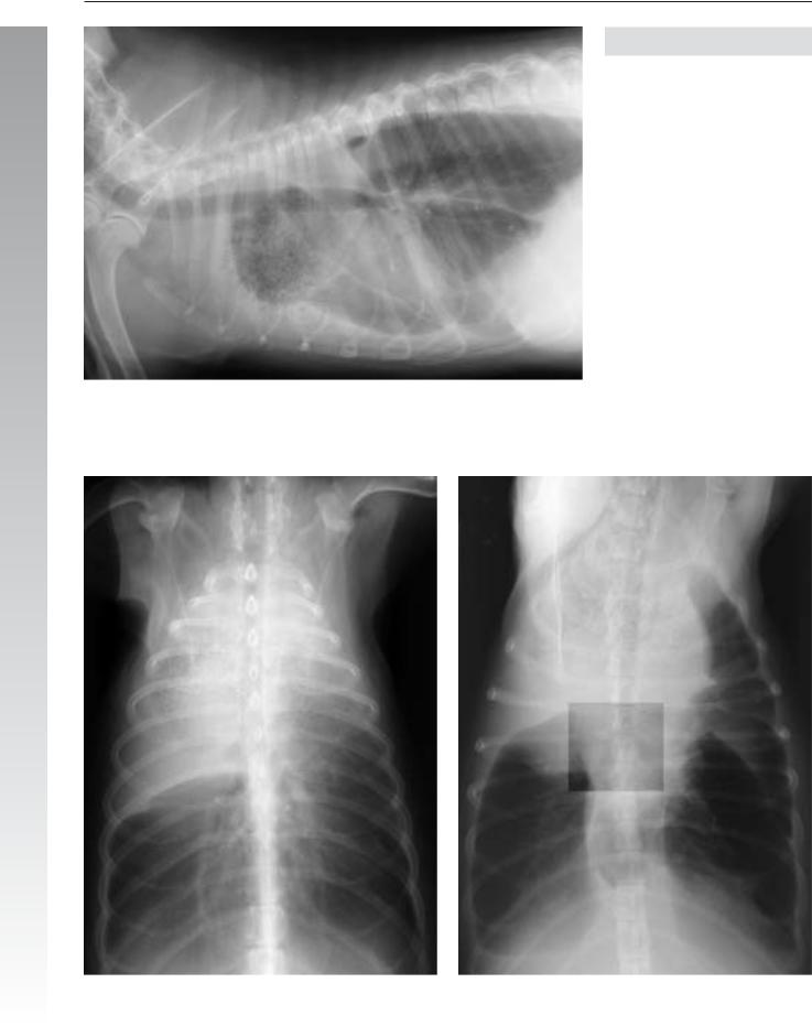
Signalment/History: “Snagglepus” was a 4-month-old, female Doberman Pinscher who had been hit by a car and was brought immediately to the clinic.
Physical examination: Breathing was labored.
Radiographic procedure: Radiographs of the thorax were made.
Radiographic diagnosis (day 1): Severe pulmonary hemorrhage affected all the lung lobes, but was more severe on the right. Generalized pleural fluid was also more evident on the right. Both the thoracic wall and the diaphragm were intact.

Lung injury 73
2
Day 3
Radiographic diagnosis (day 3): These radiographs showed a marked clearing of the pulmonary edema and hemorrhage from all but the right cranial lobe. Pleural fluid was still present. Note that the thoracic cavity remained as distended as at the time of presentation.
Treatment/Management: It was recommended that the dog remain hospitalized to await the diagnosis of why the right cranial lobe was failing to re-aerate. This was especially worrisome because the cranial lung lobes are normally well protected from trauma by the shoulder muscles. The possibility of either secondary pneumonia or a bronchial blockage from a mucous plug causing an obstructive atelectasis was considered. The normal anatomical location of the airways in the lobe tended to rule out torsion.
The puppy was discharged several days later in good health. As in most patients, the cause for the delay in healing of the cranial lobe could not be determined absolutely.

74 Radiology of Thoracic Trauma
Case 2.30
2
Day 1
Signalment/History: A stray female cat was observed being struck by a car and was brought to the clinic.
Physical examination: A limited examination indicated dyspnea and abnormal lung sounds.
Radiographic procedure: Thoracic radiographs were made.
Radiographic diagnosis (day 1): The left thoracic wall had minimal subcutaneous emphysema. The adjacent left lung lobes were increased in fluid density, suggesting pulmonary contusion and hemorrhage. A minimal pneumothorax was present and of a closed nature. On the lateral view, pleural fluid could be seen on the left trapped in the fissure between the cranial and caudal lobes. A single non-displaced fracture was noted in the left 8th rib.

Lung injury 75
2
Day 2
Radiographic diagnosis (day 2): Radiographs made the next day showed a marked increase in liquid density within the right cranial lobe and both parts of the left cranial lobe, with accentuation of the air-bronchogram pattern. The volume of the pleural fluid was increased and silhouetted with the cardiac silhouette. Note in particular the fluid between the cardiac silhouette and the sternum. The subcutaneous air had also increased in volume.
Treatment/Management: The increase in severity of the radiographic changes matched the increase in severity of the cat’s clinical signs, especially the dyspnea. The lungs showed an increasing fluid density that could be explained either by continued hemorrhage or a secondary pneumonia. The stress aerophagia continued to demonstrate something of the clinical status of the cat.
The cat was treated medically and finally recovered. She was later adopted. The amount of body fat suggested that for a stray cat, she had been eating rather well.
76 Radiology of Thoracic Trauma
Case 2.31
2

|
Lung injury 77 |
|
|
Signalment/History: “Teddy Bear” was a 3-year-old, fe- |
ing a lung torsion. A rim surrounding the mass had a homo- |
|
|
male Chow Chow who had been in chronic renal failure for |
geneous soft tissue/fluid density. The right cranial lobe |
|
|
the previous 18 months. She had been undergoing dialysis and |
bronchus terminated just distal to the carina. The right middle |
|
|
was a frequent patient in the hospital. She had chewed out a |
lung lobe was also airless with bronchial termination. Exten- |
|
|
PEG tube placed earlier and even proceeded to pull out a sec- |
sive freely moving pleural fluid was noted on both the DV and |
|
|
ond tube. The latest admission was because of persistent pleu- |
VD views. The chest wall was expanded and the diaphragm |
2 |
|
ral fluid and having suddenly developed dyspnea. |
was caudal and flattened. The trachea was on the midline sug- |
||
|
gesting there was no mediastinal mass. Chronic secondary |
|
|
Radiographic procedure: The thoracic studies were made |
joint disease was evident in both shoulders |
|
|
because of the dyspnea. |
Treatment/Management: A right cranial and middle lung |
|
|
|
|
||
Radiographic diagnosis: An area with a mottled, granular |
lobectomy was performed to correct the chronic lung torsion. |
|
|
appearance was noted lying within a fluid dense mass in the |
The history of repeated anesthesia in which the patient was |
|
|
right cranial hemithorax. This mass had an intermixed lucent |
placed in a unusual body position plus the presence of pleural |
|
|
gas pattern that suggested necrotic tissue often present follow- |
fluid were probable causes of the torsion of the lung lobes. |
|
|

78 Radiology of Thoracic Trauma
Case 2.32
2
Signalment/History: “Pal” was a 1-year-old, male Cocker
Spaniel who was severely dyspneic after being struck by a car.
Physical examination: The dog had a swollen abdomen and was comatose.
Radiographic procedure: The thorax was radiographed.
Radiographic diagnosis: The right lung lobes and the left caudal lobe showed a marked increase in fluid density, probably a result of hemorrhage from lung contusion. Only the left cranial lobe was fully aerated, while the others had an increased fluid density that silhouetted with the cardiac silhouette. The marked increase in fluid density in the lung lobes indicated atelectasis plus pulmonary hemorrhage. The pulmonary vessels were small suggesting hypovolemia.
The bilateral pneumothorax was easily identified because of the air contrasting with the fluid content in the lungs. A minimal amount of pleural fluid was pocketed caudally, adjacent to the diaphragm at the costophrenic angles. The cardiac silhouette was rounded with increased sternal contact suggesting a hemopericardium. A mediastinal shift to the left was noted. The dilated gas-filled stomach suggested panic breathing and the severity of the respiratory distress.
Treatment/Management: “Pal” died shortly after radiography due to a ruptured liver with peritoneal bleeding, pulmonary hemorrhage, pericardial hemorrhage, and cerebral hemorrhage.
Comments: It requires the combination of atelectasis plus pulmonary contusion to obtain a lung density of this severity.

Lung injury 79
Case 2.33
2
Signalment/History: “Shadow” was a 1-year-old, female Great Dane who had sustained head trauma.
Physical examination: On physical examination, the left pupil was not responsive to light and depressed frontal bone fractures were noted.
Radiographic procedure: Because of the unknown nature of the trauma, thoracic radiographs were made.
Radiographic diagnosis: A perihilar pattern of increased pulmonary density unusual in a young dog was present and was thought, because of the clinical history, to represent neurogenic pulmonary edema. Pulmonary congestion was present in the right lung lobes; perhaps a post-traumatic lung edema/hemorrhage. Pneumatoceles were present in the right middle lobe (DV enlargement, arrows).
The pulmonary vessels could not be identified due to shock. The thorax was expanded with scalloping of the lung borders. The diaphragm was caudal and flattened.
Treatment/Management: Fortunately, members of this breed possess massive frontal bones that provide good protection for the brain from direct trauma. “Shadow” recovered and was ultimately released to her owners.

80 Radiology of Thoracic Trauma
Case 2.34
2
Day 1
Signalment/History: “Wojo”, a 1-year-old, male DSH cat, had been caught in a garage door and was trapped in that position for 30 minutes, enduring great pressure on his thorax.
Physical examination: Dyspnea was severe.
Radiographic procedure: Thoracic radiographs were made.
Radiographic diagnosis (day 1): Pulmonary infiltrate was noted throughout all the lung lobes, being most prominent caudally. Minimal pleural fluid was present. No injury to the thoracic wall was detected. No peritoneal fluid was noted.

Lung injury 81
2
Day 5
Radiographic diagnosis (day 5): Radiographs made four days later showed resolution of both the pulmonary and pleural fluid.
Treatment/Management: The pulmonary contusion was probably the result of a rupture of pulmonary alveoli due to the supreme effort required at inspiration against the great external pressure on the thorax caused by the door. However, an air-bronchogram pattern was not prominent. The rather rapid healing suggested that there was no direct trauma to the lungs from the door closing on the cat.