

Essentials of Orthopedic Surgery, third edition / 01-Basic Science of Bone and Cartilage Metabolism
.pdf1. Basic Science of Bone and Cartilage Metabolism |
11 |
Normal Bone
MINERAL
MATRIX
Osteoporosis
MINERAL
MATRIX
Osteomalacia
MINERAL
MATRIX
mineral = mineral > mineral matrix matrix matrix
FIGURE 1-10. Ratio of mineral to matrix in certain disease states. In osteoporosis, the ratio remains constant despite an overall decrease in bone mass. However, in osteomalacia there is a decrease in the ratio of mineral to matrix as a result of skeletal demineralization; in addition, there is an overall decrease in bone mass.
The relationship (ratio) of mineral to matrix may be affected in abnormal metabolic states (Fig. 1-10). For example, osteoporosis is a loss of bone mass, but there is an equivalent loss of matrix and mineral; therefore, the ratio remains normal. In contrast, osteomalacia is a relative loss of mineral resulting in a predominance of matrix, hence decreasing the ratio of mineral to matrix. Serum calcium is rarely representative of skeletal activity. Considering that more than 95% of the body’s calcium is stored in bone apatite, it is understandable that the 180 mg of ionized plasma calcium represents literally the “tip of the iceberg.” Peripheral sampling of the serum calcium provides only a remote clue to the true content of skeletal apatite. It does, however, provide a convenient way to think about and classify metabolic bone disease.
Eucalcemic States: Osteoporosis
As mentioned, osteoporosis is a predominance of bone resorption over bone formation, with the net effect being bone loss (Fig. 1-11). There is a parallel loss of mineral and matrix, so their ratio remains normal. Essentially, osteoporosis is a decrease in bone mass with an increase in cortical porosity and in diaphyseal bone diameter. This latter phenomenon is an attempt by the organism to use what limited bone there is and to disperse it as far as possible from the neutral axis of the long bone. Mechanically, this increases the torsional rigidity of the bone. Numerous etiologies of osteoporosis have been identified (Table 1-1), but clinically most significant is the postmenopausal type, which occurs shortly after the withdrawal of estrogen (naturally or surgically) from the predisposed

12 J.N. Delahay
% bone
100
|
|
|
|
|
|
|
Cortical |
|
80 |
|
|
|
|
|
|
Trabecular |
|
|
|
|
|
|
|
Mean ± 2 S.E. |
||
|
|
|
|
|
|
|
||
60 |
|
|
|
|
|
|
|
|
40 |
|
|
|
|
|
|
|
|
20 |
|
|
|
|
|
|
|
|
0 |
|
|
|
|
|
|
|
|
10 |
20 |
30 |
40 |
50 |
60 |
70 |
80 |
90 |
Age in years
FIGURE 1-11. The relative decrease in cortical and trabecular bone with age in apparently normal persons. Note the relatively rapid loss early in life in trabecular bone and comparatively little loss at this age in cortical bone. The situation is reversed after age 55. (From Jowsey J. Metabolic Diseases of Bone. Philadelphia: Saunders, 1977. Reprinted by permission.)
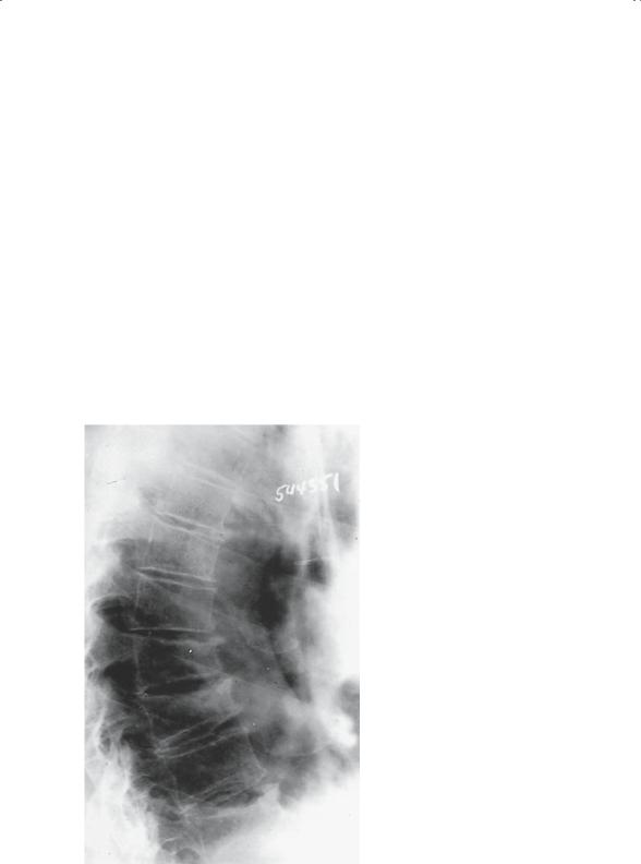
female (Table 1-2). The yearly cost in dollars, as well as pain and suffering, is overwhelming. Women with this affliction frequently sustain classic osteoporotic fractures. These fractures typically involve the vertebrae, the wrist, the proximal femur, and/or the proximal humerus. In addition to the pathologic fractures, there is frequently a loss of height as a result of the cumulative effect of multiple vertebral fractures, as well as the progressive development of a kyphotic deformity in the thoracic spine, which is referred to as a “dowager’s hump” (Fig. 1-12).
Patients present with a history of pain and/or repeated fractures. Occasionally they complain of early satiety because of some abdominal compression resulting from loss of height of the vertebral column. Similarly, the increasing kyphosis in the thoracic region may be responsible for some shortness of breath. On examination, typically one finds the prominent dowager’s hump, a barrel chest, a protuberant abdomen, and generalized bone pain with percussion tenderness.
One of the most difficult problems in the past has been to determine bone mass. Typically, a crude estimate of bone density determined by plain radiograph has been used to extrapolate to the amount of bone previously lost. Classically, once osteopenia is noticeable radiographically, it has been estimated that the bone density is decreased by 30% to 50%.
Recently, additional diagnostic techniques have become available to more carefully estimate the amount of bone loss and, therefore, the amount of bone that remains. Isotope measurements, specifically single-photon

1. Basic Science of Bone and Cartilage Metabolism |
13 |
absorptiometry, using an iodine compound, or dual-photon absorptiometry, using a gadolinium compound, have been developed. They have significant technical limitations. The single-photon technique, measuring peripheral sites, such as the forearm and heel, is rarely an adequate reflection of the true bone mineral density in the axial skeleton. The dual-photon study, although providing more reliable information about the bone mineral density of the axial skeleton, continues to have some technical limitations.
TABLE 1-1. Causes of osteoporosis.
Primary
Involutional (postmenopausal or senile)
Idiopathic (juvenile or adult)
Secondary
Endocrine
Hypogonadism
Adrenocortical hormone excess (primary or iatrogenic)
Hyperthyroidism
Hyperparathyroidism
Diabetes mellitus
Growth hormone deficiency
Nutritional
Calcium deficiency
Phosphate deficiency
Phosphate excess
Vitamin D deficiency
Protein deficiency
Vitamin C deficiency
Intestinal malabsorption
Drug
Heparin
Anticonvulsants
Ethanol
Methotrexate
Genetic
Osteogenesis imperfecta
Homocystinuria
Miscellaneous
Rheumatoid arthritis
Chronic liver disease
Chronic renal disease
Immobilization
Malignancy (multiple myeloma)
Metabolic acidosis
Cigarette smoking
Source: From Borenstein D, Wiesel SW. Low Back Pain: Medical Diagnosis and Comprehensive Management. Philadelphia: Saunders, 1989:329. Reprinted with permission.
14 |
J.N. Delahay |
|
|
TABLE 1-2. Types of involutional osteoporosis. |
|
||
|
|
Type 1 |
Type 2 |
|
|
(Postmenopausal) |
(Senile) |
|
|
|
|
Age (years) |
51–75 |
Over 70 |
|
Sex ratio (M/F) |
1 : 6 |
1 : 2 |
|
Type of bone loss |
Trabecular |
Trabecular and cortical |
|
Fracture site |
Vertebrae (crush) |
Vertebrae (multiple wedge) |
|
|
|
Distal radius |
Hip |
Main causes |
Menopause |
Aging |
|
Calcium absorption |
Decreased |
Decreased |
|
1,25-(OH)2-vitamin D synthesis |
Secondary decrease |
Primary decrease |
|
from 25-(OH) vitamin D |
|
|
|
Parathyroid function |
Decreased |
Increased |
|
|
|
|
|
Source: Modified from Riggs BL, Melton LJ III. Involutional osteoporosis. N Engl J Med 1986;314:1676.
FIGURE 1-12. Radiograph of spine showing osteoporosis. Cortical bone appears accentuated by contrast with osteopenic marrow. Longitudinal trabeculae also appear accentuated because smaller transverse trabeculae are absent. Anterior wedging and endplate compression are present. (From Bogumill GP. Orthopaedic Pathology: A Synopsis with Clinical Radiographic Correlation. Philadelphia: Saunders, 1984. Reprinted by permission.)

1. Basic Science of Bone and Cartilage Metabolism |
15 |
As of this writing, it is probably fair to say that both these techniques have been replaced by dual-energy X-ray absorptiometry (DEXA) scanning. The DEXA technique is currently the standard, used in the evaluation of bone mineral density (BMD) in women approaching or following their menopause. This technique allows accurate and reproducible measures of density of the spine and the hip and does so with a minimal amount of radiation exposure. Current guidelines as recommended by the National Osteoporosis Foundation and the World Health Organization allow comparison of an individual’s bone density to that of healthy normals. The difference is expressed as a T-score, which essentially represents one standard deviation above or below ideal bone mass. The definitions based on T-scores are as follows:
Normal |
0 to −1 |
Osteopenia |
−1 to −2.5 |
Osteoporosis |
Less than −2.5 |
The unfortunate result of DEXA scanning, however, has been to adulterate the use of the term osteopenia. For many years, this term was defined as a generalized decrease in radiographic bone density. As such, it was nonpejorative and did not speak to a specific metabolic bone disease. In its present accepted context, the implication of using the term osteopenia is to imply a mild form of postmenopausal osteoporosis, which was certainly not the original connotation of the term. Diseases other than osteoporosis, such as hyperthyroidism and multiple myeloma, are characterized by observed decreases in radiographic bone density, hence osteopenia.
Without question, the most definitive diagnostic technique is direct bone biopsy with or without tetracycline labeling. It can clearly give the most reliable information regarding the presence of osteoporosis, its degree, and whether a superimposed osteomalacic state exists. Once the diagnosis has been confirmed and the risk analysis carried out, a treatment protocol can be tailored for the individual patient.
Most treatment regimens are considered either prophylactic or therapeutic. Prophylactic regimens include regular weight-bearing exercise, such as walking or jogging, supplemental calcium administration, and vitamin D administration with or without the administration of postmenopausal estrogen substitutes. The complications of oral estrogen administration, such as its relation to breast and cervical cancer and to heart disease and the incidence of deep venous thrombosis (DVT), make its general use controversial; however, its efficacy in maintaining skeletal mass is beyond question.
Therapeutic regimens, in contrast, are much more debatable. Current therapeutic regimens include the use of any or all of several different pharmacologic agents. Selective estrogen receptor modulators (SERMs) are drugs that behave either as an agonist or antagonist of estrogen. They have been shown, in selective populations, to decrease or minimize bone loss.

16 J.N. Delahay
These drugs theoretically have an estrogen-like protective effect on bone. It has also been suggested that they have inhibitory (protective) effects on the breast and the endometrium.
Bisphosphonates are structurally similar to naturally occurring pyrophosphates. Because they have a strong chemical affinity for hydroxyapatite, they are potent inhibitors of bone resorption. They, therefore, are able to decrease the rate at which bone remodeling occurs and, as a result, to reduce the amount of bone resorption. It has been said that bisphosphonates are able to “freeze the skeleton.” It is hoped that the consequence of decreasing bony resorption will be a coincident increase in bone mass. At the present time, the most popular bisphosphonate in current use is Fosamax, which has been approved for both the prevention and treatment of osteoporosis.
Calcitonin, a naturally occurring polypeptide hormone, is currently being administered in an effort to also decrease the rate of bony resorption by decreasing the number and activity of osteoclasts. The drug is currently being administered in the form of a nasal spray.
The current regimens used for the therapeutic management of osteoporosis include one or more of these drugs in addition to the standard prophylactic measures. Not infrequently, these agents are used cyclically or in an alternating fashion. Because the true measure of any therapeutic regimen for osteoporosis is an increase in bone density and a reduction in fracture risk or in the number of fractures, the true efficacy of these agents and various therapeutic regimens must be evaluated over the long term. As of this writing, the use of SERMs, bisphosphonates, and calcitonin all have shown early promise in this context.
Hypercalcemic States: Hyperparathyroidism
The effect of parathormone on bone is the same whether it is released as a result of a parathyroid adenoma (primary hyperparathyroidism) or by one of several secondary causes. In essence, parathormone stimulates osteoclastic activity, causing an intense resorption of bone (Fig. 1-13). The cavities resulting from this clastic activity fill with vascular fibrous tissue, resulting in the classic “osteitis fibrosa cystica.” As the cavities coalesce, they form a single large cyst called a “brown tumor” because of the hemosiderin staining one sees within them. Clinical and radiographic changes result from this cavitation as well as from the erosive changes occurring under the periosteum.
Hypocalcemic States: Rickets and Osteomalacia
The same underlying mechanism accounts for rickets and osteomalacia: there is a general failure to mineralize bony matrix, resulting in the presence of unmineralized osteoid about bony trabeculae. This lack of mineral

1. Basic Science of Bone and Cartilage Metabolism |
17 |
FIGURE 1-13. “Cutting cone.” Successive relays of osteoclasts on the right resorb a tunnel of bone, making it longer and wider with each relay. Behind the cutting cone is a “filling cone” of successive relays of osteoblasts secreting osteoid. Resorption is facilitated by high-speed flow of well-oxygenated blood in small vessels, whereas refill is accompanied by dilated sinusoidal vessels with sluggish flow and low oxygen content. (From Bogumill GP. Orthopaedic Pathology: A Synopsis with Clinical Radiographic Correlation. Philadelphia: Saunders, 1984. Reprinted by permission.)
for adequate mineralization can be caused by a number of different etiologies: nutritional deficiency, malabsorption states, or renal disease (Table
1-3) are some of the more common. Despite the etiology, the metabolic effects on the skeleton are similar.
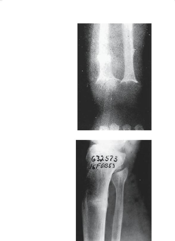
If the failure of mineralization impacts the skeleton before physeal closure, the result is rickets. The affected child will demonstrate the characteristic hallmarks of the disease: bowlegs, frontal bossing, ricketic rosary, and knobby joints (Fig. 1-14). All these findings are due to the presence of large masses of unmineralized osteoid. In addition, abnormalities of the physis and abnormal physeal growth can be anticipated.
If the process impacts the skeleton after physeal closure, the disease that results is osteomalacia. As noted earlier, the ratio of mineral to matrix decreases as a result of the paucity of mineral available to the skeleton. In the adult, these areas of unmineralized osteoid present as radiographically lucent areas in the bone, frequently referred to Looser’s lines (Fig. 1-15). In addition, the bones themselves tend to be somewhat malleable and can bow under load; this is in contradistinction to osteoporotic bone, which is very brittle.

18 |
J.N. Delahay |
|
TABLE 1-3. Diseases associated with osteomalacia. |
||
Disorder |
Metabolic defect |
|
Vitamin D: |
Decreased generation of vitamin D3 |
|
Deficiency |
|
|
|
Dietary |
|
|
Ultraviolet light exposure |
|
Malabsorption |
Decreased absorption of vitamins D2 |
|
|
Small intestine |
and D3 |
|
Inadequate bile salts |
|
|
Pancreatic insufficiency |
|
Abnormal metabolism |
|
|
|
Hereditary enzyme deficiency |
Decreased 1-alpha-hydroxylation of |
|
|
25-(OH)-vitamin D |
|
D-dependent rickets (type I) |
|
|
Chronic renal failure |
Decreased 25-hydroxylation of |
|
Mesenchymal tumors |
vitamin D |
|
Systemic acidosis |
|
|
Hepatic failure |
|
Anticonvulsant drugs |
|
|
Peripheral resistance |
Absent or abnormal 1,25-(OH)2- |
|
|
Vitamin D-dependent rickets (type II) |
Vitamin D receptors |
Phosphate depletion: |
|
|
Dietary |
Inadequate bone mineralization |
|
|
Malnutrition (rare)? |
secondary to low serum |
|
Aluminum hydroxide ingestion |
concentrations |
Renal tubular wasting |
|
|
Hereditary |
Decreased serum phosphate |
|
|
X-linked hypophosphatemic osteomalacia |
concentrations |
Acquired |
|
|
|
Hypophosphatemic osteomalacia |
|
|
Renal disorders |
|
|
Fanconi’s syndrome |
|
|
Mesenchymal tumors |
|
|
Fibrous dysplasia |
|
Mineralization defects: |
|
|
Hereditary |
Abnormal alkaline phosphatase |
|
|
Hypophosphatasia |
activity |
Acquired |
|
|
|
Sodium fluoride |
Inhibition of bone mineralization |
|
Disodium etidronate |
|
Miscellaneous: |
|
|
Osteopetrosis |
Abnormal osteoclast activity |
|
Fibrogenesis imperfecta |
Unknown |
|
Axial osteomalacia |
Unknown |
|
Calcium deficiency |
Inadequate bone mineralization |
|
|
|
Secondary to low serum calcium |
|
|
concentration |
|
|
|
Source: From Borenstein D, Wiesel SW. Low Back Pain: Medical Diagnosis and Comprehensive Management. Philadelphia: Saunders, 1989:339. Reprinted with permission.
1. Basic Science of Bone and Cartilage Metabolism |
19 |
FIGURE 1-14. Radiograph of wrist of child with active rickets exhibiting the irregular widened zone of provisional calcification that is replaced by abnormal osteoid. The cartilage masses are not visible, but the widened epiphyseal growth plate and irregular calcification are readily seen. Note pathologic fracture of radial shaft. (From Bogumill GP. Orthopaedic Pathology: A Synopsis with Clinical Radiographic Correlation. Philadelphia: Saunders, 1984. Reprinted by permission.)
FIGURE 1-15. Radiograph of osteomalacia showing a Looser’s transformation zone. These lines appear at sites in which stress fractures would occur. Stress of normal use incites remodeling with removal of bone. In normal individuals, the removed bone is replaced by normal osteons. In persons with osteomalacia, the removed bone is replaced with abnormal osteoid, which fails to mineralize and leaves a linear radiolucency that may persist for years. (From Bogumill GP. Orthopaedic Pathology: A Synopsis with Clinical Radiographic Correlation. Philadelphia: Saunders, 1984. Reprinted by permission.)

20 J.N. Delahay
Miscellaneous Metabolic Bone Disease:
Renal Osteodystrophy
Renal osteodystrophy encompasses the skeletal changes that result from chronic, acquired renal disease. These changes are truly a “collage” of the other metabolic bone diseases. To understand the pathogenesis of renal osteodystrophy is to understand the basis of all the metabolic afflictions of the skeleton (Fig. 1-16). Chronic uremia allows a twofold drive to depress the serum calcium. First, the kidney is unable to excrete phosphate, hence the serum phosphate level rises. The serum calcium level is then of necessity driven down to maintain the fixed solubility product. Coincidentally, because the absence of a functional renal parenchyma stops the output of significant amounts of activated vitamin D, intestinal absorption of calcium is retarded, further depressing serum calcium. This dual mechanism profoundly depresses serum calcium and thus in turn mandates a parathormone response. The changes in the bone reflect the metabolic drives. The vitamin D deficiency is demonstrated by the presence of unmineralized osteoid (Fig. 1-17). The elevated levels of parathormone cause osteitis fibrosis cystica. Unique to this syndrome, the hyperphosphatemia results in a diffuse osteosclerosis. The latter finding causes one of the most pathognomic radiographic findings (Fig. 1-18), the “rugger jersey” spine.
PO4 |
|
|
|
Renal |
||
Uremia |
||||||
|
|
|||||
Retention |
|
|
parenchymal |
|||
|
|
|||||
|
|
|
|
|
||
|
|
|
|
|
damage |
|
|
|
|
|
|
|
|
↑(PO4)s
↓Vit. D synthesis
↓Absorption of Ca
Maintain |
|
|
|
|
(Ca)s↓↓ |
||||
solubility product |
||||
|
|
|||
|
|
|
|
|
↑ PTH
FIGURE 1-16. Pathogenesis of renal osteodystrophy.
FIGURE 1-18. Radiograph of patient with long-standing renal osteodystrophy. Marked osteoporosis attributable to secondary hyperparathyroidism is evident. There is bowing of the proximal femurs, marked lordosis, and pelvic tilt. The deformity of the pelvis is commonly seen in osteomalacia, but it does not usually occur in primary hyperparathyroidism. (From Bogumill GP. Orthopaedic Pathology: A Synopsis with Clinical Radiographic Correlation. Philadelphia: Saunders, 1984. Reprinted by permission.)