

МРТ - в диагностике болезней суставов
.pdf
CHAPTER 4
THE WRIST
William D. Middleton, M.D
The human wrist is composed of a plethora of anatomic structures including bones, tendons, ligaments, muscles, and neurovascular structures. Because there are so many anatomic structures located in a relatively confined area, the anatomy of the wrist can be intimidating when seen in cross section. However, careful analysis of cross-sectional images and anatomic sections reveals a relatively detailed, yet understandable, portrayal of articular and periarticular structures.
The carpal bones are arranged in a proximal and distal row. The proximal row is composed of the scaphoid, lunate, triquetrum, and pisiform bone and the distal row is composed of the trapezium, trapezoid, capitate, and hamate. With the exception of the pisiform, the bones of the proximal carpal row articulate with the radius, the distal carpal row, and each other. The pisiform articulates only with the triquetrum. The bones of the distal carpal row articulate with the proximal carpal row, each other, and the metacarpals. Their complex interrelations are shown in Fig. 4-1.
The triangular fibrocartilage separates the scaphoid and lunate from the ulna and attaches the distal radius and ulna to each other. It is ligamentous peripherally and this ligamentous portion extends from the ulnar styloid to the dorsal and palmar aspect of the radius.
A number of ligaments support the carpus.'The radiocarpal articulations are supported on their palmar and dorsal aspects by broad, thin ligaments extending from the distal radius and ulna to the scaphoid, lunate, and triquetrum. These are called the palmar and dorsal radial carpal ligaments. The radial and ulnar collateral ligaments extend from the radial styloid to the scaphoid and from the ulnar styloid to the triquetrum and pisiform, respectively.
In addition to these ligaments, a complex arrangement of ligaments connect the carpal bones to each other. Numerous palmar and dorsal ligaments connect the superficial surfaces of the carpal bones and multiple interosseous ligaments connect the deep surfaces of the carpal bones.
The flexor retinaculum is a thick ligamentous band that attaches primarily to the hook of the hamate and the tubercle of the trapezium. It is thinner proximally where attenuated strands attach to the pisiform and the tubercle of the scaphoid. The flexor retinaculum forms the superficial border of the carpal tunnel, with the carpal bones forming the deep border.
83

Anatomy and MRI of the Joints |
84 |
The extensor retinaculum is much thinner than the flexor retinaculum. It forms multiple fibro-osseous tunnels along the dorsal aspect of the wrist, through which pass the extensor tendons.
The major muscles of the wrist are limited to those of the thenar and hypothenar eminence, shown in Fig. 4-2. The three thenar muscles all arise from the flexor retinaculum. The abductor pollicis brevis and flexor pollicis brevis are superficial and both insert at the base of the first phalanx. The opponens pollicis is deep and has a broad insertion on the shaft of the first metacarpal. The origins and insertions of the three hypothenar muscles are similar to those of the thenar muscles, except the insertions are onto the fifth digit.
The adductor pollicis is located deep to the thenar muscles. It arises from the capitate and the third metacarpal and inserts at the base of the first phalanx. It is divided into an oblique (proximal) and transverse (distal) portion by the radial artery. There is no adductor muscle to the fifth digit.
Other muscles which are not strictly part of the wrist but which are seen in images of the wrist are the pronator quadratus and the lumbrical muscles. The pronator quadratus arises from the palmar aspect of the distal ulna and inserts on the palmar aspect of the distal radius. It is the major pronator of the wrist. The four lumbricals arise from the tendons of the flexor digitorum profundus and insert on the radial aspect of the dorsal extensor tendon expansion.
Normally all of the muscles of the forearm become tendinous prior to the wrist. Because of their number, these tendons can become confusing when seen in cross section. However, their arrangement is relatively constant and they are actually fairly easy to identify and separate from each other. They are best understood by dividing them into dorsal and palmar groups.
The palmar group includes the flexors of the wrist and fingers. The superficial flexors of the forearm include the flexor carpi radialis, flexor carpi ulnaris, and palmaris longus. The tendon of the flexor carpi radialis travels su-
85 |
THE WRIST |
perficial to the distal scaphoid and pierces the radial aspect of the flexor retinaculum. It then runs in its own fibroosseous tunnel formed by two layers of the flexor retinaculum and the vertical groove of the trapezium. It inserts primarily on the base of the second metacarpal, although some fibers do extend to the base of the third metacarpal. The flexor carpi ulnaris tendon inserts onto the pisiform bone and continues beyond the pisiform to the hamate and fifth metacarpal as the pisohamate and pisometacarpal ligaments. The palmaris longus tendon is flattened and less bulky than the other flexor tendons. It runs superficial to the flexor retinaculum and eventually blends into the palmar aponeurosis.
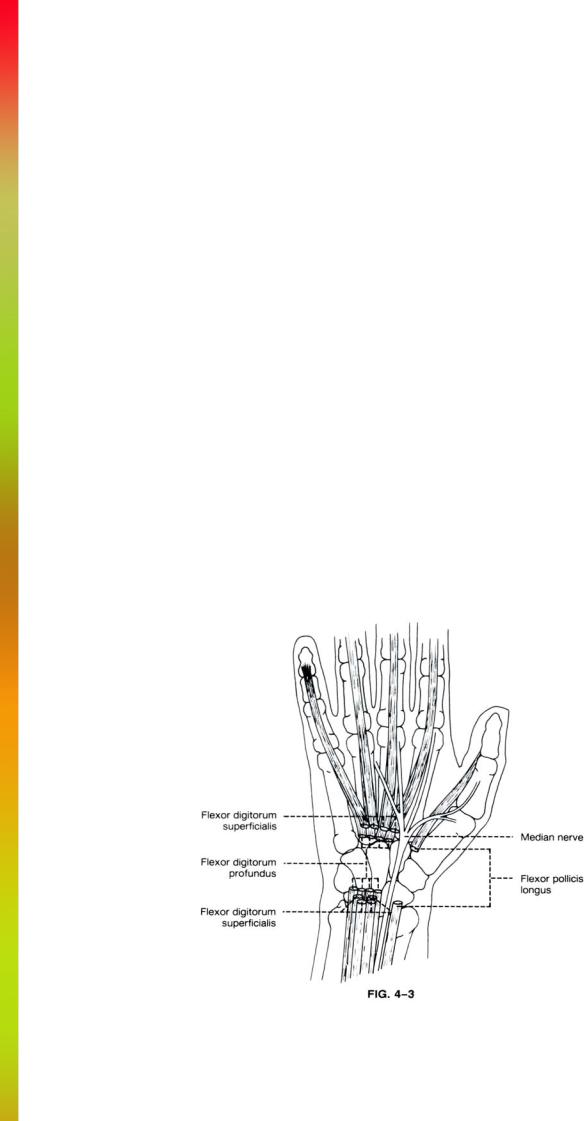
The intermediate flexor of the forearm is the flexor digitorum superficialis. This muscle divides into four tendons prior to reaching the wrist. These four tendons pass just beneath the flexor retinaculum and eventually extend to the sides of the second phalanges. Within the carpal tunnel, the tendons of the third and fourth digits are relatively superficial, while those to the second and fifth digits are deeper. Because of this arrangement, the four tendons are situated in an arc just deep to the flexor retinaculum. This arrangement is shown in Fig. 4-3.
The deep flexors of the forearm are the flexor digitorum profundus and flexor pollicis longus. Similar to the flexor digitorum superficialis, the flexor digitorum profundus divides into four tendons prior to reaching the wrist. These tendons occupy the deepest portion of the carpal tunnel. As they extend distally to insert at the distal phalanges, they diverge from one another and pair up with the corresponding tendons of the flexor digitorum superficialis (Fig. 4-3).
The flexor pollicis longus tendon occupies the most radial aspect of the carpal tunnel (Fig. 4-3). Beyond the carpal tunnel it runs between the flexor pollicis brevis and the adductor pollicis brevis muscles. It inserts onto the base of the first distal phalanx.
The dorsal tendon group includes the extensors of the digits and wrist and the long abductor tendon of the thumb. All of the tendons pass beneath the extensor retinaculum and are divided into groups by six fibro-osseous tunnels formed by the retinaculum and grooves within the distal radius. The overall arrangement of the dorsal tendons is shown in Fig. 4-4
The most lateral of these groups are the abductor pollicis longus and extensor pollicis brevis. The extensor pollicis brevis runs just dorsal to the abductor pollicis longus. The extensor pollicis brevis inserts on the base of the first proximal phalanx and the abductor pollicis longus inserts at the base of the first metacarpal.
Just medial to the extensor pollicis brevis and abductor pollicis longus are the extensor carpi radialis longus and brevis. These tendons run together along the radial border of the radius with the brevis position medially and dorsally. After they run beneath the thin extensor retinaculum, they separate from each other and proceed to their insertions on the base of the second (longus) and third (brevis) metacarpals.
The extensor pollicis longus originates medial to the extensor carpi radialis longus and brevis. After passing through its own tunnel in the extensor retinaculum, it passes over the extensor carpi radialis longus and brevis and eventually lies lateral to these tendons. The extensor pollicis longus inserts on the base of the first distal phalanx.

Anatomy and MRI of the Joints |
86 |
The extensor digitorum communis lies medial to the extensor pollicis longus. Like the flexor tendons of the fingers, it divides into four tendons prior to reaching the wrist. These four tendons pass within their own compartment (along with the extensor indicis) through the extensor retinaculum. Distally the four tendons contribute to the dorsal extensor tendon expansion of the second through fifth digits. The separate tendons of the extensor digiti minimi and extensor indices accompany the tendon of the extensor digitorum communis to the index and small fingers. The extensor digiti minimi travels in its own fibro-osseous tunnel medial to that of the extensor digitorum communis and extensor indicis.
The most medial of the extensor tendons is the extensor carpi ulnaris. It runs in a separate compartment of the extensor retinaculum within a groove formed between the styloid process and head of the ulna. Distally it inserts at the base of the fifth metacarpal.
The blood supply to the hand and wrist comes from the radial and ulnar arteries. The radial artery travels along the radial aspect of the forearm to reach the wrist, where it is located lateral to the flexor carpi radialis tendon. At the wrist, the radial artery descends under the abductor pollicis longus and extensor pollicis brevis and longus into the "anatomical snuff box." As it reaches the interosseous space between the thumb and index finger, it perforates
the first interosseous muscle to enter the palm of the hand deep to the adductor pollicis longus and lumbrical muscle. There it anastomoses with the deep palmar branch of the ulnar artery to form the deep palmar arch. At the level of the wrist the radial artery also gives off a small superficial branch which anastomoses with the ulnar artery to form the superficial palmar arch.
The ulnar artery travels in the forearm deep to the flexor carpi ulnaris muscle. At the wrist the ulnar artery remains superficial to the flexor retinaculum. It extends beyond the wrist to anastomose with the superficial branch of the radial artery to form the superficial palmar arch.
The ulnar nerve travels medial to the ulnar artery throughout most of its course. At the wrist it remains closely associated with the ulnar artery and travels superficially, sending branches to the fifth and fourth digits. It also has a deep branch that travels adjacent to the deep palmar branch of the ulnar artery.
Unlike the ulnar nerve, the median nerve is not associated with an artery in the wrist. In the forearm it travels deep to the muscle of the flexor digitorum superficialis. At the wrist it extends deep to the flexor retinaculum within the carpal tunnel (Fig. 4-3). As it exits the carpal tunnel, it divides into three branches that extend to the interosseous spaces between the first four digits.

THE WRIST
AXIAL |
|
Cryomicrotomes ........................................................ |
FIGS. 4-5 to 4-10 |
MR Images.................................................................. |
FIGS. 4-11 to 4-16 |
SAGITTAL |
|
Cryomicrotomes ........................................................ |
FIGS. 4-17 to 4-22 |
MR Images.................................................................. |
FIGS. 4-23 to 4-27 |
CORONAL |
|
Cryomicrotomes ........................................................ |
FIGS. 4-28 to 4-32 |
MR Images.................................................................. |
FIGS. 4-33 to 4-37 |

Anatomy and MRI of the Joints |
88 |
Dorsal
Radial Ulnar
Palmar

89 |
THE WRIST |
Dorsal
Radial Ulnar
Palmar

Anatomy and MRI of the Joints |
90 |
Dorsal
Radial Ulnar
Palmar

91 |
THE WRIST |
Dorsal
Radial Ulnar
Palmar