

МРТ - в диагностике болезней суставов
.pdfAnatomy and MRI of the Joints |
52 |
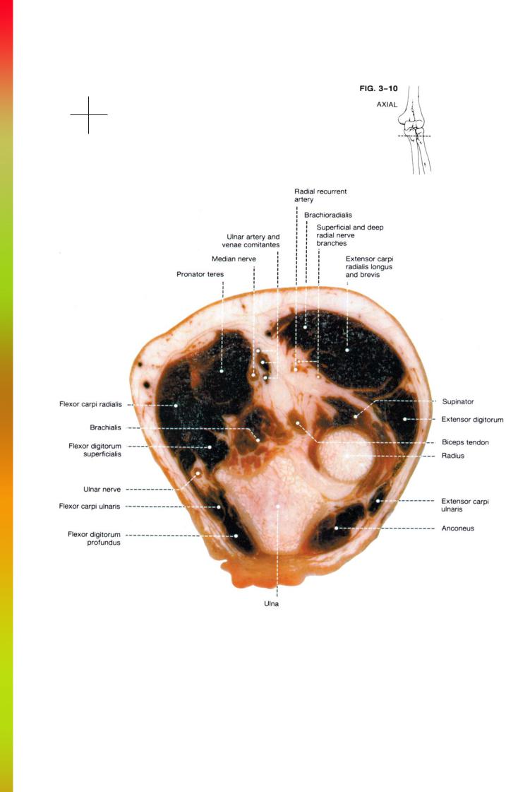
The medial muscle group includes the pronator teres, the flexors of the fingers and wrist, and the palmaris longus. These muscles are arranged in an arc along the anterior and medial aspect of the elbow. The pronator teres is the most anterior and lateral of the group. It has a superficial and deep portion arising respectively from the distal humerus just proximal to the medial epicondyle and from the proximal ulna just medial to the ulnar tuberosity. The median nerve runs between the smaller deep portion and the larger superficial portion of the pronator teres. The other muscles of the medial group arise as a group from a common flexor tendon originating on the medial epicondyle. They are arranged from anterior to posterior as the flexor carpi radialis, palmaris longus, flexor digitorum superficialis, flexor carpi ulnaris, and flexor digitorum profundus. These muscles become progressively more distinct distally. The flexor carpi ulnaris also has a head originating from the proximal ulna and the flexor digitorum superficialis has a second and third head originating from the proximal ulna and the proximal to mid radius.
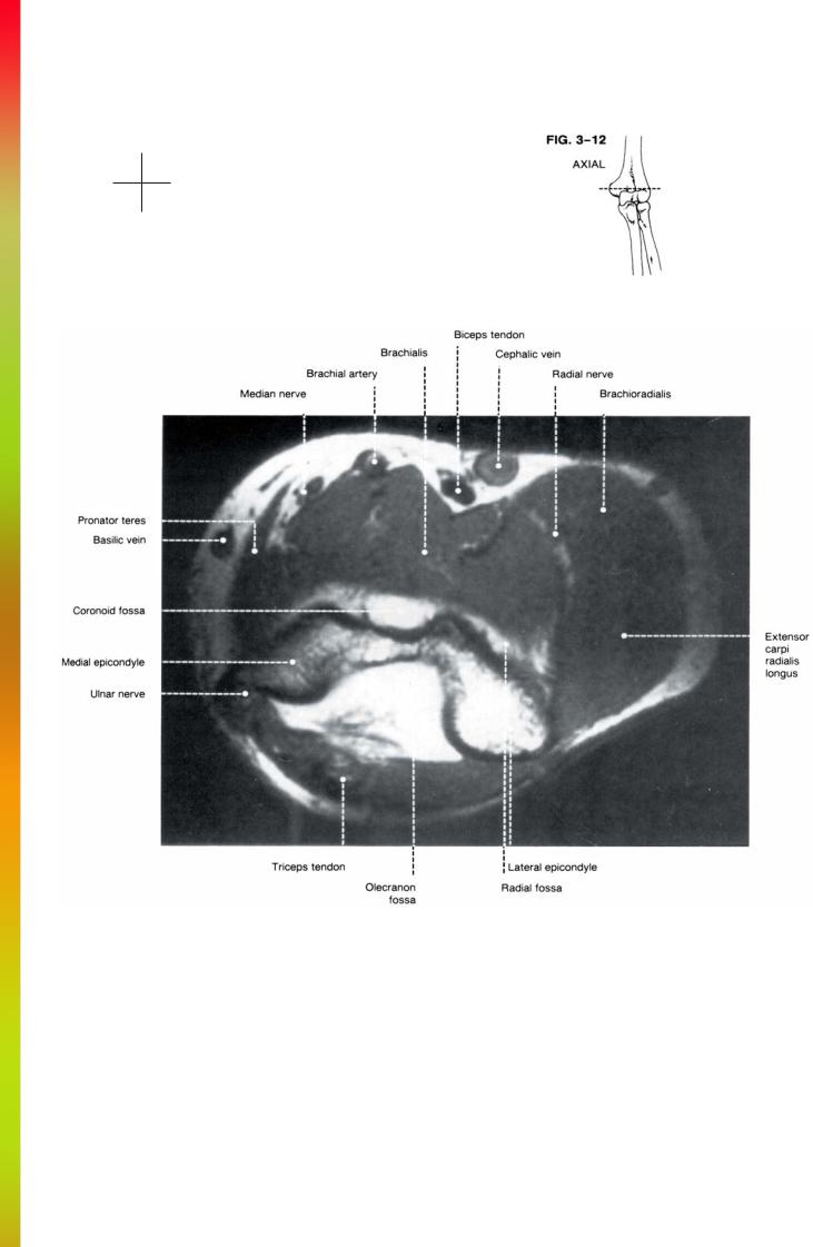
The anterior muscle group consists of the brachialis and biceps. The brachialis originates from the anterior surface of the distal humerus and crosses the elbow joint to insert on the ulnar tuberosity. The brachialis remains muscular with its central tendon embedded within the muscle belly as it inserts on the tuberosity.
The biceps muscle originates as two heads, but before reaching the elbow, these separate muscle bellies converge and become tendinous in the region of the elbow. The biceps tendon travels along the superficial surface of the brachialis, and inserts onto the radial tuberosity.
A prominent fascial band known as the biceps aponeurosis runs obliquely from the distal biceps tendon to the superficial surface of the pronator teres. The brachial ar-
tery, vein, and median nerve run under the biceps aponeurosis in a fibromuscular tunnel also bordered by the biceps tendon, the brachialis muscle, and the pronator teres muscle.
The posterior muscle group includes the triceps and anconeus muscles. The triceps muscle originates as three muscle bellies. These muscle bellies converge in the region of the elbow and fuse into a single musculotendinous unit. The central tendon of the triceps inserts onto the posterior and superior surface of the olecranon process. The anconeus is a small triangular shaped muscle which originates from the posterior aspect of the lateral epicondyle and crosses the elbow joint to insert on the posterior lateral aspect of the olecranon. It is felt by some to represent a detached portion of the triceps muscle.
Neurovascular structures: The brachial artery represents the only major arterial vessel in the region of the elbow. It runs along the superficial aspect of the brachialis muscle medial to the biceps muscle and tendon. In the region of the elbow it divides into the radial and ulnar arteries. The ulnar artery runs deep to the pronator teres whereas the radial artery runs between the brachioradialis and pronator teres. Recurrent branches arise from both the radial and ulnar arteries and travel superiorly adjacent to the radial and ulnar nerves. The brachial artery and its branches have corresponding venae comitantes which run adjacent to the arteries. The superficial veins include the medial basilic vein and the anterolateral cephalic vein.
Three major nerves are present in the region of the elbow. The radial nerve runs between the brachialis and brachioradialis. It divides into a deep and superficial branch at the elbow. These branches of the radial nerve can be distinguished from the recurrent radial artery be-

53 |
THE ELBOW |
cause they are more superficial in position. Identification of the nerves as well as the recurrent radial artery depends on the amount of fat separating these structures from the adjacent muscles. The ulnar nerve is identified just posterior to the medial epicondyle. On more distal sections, the ulnar nerve becomes difficult to identify as it runs in a triangular region formed between the flexor digitorum superficialis, the flexor digitorum profundis, and the flexor carpi ulnaris.
The median nerve runs medial to the brachial artery along the superficial surface of the brachialis muscle and goes on to pierce the pronator teres muscle between the superficial and deep heads. As with the other nerves, visualization depends on the amount of adjacent fat. Because of this, the median nerve is best identified on proximal sections. Distally as the nerve becomes surrounded by the brachioradialis and pronator teres muscles, it becomes progressively more difficult to identify.


THE ELBOW
AXIAL |
|
Cryomicrotomes ........................................................ |
FIGS. 3-7 to 3-11 |
MR Images.................................................................. |
FIGS. 3-12 to 3-16 |
SAGITTAL |
|
Cryomicrotomes ........................................................ |
FIGS. 3-17 to 3-20 |
MR Images.................................................................. |
FIGS. 3-21 to 3-24 |
CORONAL |
|
Cryomicrotomes ........................................................ |
FIGS. 3-25 to 3-28 |
MR Images................................................................. |
FIGS. 3-29 to 3-32 |

Anatomy and MRI of the Joints |
56 |
Anterior
Medial Lateral
Posterior

57 |
THE ELBOW |
Anterior
Medial Lateral
Posterior

Anatomy and MRI of the Joints |
58 |
Anterior
Medial Lateral
Posterior
59 |
THE ELBOW |
Anterior
Medial Lateral
Posterior

Anatomy and MRI of the Joints |
60 |
Anterior
Medial Lateral
Posterior
61 |
THE ELBOW |
Anterior
Medial Lateral
Posterior