

МРТ - в диагностике болезней суставов
.pdfAnatomy and MRI of the Joints |
112 |
Distal
Radial Ulnar
Proximal

113 |
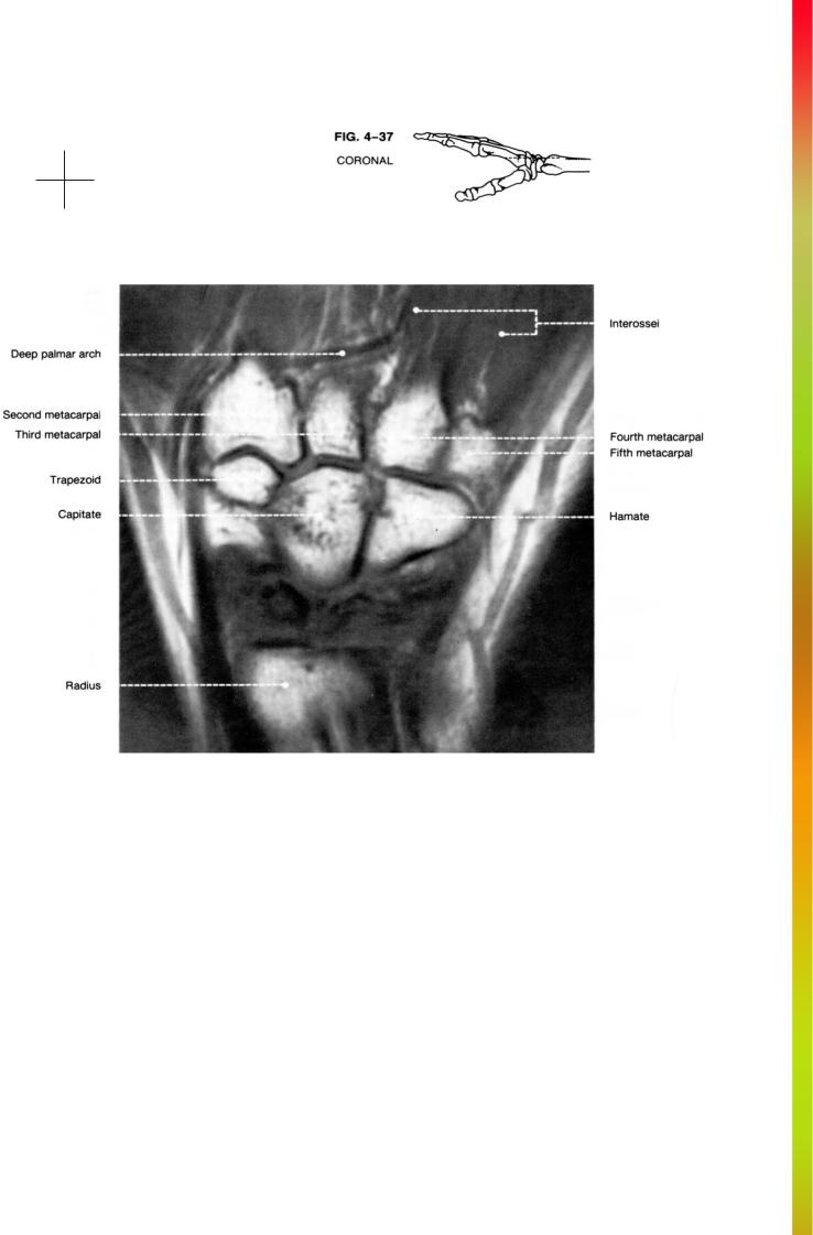
THE WRIST |
Distal
Radial Ulnar
Proximal

Anatomy and MRI of the Joints |
114 |
Distal
Radial Ulnar
Proximal

115 |
THE WRIST |
Distal
Radial Ulnar
Proximal

Anatomy and MRI of the Joints |
116 |
Distal
Radial Ulnar
Proximal

117 |
THE WRIST |
Distal
Radial Ulnar
Proximal

Anatomy and MRI of the Joints |
118 |
Distal
Radial Ulnar
Proximal

119 |
THE WRIST |
Distal
Radial Ulnar
Proximal
Anatomy and MRI of the Joints |
120 |
Distal
Radial Ulnar
Proximal

CHAPTER 5
THE FINGER
Scott Erickson, M.D.
The three articulations of the finger are the metacarpophalangeal joint, the proximal interphalangeal joint, and the distal interphalangeal joint. The metacarpophalangeal joint allows limited abduction and adduction as well as flexion and extension. The interphalangeal joints permit only flexion and extension.
A number of ligaments support the etacarpophalangeal joint. The palmar ligament, or volar plate, is a fibrocartilaginous plate extending from the base of the proximal phalanx to the head of the metacarpal. The transverse metacarpal ligament acts to unite the palmar ligaments. The collateral ligament is composed of two portions. The weaker, more proximal fanlike part acts to fix the palmar ligament to the metacarpal head. The stronger, cordlike part is the more distal component. The interphalangeal joints possess palmar and collateral ligaments having the same morphology as those of the metacarpophalangeal joint (Fig. 5-1).
The flexor digitorum tendons pass along the palmar surface of the digits. The flexor digitorum superficialis tendon is superficial to the flexor digitorum profundus tendon at the level of the metacarpophalangeal joint (Fig.
5-1). The superficialis tendon subsequently splits to lie first on either side of the profundus tendon at the level of the mid-proximal phalanx, then deep to the profundus at the level of the proximal interphalangeal joint, and finally deep and lateral to the profundus as it inserts onto the mid-portion of the middle phalanx. The flexor digitorum profundus tendon inserts onto the base of the distal phalanx (Fig. 5-1). The superficialis and profundus tendons each have their own synovial sheaths and also are surrounded by a common synovial sheath. This common sheath is then housed in a fibrous digital sheath, or fibrosseous tunnel.
The extensor expansion arises from the distal portion of the extensor digitorum tendon (Fig. 5-1). The central portion inserts onto the base of the middle phalanx along its dorsal aspect. The medial and lateral portions insert onto the base of the distal phalanx. The four lumbrical muscles arise from the flexor digitorum profundus tendons and insert onto the extensor expansion (Fig. 5-1). The dorsal and palmar interosseous muscles insert onto the expansion and also onto the bases of the proximal phalanges (Fig. 5-1).
121