

МРТ - в диагностике болезней суставов
.pdfAnatomy and MRI of the Joints |
42 |
Superior
Lateral Medial
Inferior

43 |
THE SHOULDER |
Superior
Lateral Medial
Inferior

Anatomy and MRI of the Joints |
44 |
Superior
Lateral Medial
Inferior

45 |
THE SHOULDER |
Superior
Lateral Medial
Inferior

Anatomy and MRI of the Joints |
46 |
Superior
Lateral Medial
Inferior

47 |
THE SHOULDER |
Superior
Lateral Medial
Inferior

Anatomy and MRI of the Joints |
48 |
Superior
Anterior Posterior
Inferior

CHAPTER 3
THE ELBOW
Stephan J. Macrander, M.D.
The elbow is a hinged joint joining the distal humerus to the proximal radius and ulna. Intimately associated with the elbow joint proper is the superior radial ulnar joint. These two joints share a common joint capsule and both will be discussed in this chapter. In addition, all
of the supporting ligaments, musculotendinous structures, and neurovascular structures will be described andillustrated.
Bones and articulations (Figs. 3-1 and 3-2):
The distal articular surface of the humerus is contoured into two components toaccommodate the proximal ulna and radial head. The medial surface articulating with the ulna is the trochlea, which is shaped similar to an hourglass laid on its side. The lateral surface is the capitellum that articulates with the fovea of the radial head. The capitellum has a rounded convex surface.
Important non-articulating processes of the distal humerus are the medial and lateral epicondyles. The medial epicondyle is the most prominent of the two. It provides the surface of origin for the pronator teres and common tendon of origin for the flexor muscles of the forearm. The lateral epicondyle affords the surface of attachment for the supinator and the common tendon of origin for the forearm extensor muscles.
The olecranon fossa is a prominent concavity on the posterior surface of the distal humerus, just proximal to the trochlea. It accommodates the olecranon process when the elbow is extended. The coronoid fossa is a similar, but shallower depression located anteriorly that accommodates the coronoid process of the ulna during elbow flexion. A small radial fossa is also present above the capitellum.
The articular surface of the proximal ulna is
composed of trochlear and radial notches. The trochlear notch is a rounded concavity situated between the olecranon and coronoid process. The radial notch is a concavity articulating with the radial head.
49

Anatomy and MRI of the Joints |
50 |
The radial head is the round pill-shaped portion of the proximal radius. It has a proximal concave surface called the fovea that articulates with the capitellum and a circumferential portion that articulates with the radial notch.
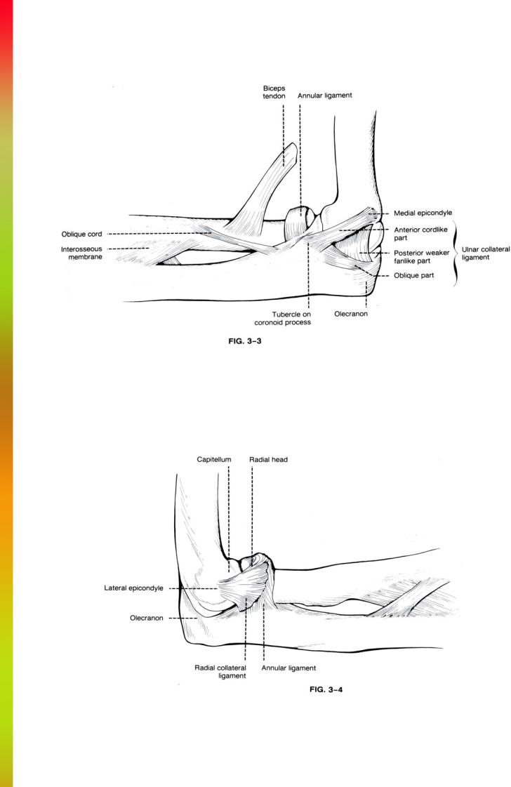
Ligaments (Figs. 3-3 and 3-4): The entire joint is surrounded by a loose articular capsule allowing for flexion and extension. Medial and lateral thickening of this capsule represent the collateral ligaments. The ulnar ollateral ligament originates from the medial epicondyle and fans out to attach to the medial aspect of the trochlear notch.
The anterior and oblique portions are thicker than the intermediate posterior fanlike portion.
The radial collateral ligament is triangular shaped with the apex arising from the lateral epicondyle and the base attaching to the annular ligament.
The annular ligament surrounds the circumference of the radial head and secures it in the radial notch. The ligament is attached to the anterior and posterior aspect of the radial notch.
Muscles and tendons (Figs. 3-5 and 3-6): A large number of muscles and tendons originate and insert at the elbow. This makes the cross-sectional anatomy of the muscles and tendons of the elbow rather confusing. Understanding the spatial relations of the musculotendinous structures is best accomplished by dividing them into posterior, anterior, lateral, and medial groups. These groups are best identified on axial images.
The lateral muscle group includes the brachioradialis, the extensors of the fingers and wrist, and the supinator.
The most superficial and anterior muscle of this group is the brachioradialis. It originates from the lateral supracondylar ridge of the distal humerus and extends to insert on the lateral aspect of the radial styloid. Just posterior and deep to the brachioradialis is the extensor carpi radialis longus. The extensor carpi radialis longus originates from the supracondylar ridge inferior to the brachioradialis and inserts on the base of the second metacarpal. The extensor carpi radialis brevis, extensor digitorum, ex-
51 |
THE ELBOW |
tensor carpi ulnaris, and extensor digiti minimi all arise from a common extensor tendon originating on the lateral epicondyle. Because of their common origin, these latter four muscles are difficult to separate from each other in the region of the elbow. They become progressively more distinct distally.
The supinator is the deepest of the lateral muscle group. It arises primarily from the posterolateral aspect of the ulna. However, portions of the muscle arise from the lateral epicondyle. It runs inferiorly and laterally to insert on the lateral aspect of the proximal radius. With the arm pronated, the supinator completely surrounds the radius.