

МРТ - в диагностике болезней суставов
.pdfAnatomy and MRI of the Joints |
122 |
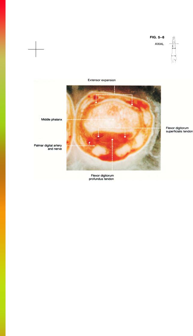
The neurovascular supply to the finger consists of both palmar and dorsal digital branches of nerves, veins, and arteries (Fig. 5-2). The palmar digital arteries and nerves
are larger than the dorsal arteries and nerves and are more easily seen on distal axial sections. The converse is true of the igital veins.

THE FINGER
AXIAL |
|
|
|
Cryomicrotomes |
.............................................................FIGS. 5 |
-3 |
to 5-6 |
MR Images ...................................................................... |
FIGS. 5 |
-7 |
to 5-10 |
SAGITTAL |
|
|
|
Cryomicrotomes ............................................................. |
FIGS. 5-11 to 5-12 |
||
MR Images ...................................................................... |
FIGS. 5 |
-13 to 5-14 |
|
CORONAL |
|
|
|
Cryomicrotome ............................................................... |
FIG. 5-15 |
|
|
MR Image ....................................................................... |
FIG. 5-16 |
|
|

Anatomy and MRI of the Joints |
124 |
Dorsal
Radial Ulnar
Palmar

125 |
THE FINGER |
Dorsal
Radial Ulnar
Palmar

Anatomy and MRI of the Joints |
126 |
Dorsal
Radial Ulnar
Palmar
127 |
THE FINGER |
Dorsal
Radial Ulnar
Palmar

Anatomy and MRI of the Joints |
128 |
Dorsal
Radial Ulnar
Palmar

129 |
THE FINGER |
Dorsal
Radial Ulnar
Palmar

Anatomy and MRI of the Joints |
130 |
Dorsal
Radial Ulnar
Palmar

131 |
THE FINGER |
Dorsal
Radial Ulnar
Palmar