

Micro-Nano Technology for Genomics and Proteomics BioMEMs - Ozkan
.pdfxxiv |
PREFACE |
generation of bioresearch and biomedical micro/nano biodevices will be the corresponding development of both viable and efficient nanofabrication and micro/nano integration processes.
The Volume II: Micro/Nano Technologies for Genomics and Proteomics presents a wide range of exciting new science and technology, and includes key sections on DNA micro/nanoarrays which additional chapters on peptide arrays for proteomics and drug discovery, new dielectrophoretic cell separation systems and new nanofabrication and integration processes; advanced microfluidic devices for the human genome project (whole genome sequencing); and final section on nanoprobes for imaging and sensing. Overall this volume should be of considerable value for a wide range of multidisciplinary scientists and engineers who are either working in or interested in bionanotechnology and the next generation of micro/nano biomedical and clinical diagnostic devices.
Mihrimah Ozkan
Dept. of Electrical Engineering,
University of California, Riverside,
Riverside, California USA
Michael J. Heller
Dept. of Bioengineering,
University of California, San Diego,
La Jolla, California USA
Mauro Ferrari
Professor, Brown Institute of Molecular Medicine Chairman
Department of Biomedical Engineering
University of Texas Health Science Center, Houston, TX
Professor of Experimental Therapeutics
University of Texas M.D. Anderson Cancer Center, Houston, TX
Professor of Bioengineering, Rice University, Houston, TX
Professor of Biochemistry and Molecular Biology
University of Texas Medical Branch, Galveston, TX
President, the Texas Alliance for NanoHealth, Houston, TX
I
Application of Microarray
Technologies
1
Electronic Microarray Technology
and Applications in Genomics
and Proteomics
Ying Huang, Dalibor Hodko, Daniel Smolko, and Graham Lidgard
Nanogen Inc., 10398 Pacific Center Court, San Diego, CA 92121, USA.
Keywords: Electronic microarray/ Miniaturization/ Single nucleotide polymorphism/ Gene expression profiling/ Cell separation/ Protein kinase/ Forensic detection/ Biological warfare
Electronic microarrays that contain planar arrays of microelectrodes have been developed to provide unique features of speed, accuracy and multiplexing for genomic and proteomic applications through utilizing electric field control to facilitate analytes concentration, DNA hybridization, stringency and multiplexing. An overview of electronic microarray technology is presented followed by its variety applications in genomic research and DNA diagnostics, forensic detection, biologic warfare, and proteomics.
1.1. INTRODUCTION
DNA microarrays have provided a new and powerful tool to perform important molecular biology and clinical diagnostic assays. The basic idea behind DNA microarray technology has been to immobilize known DNA sequences referred to as probes in micrometer-sized spots on a solid surface (microarray) and specifically hybridize a complementary sequence of the analyte DNA or a target. A fluorescently labeled reporter facilitates fluorescent detection of the presence or absence of a particular target or gene in the sample. By using laser-scanning and fluorescence detection devices such as CCD cameras, different target hybridization patterns can be read on the microarray and the results quantitatively analyzed. This chapter describes a specific microarray technology where an electric field and
4 |
YING HUANG ET AL. |
phenomena induced by the application of the electric field are used to direct and concentrate the DNA molecules through permeation layer [25] on the array.
Whereas, in the past, different technologies have been used to immobilize DNA probes including physical deposition [13, 40], photolithographic synthesis [Fodor et al., 1993, 7], and utilization of electric field [25]. Accordingly, several substrates have been used to generate different DNA microarrays ranging from glass slides, membrane to silicon. High density microarrays have been used to identify disease outcomes through relevant RNA expression patterns on thousands of genes [1] and for gene sequencing [33]. However, focused arrays, which often consists of 100–1,000 test sites, are better suited to detect a panel of genes for applications in point of care diagnostics, detection of infectious diseases, as well as identification of biological warfare agents. In these particular applications, speed, accuracy and multiplexing are basic requirements. Electronic microarrays, one type of the focused arrays, can meet all these requirements through utilizing electric field control to facilitate analytes concentration, DNA hybridization, stringency and multiplexing [17, 23–25, 45]. In this chapter, an overview of electronic microarray technology is presented followed by its applications in genomics and proteomics.
1.2. OVERVIEW OF ELECTRONIC MICROARRAY TECHNOLOGY
Nanogen, Inc. has developed an electronic micro-array based technology (NanoChip R Electronic Microarray) for manipulation, concentration and hybridization of biomolecules on the chip array (Figure 1.1). This approach extends the power of microarrays through the use of electronics by connecting each test site on the NanoChip R array to an electrode.
|
Charged |
|
biomolecules |
|
Electrode |
Electronic |
site |
array |
energized |
|
Charged biomolecules
Negatively charged
DNA Analyte molecules concentrate
+
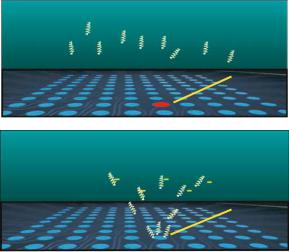
FIGURE 1.1. Nanochip R micro-array technology uses electronic addressing of charged biomolecules on the electrode array to separate and concentrate analyte targets. Negatively charged DNA targets and molecular probes (top) are moved to a particular site by energizing the electrodes at a reverse potential (bottom). Targeted molecules concentrate at the array site where they can be bound chemically or hybridized to a DNA probe. Fluorescent signal is obtained from the reporter probes hybridized to the target DNA and signal proportional to the concentration of analyte DNA is measured.
ELECTRONIC MICROARRAY TECHNOLOGY AND APPLICATIONS |
5 |
Most biological molecules have a natural positive or negative charge. When biological molecules are exposed to an electric field (Figure 1.1), molecules with a positive charge move to electrodes with a negative potential, and molecules with a negative charge move to electrodes with a positive potential. Current and voltages are applied to the test sites via individual electrode activation to facilitate the rapid and controlled transport of charged molecules to any test sites. Additional advantages of electrically facilitated transport include
(1) the ability to produce reconfigurable electric fields on the microarray surface that allows the rapid and controlled transport of charged molecules to any test sites [22, 25]; (2) the ability to carry out site selective DNA or oligonucleotide addressing and hybridization [11];
(3) the ability to significantly increase DNA hybridization rate by concentration of target at the test sites (Kassegne et al., 2003); and (4) the ability to use electronic stringency to improve hybridization specificity (Sosnowski, et al., 1997).
1.2.1.NanoChip R Array and NanoChip R Workstation
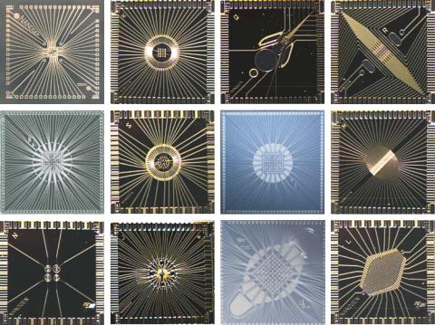
1.2.1.1.Fabrication Electronic microarrays consist of an array of electrodes that have been fabricated on silicon with array sizes ranging from 5 to 10,000 individual electrodes or test sites [24, 25, 42]. Figure 1.2 shows a number of electronic microarrays with arrays ranging from 4 to 100 electrodes or test sites. These arrays have been designed
FIGURE 1.2. A series of designs of Nanogen’s silicon microarray chips. Chip sizes shown range from 4–100 sites. These include different electrode geometries as well as chips designed for particles and or biomolecular microseparations (last column of chips).
6 |
YING HUANG ET AL. |
80 m diameter test site
Hydrogel Permeation Layer
Containing Streptavidin
Platinum
Silicon Microelectrode
Base
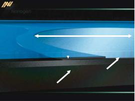
FIGURE 1.3. Cross-section of a single platinum micro-electrode pad on the NanoChip R microarray. A hydrogel permeation layer loaded with streptavidin covers the electrode array and serves for capturing electronically addressed molecules.
for both microassays and microseparations. The current commercialized NanoChip R array comprising 100 platinum microelectrodes with additional 20 outer microelectrodes acting as counter-electrodes.
The electrodes on the NanoChip R array are fabricated on a silicon substrate using standard photolithography and deposition processes. Each electrode is 80 µm in diameter with 200 µm center-to-center space between and is connected to the outside edge of the chip by a platinum wire trace. Figure 1.3 shows a cross section of a single electrode pad on the NanoChip R array. The base structure of the array consists of silicon over which an insulating layer of silicon dioxide is applied. Platinum is deposited and selectively held in place to form electrodes and accompanying electrical traces. These wire traces terminate at the edges of the chip forming electrical contact pads. Additional layers of silicon dioxide and silicon nitride are deposited to electrically insulate the platinum electrical traces, leaving the central array of 80 µm diameter microelectrodes, outer microelectrodes and contacts pads exposed. The chips are flip-chip bonded to a ceramic substrate which contains contacts to pogo-pins.
1.2.1.2. Permeation Layer Typically, on the surface of the array, a 10 µm thick hydrogel permeation layer (Figure 1.3). containing co-polymerized streptavidin is deposited by microreaction molding. This permeation layer serves two main functions [24]. First it protects the sensitive analytes from the adverse electrochemical effects at the platinum electrode surface during active operation. These electrochemical products include the generation of hydrogen ions (H+) and oxygen at the positively biased (anode) microelectrodes and hydroxyl ions (OH−) and hydrogen at the negatively biased electrodes (cathode), as well as various free radical entities. Secondly, the permeation layer also serves as a matrix for the attachment of biotinylated molecules e.g., analytes capture oligos, antibodies, and primers through biotin and streptavidin binding [24, 25]. Figure 1.4 is a close-up photograph of the 100-site array covered with the permeation layer.
ELECTRONIC MICROARRAY TECHNOLOGY AND APPLICATIONS |
7 |
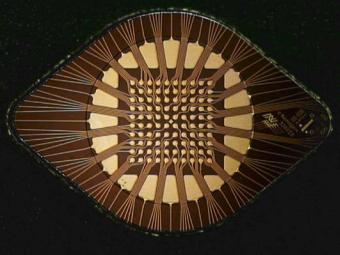
FIGURE 1.4. Photograph of the 100-site NanoChip R microarray. A hydrogel permeation layer covers the electrode array including working and counter electrodes. The hydrogel layer is visible as a circle surrounding counter electrodes (yellow pads).
1.2.1.3.NanoChip R Cartridge The 100-site array is assembled into a complete NanoChip R cartridge (Figure 1.5a) by ultrasonically welding two molded polymethyl methacrylate (PMMA) cartridge bodies that contain fluidic channels and inlet and out-
let ports. The cartridge eliminates sample evaporation, prevents sample contamination and provides a fluidic interface to the NanoChip R Workstation.
1.2.1.4.NanoChip R Workstation The NanoChip R electronic microarrays are operated through a fully integrated and automated NanoChip R Workstation (Figure 1.5b). The system consists of three major subsystems: (1) the NanoChip R Loader for loading patient samples on one to four NanoChip R Cartridges, (2) the NanoChip R Reader, a highly sensitive, laser-based fluorescence scanner for detection of assay results and (3) computer hardware and software which automates import, analysis and export of sample information making data analysis simple.
1.2.2.Capabilities of the NanoChip R Electronic Microarrays
Using electric field, Nanochip R electronic microarrays have provided many unique features over other passive microarrays (Table 1.1). Electronic addressing allows users to quickly customize arrays in their own laboratory. Electronic hybridization provides an extremely accurate and specific hybridization process by creating optimal electric and pH conditions at the hybridization sites [11, 25]. Electronic stringency, in combination with thermal control, enables researchers to remove unbound and nonspecifically-bound DNA quickly and easily after hybridization at the microarrays, achieving rapid determination of single base mismatch mutations in DNA hybrids [44].

8 |
YING HUANG ET AL. |
NanoChip*
(a)Electronic Microarray
NanoChip*
Cartridge
(b)
Nanochip®
Reader
Nanochip®
Loader
FIGURE 1.5. Photograph of the Nanochip R cartridge containing the electronic microarray, and b) Nanogen’s Nanochip R Workstation which allows fully automated processing of 4 cartridges simultaneously in the loader and fluorescent detection in the reader.
1.2.2.1. Assay Formats on NanoChip R Electronic Microarrays The open architecture of electronic microarray enables flexibility in assay design. Since each individual electrode can be selectively activated, different assays can be generated depending on different analytes to be addressed (Figure 1.6). For example, a dot blot assay [19] is conducted when biotinylated PCR amplicon is addressed to the selected electrodes and remained embedded through interaction with strepavidin in the permeation layer. The DNA at each electrode is then hybridized to mixtures of allelespecific oligonucleotides (discriminators) and fluorescently labeled oligonucleotides. The thermal or electronic stringency is used to discriminate
TABLE 1.1. Comparison between NanoChip R microarray active hybridization technology and passive hybridization technologies.
|
|
|
Concentration |
Stringency |
|
Hybridization time |
Concentration of targets |
factor at a site |
control |
|
|
|
|
|
NanoChip R active |
10–100 seconds |
Directed and localized at the |
>1000 times |
Electronic, |
hybridization |
|
array sites; Ability to |
|
Thermal |
|
|
control individual sites |
|
Chemical |
Passive |
1–2 hours |
Non-directed; Sites cannot be |
Low, diffusion |
Thermal, |
hybridization |
|
controlled independently |
dependent |
Chemical |
|
|
|
|
|
ELECTRONIC MICROARRAY TECHNOLOGY AND APPLICATIONS |
9 |
|
Electronic addressing of Electronic hybridization |
Report and stringency |
|
biotinylated analytes
Green
Red
• Denatured PCR product |
• Helper oligos |
• Dot blot SNP |
• Capture oligos |
• Denatured PCR product |
• Sandwiched SNP |
• Capture oligos |
• RNA (or cDNA) |
• Gene expression profiling |
• SDA primer |
• Denatured genomic DNA |
• Anchored SDA |
• Antibody |
• Antigen |
• Immunoassay |
FIGURE 1.6. The open platform of the electronic microarray enables flexibility in assay design. Depending on the nature of the biotinylated analytes (denatured PCR products, capture oligos, SDA primers or antibody) in electronic addressing and on the targets (helper oligos, denatured PCR products, RNA, denatured genomic DNA or antigen), different assays (dot blot SNP, sandwiched SNP, gene expression profiling, anchored SDA or immunoassay) can be performed on the electronic microarrays.
the SNP. In this assay format, multiple samples can be analyzed at different electrodes on a single array.
A “sandwich” assay is created when biotinylated capture probes are addressed to selective electrodes. The capture probes can be sequence specific oligos or antibodies. The embedded capture probes are then electronically hybridized to the targets. Depending on the specific targets (PCR amplicons, or RNA, or antigens), the assays can be SNP analysis, gene expression profiling, or immunoassay.
Most recently, a special assay, anchored strand displacement amplification (aSDA), which integrates the amplification and discrimination, is demonstrated on electronic microarrays [12, 29, 50, 51]. In this assay format, biotinylated SDA primers were addressed and anchored on selective electrodes. These anchored primers were then electronically hybridized to the denatured genomic DNA. Target DNA was amplified over the electrodes when an enzyme mix containing restriction endonuclease and DNA polymerase and dNTPs was introduced to the array. After amplification the final discrimination was determined using same base-stacking principle [37]. Using aSDA, multiple genes from one sample or one gene from multiple samples can be simultaneously amplified and detected on a single electronic microarray.
1.2.2.2. Electronic Multiplexing By electronically controlling each test site, the electronic microarrays also provide a platform to perform different types of multiplexed assays:
(1)single array multiplexing where multiple genes from one sample can be analyzed;
(2)single array multiplexing where multiple samples with one gene of interest can be analyzed;
(3)single array multiplexing where multiple samples with multiple genes of interest can be analyzed;
10 |
YING HUANG ET AL. |
(4)single site multiplexing where several targets are discriminated on the same site using different fluorescent probes;
(5)single site multiplexing where several targets are addressed, different discriminator oligonucleotides hybridized and reporter addressed—this method allows the use of a universal reporter for different targets.
The ability to electronically control individual test sites permits biochemically unrelated molecules to be used simultaneously on the same microchip. In contrast, sites on a conventional DNA array cannot be controlled separately, and all process steps must be performed on an entire array. Nanogen’s electronic microarray technology delivers increased versatility over such conventional methods. This is particularly important in applications such as biological warfare and infectious disease detection since the accurate identification of biological agents requires determination of two or more (often five) characteristic genes of a particular agent.
1.3. APPLICATIONS
1.3.1. Single Nucleotide Polymorphisms (SNPs)—Based Diagnostics
Given the advances in genomic studies, more and more single nucleotide polymophisms (SNPs) are found to be contributory factors for human disease and can be used as genetic markers for molecular diagnostics. The speed, accuracy and flexibility provided by electronic microarrays have received great interest from clinical diagnostic researchers for a variety of genotyping applications (Table 1.2). Using the Nanochip R system, researchers at American Medical Laboratories analyzed 635 clinical samples for the G1691A mutation on the factor V Leiden, associated with thrombosis with 100% accurate in characterizing wildtype, heterozygous, and homozygous samples [15]. Researchers at ARUP Laboratories have evaluated 3 thrombosis associated SNPs, factor V (Leiden), factor II (prothrombin), and methylenetetrhydrofolate reductase (MTHFR), on 225 samples with 100% accuracy [14]. Schrijver et al at Stanford University Medical Center found that the SNP analysis based on Nanogen electronic microarray for factor V (Ledein) and factor II (prothrombin) on 800 samples were comparable with other SNP analysis methods such as restriction enzyme digestion (RFLP) and the Roche LightCycler [41]. Researchers at Children’s Nantional Medical Center, Washington DC, genotyped 8 common MeCP2 mutations associated to Rett syndrome on 362 samples with 100% specificity [47]. Using electronic microarray, researchers at Mayo Clinic Cancer Center have developed genotyping assays for 5 different cytokine polymorphisms [43]. Moreover, 8 SNPs distributed within a highly variable region of the polC gene from six isolates of Staphylococcus aureus were analyzed on electronic microarrays [9].
1.3.2. Forensic Detection
Short tandem repeats (STRs) represent another type of polymorphism with important applications in forensic DNA identification. In 1990, the FBI created a combined DNA index system (CODIS), which consist of 13 polymorphic STR loci, to provide a database of forensic DNA profile for nearly all forensic laboratories in the United States [5, 6]. The