


Alveolar-Blood Gas Exchange |
2 |
THE NORMAL LUNG
Partial Pressure ofa Gas in AmbientAir
Pgas = Fgas X Patin
By convention, the partial pressure of the gas is expressed in terms of its dry gas concentration. For example, the P02 in ambient air is:
P02 = 0.21 x 760 = 160 mm Hg
PartialPressure ofa Gas in Inspired Air
Inspired air is defined as air that has been inhaled, warmed to 37°C, and com pletely humidified, but has not yet engaged in gas exchange. It is the fresh air in the anatVD that is about to enter the respiratory zone.
The partial pressure of H20 is dependent only on temperature and at 37°C is 47 mm Hg. Humidifying the air reduces the partial pressure of the other gases present.
Pigas = Fgas (Patm - PH20)
For example, the P02 ofinspired air is:
PI02 = 0.21(760 - 47) = 150 mm Hg
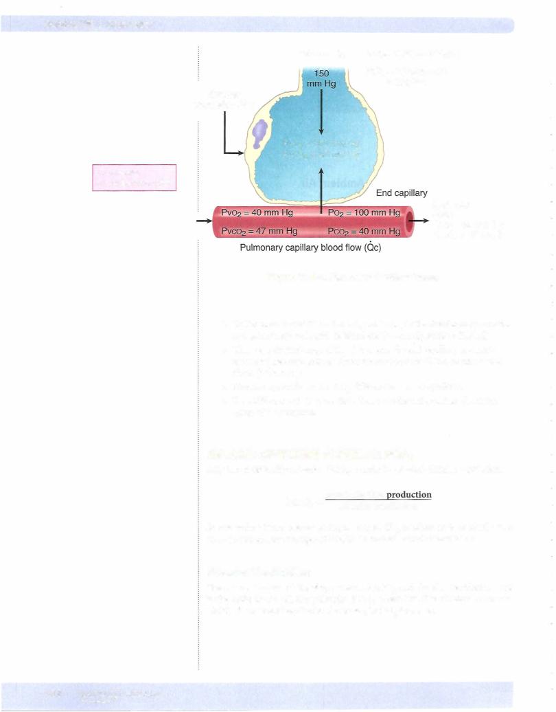
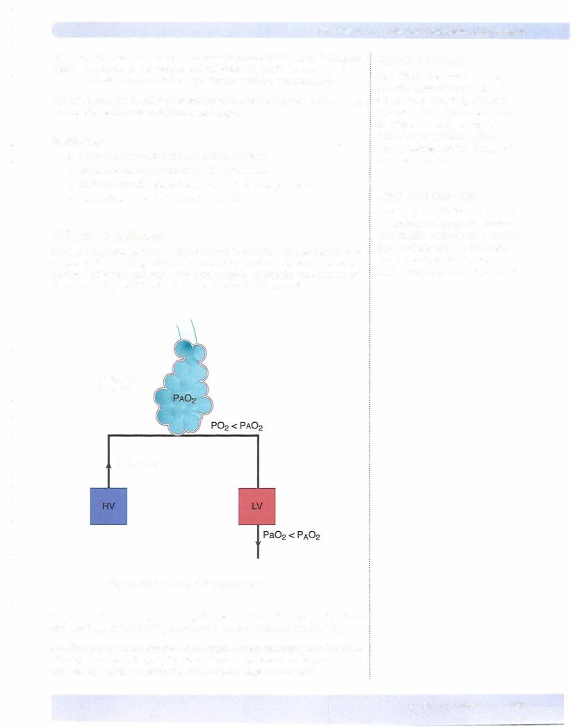
Figure VIl-2-1 shows the pressures ofoxygen and carbon dioxide in the alveolar, pulmonary end capillary, and systemic arterial blood.
Patm = atmospheric pressure
Pgas = partial pressure of a gas
Fgas = concentration of a gas
Plgas = partial pressure ofinspired gas
PH20 = partial pressure of H20 vapor
In a Nutshell
Dalton's law of partial pressures states that the total pressure exerted by a mixture of gases is the sum of the pressures exerted independently by each gas in the mixture. Also, the
pressure exerted by each gas (its partial pressure) is directly proportional to its percentage in the total gas mixture.
MEDICAL 1 59

Chapter 2 • Alveolar-Blood Gas Exchange
Hyperventilation
During hyperventilation, there is an inappropriately elevated level of alveolar ventilation, and PAC02 is depressed.
IfVA is doubled, then PAC02 is decreased by half.
e.g., PAC02 = 40 mm Hg 2 X VA; PAC02
Hypoventilation
During hypoventilation, there is an inappropriately depressed level of alveolar ventilation, and PAC02 is elevated.
IfVA is halved, then PAC02 is doubled. e.g., PAC02
1/2 VA; PAC02
Metabolic Rate
There is a direct relationship between alveolar PC02 and body metabolism. For PAC02 to remain constant, changes in body metabolism must be matched with equivalent changes in alveolar ventilation.
•IfVA matches metabolism, then PAC02 remains constant.
•For example, during exercise, if body metabolism doubles, then VA must PAC02 is to remain constant.double if
• If body temperature decreases and there is no change in ventilation, PAC02 decreases, and the individual can be considered to be hyperventi lating.
FACTORS AFFECTING ALVEOLAR P02
AlveolarAir Equation
The alveolar air equation includes allthe factors that can affect alveolar P02.
PAC02
PA02 = (Patm - 47)Fr02 - liQ
Practical application ofthe equation includes differential diagnosis ofhypoxemia by evaluating the alveolar arterial (A-a) gradient of oxygen.
Three important factors can affect PA02:
Patm = atmospheric pressure, at sea level 760 mm Hg
An increase in atmospheric pressure (hyperbaric chamber) increases alveolar P02, and a decrease (high altitude) decreases alveolar P02•
F102 = fractional concentration of oxygen, room air 0.21
In a Nutshell
What is the difference between respiratory quotient (RQ) and
respiratory exchange ratio (RER)? Answer: RQ is the ratio between C02 production and 02 consumption at the cellular level. RER is the ratio of C02 output and oxygen uptake occurring in the lung. In a steady state, RQ and RER are equal.
MEDICAL 1 61
Chapter :z • Alveolar-Blood Gas Exchange
A structural problem in the lungs is any situation in which there is a loss ofsur face area and/or an increase in the thickness of the membrane system between the alveolar air and the pulmonary capillary blood. In all cases, therate ofoxygen and carbon dioxide diffusion decreases. The greater the structural problem, the greater the effect on diffusion rate.
Factors Specific to Each Gas Present
D (diffusion constant) = main factoris solubility
The only clinically significant feature ofD is solubility. The more soluble the gas, the faster it diffuses across the membranes. co2 is the most soluble gas with which we willbe dealing. The great solubility of C02 is the main reason why it diffuses faster across the alveolar membranes than 02.
Gradientacross the membrane
(P1 - P2): This is the gas partial pressure difference across the alveolar membrane. The greater the partial pressure difference, the greater the rate ofdiffusion.
Under resting conditions, when blood first enters the pulmonary capillary, the gra dient for 02 is:
100 - 40 = 60 mm Hg
An increasein the P02gradientacross the lung membranes helps compensate for a structural problem. Ifsupplemental 02 is administered, alveolar P02 increases, because of the elevated gradient. However, supplemental Oz does not improve the ability ofthe lungs to remove COz from blood. This increased gradient helps return the rate of02diffusiontowardnormal.The greater the structural problem, the greater the gradient necessary for a normal rate ofOz diffusion.
The gradient for C02 is 47 - 40 = 7 mm Hg.
Even though the gradient for COz is less than for 02, COz still diffuses faster because ofits greater solubility.
DIFFUSING CAPACITY OF THE LUNG (DLCO)
There are 2 terms that describe the dynamics of the transfer of individual sub stances between the interstitium and the capillary:
•Ifthe substance equilibrates between the capillary and interstitium, it is said to be in a perfusion-limited situation.
•Ifthe substance does not equilibrate between the capillary and intersti tium, it is said to be in a diffusion-limited situation.
Carbon monoxide is a unique gas in that it typically doesn't equilibrate between the alveolar air and the capillary blood. Thus, it is a diffusion-limited gas. This is taken advantage of clinically, and the measurement ofthe uptake of CO in mL/ min/mm Hg is referred to as the diffusing capacity ofthe lung. It is an index of the lung's structural features.
MEDICAL 163

Section VII • Respiration
Carbon Monoxide: A Gasthat is Always Diffusion Limited
Carbon monoxide has an extremely high affinity for hemoglobin. When it is present in the blood, it rapidly combines with hemoglobin, and the amount dis solved in the plasma is close to zero (therefore, partial pressure in the plasma is considered zero). Thus, the alveolar partial pressure gradient (P1 - P2) is simply P1 (alveolar partial pressure), since P2 is considered to be zero. At a constant and known alveolar partial pressure, the uptake ofcarbon monoxide depends only on the structural features of the lung, as illustrated in Figure VIl-2-2.
Figure Vll-2-2. Carbon Monoxide
•This measured uptake of carbon monoxide is called the diffusing capacity of the lung (DL; rnL/min/mm Hg).
•It is an index of overall surface area and membrane thickness.
•With a structural problem, it correlates with the extent of lung damage and is particularly useful when measured serially over time.
•DL (rate of CO diffusion) decreases in emphysema and fibrosis but increases during exercise.
164 MEDICAL

Chapter 2 • Alveolar-Blood Gas Exchange
Chapter Summary
•In a normal resting individual at sea level, the partial pressures of oxygen
and carbon dioxide are not different between the alveolar and pulmonary end capillary compartments (P02 = 100 mm Hg and PC02 = 40 mm Hg).
•PaC02 is the same as alveolar, but Pa02 is less than alveolar (A-a gradient), which is primarily the result of shunt blood flow.
•Only two factors affect alveolar PC02: body metabolism and alveolar ventilation. If body metabolism is constant, there is an inverse relationship between alveolar ventilation and alveolar PC02•
•Three important factors affect alveolar P02: atmospheric pressure, oxygen concentration in the inspired air, and alveolar PC02•
•A change in alveolar PC02 causes a change in alveolar P02• They change in opposite directions by approximately the same amount in mm Hg.
•Two structural factors and two gas factors affect the rate of gas diffusion across lung membranes.
•Diffusing capacity is directly proportional to membrane surface area and inversely proportional to membrane thickness.
•The partial pressure gradient is the driving force for diffusion.
•Because C02 is an extremely soluble gas, it diffuses across the lung membranes faster than oxygen even though it has a small gradient.
•Supplemental oxygen raises the oxygen gradient and can compensate fora diffusion problem.
•Diffusing capacity ofthe lung is an index ofthe overall surface area and membrane thickness. It is measured as the uptake ofCO from the alveolar air to the blood in ml/min/mm Hg.
MEDICAL 165


Transport of 02 and C02 and |
3 |
the Regulation of Ventilation |
TRANSPORT OF OXYGEN
Units ofOxygen Content
Oxygen content = concentration ofoxygen in the blood, e.g., arterial blood
=20 volumes %
=20 volumes ofoxygen per 100 volumes ofblood
=20 mL ofoxygen per 100 mL ofblood
=0.2 mL ofoxygen per mL ofblood
Dissolved Oxygen
Oxygen dissolves in blood and this dissolved oxygen exerts a pressure. Thus, P02 ofthe blood represents the pressure exerted by the dissolved gas, and this P02 is directly related to the amount dissolved (Figure VII-3-1).
The amount dissolved (P02) is the primary determinant forthe amount of oxy gen bound to hemoglobin (Hb).
There is a direct linear relationship between P02 and dissolved oxygen (Figure VII-3-1). When P02 is 100 mm Hg, 0.3 mL 02 is dissolved in each 100 mL of
blood (0.3 vol%). Maximal hyperventilation can increase the P02 in blood to 130 mm Hg (0.4 vol%).
'::!!.. |
|
|
|
|
|
|
|
g |
0.3 |
|
|
|
|
|
|
|
|
|
|
|
|
||
0> |
|
|
|
|
|
|
|
'O0Q) |
0.2 |
|
|
|
|
|
|
|
|
|
|
|
|
|
|
Ul |
0.1 |
|
|
|
|
|
|
Ul |
|
|
|
|
|
|
|
..... |
|
|
|
|
|
|
|
c |
|
|
|
|
|
|
|
: |
|
|
|
|
|
|
|
Q) |
|
|
|
|
|
|
|
0 |
|
|
20 |
40 |
60 |
80 |
1 00 |
0C\I |
|
|
|||||
|
|
||||||
() |
|
|
|
|
|
|
|
P02 in Blood (mm Hg)
Figure Vll-3-1 . Dissolved Oxygen in Plasma
MEDICAL 167

Section VII • Respiration
Oxyhemoglobin
Each Hb molecule can attach and carry up to four oxygen molecules. Binding sites on Hb have different affinities for oxygen. Also, the affinity of a site can and does change as oxygen is loaded or unloaded from the Hb molecule and as the chemical composition ofthe plasma changes.
Site4 - 02 |
systemic arterial blood = 97% saturated |
attached when the minimal |
|
P02 = 100 mm Hg |
|
Site 3 - 02 |
systemic venous blood = 75% saturated |
attached when the minimal |
(resting state) |
P02 : = 40 mm Hg |
P50 for arterial blood. P50 is the P02 |
Site 2 - 02 |
|
attached when the minimal |
required for 50% saturation |
P02 : = 26 mm Hg |
|
Site I - 02 usually remains attached under physiologic conditions. Under physi ologic conditions, only sites 2, 3, and 4 need to be considered.
Most ofthe oxygen in systemic arterial blood is oxygen attached to Hb. The only significant form in which oxygen is delivered to systemic capillaries is oxygen bound to Hb.
Hemoglobin 02 Content
The number ofmL of oxygen carried in each 100 mL ofblood in combination with Hb depends on the Hb concentration [Hb]. Each gram of Hb can combine with 1.34 mL of 02.
If the [Hb] is 15 g/100 mL (15 g%), then the maximal amount of 02 per 100 mL ( 100% saturation) in combination with Hb is:
l.34([Hb]) = 1 .34(15) = 20 mL 02/100 mL blood = 20 vol%
This volume represents the "carrying capacity" ofthe blood.
The Hb in systemic arterial blood is about 97% saturated with oxygen, which means slightly less than 20 vol% is carried by Hb.
When blood passes through a systemic capillary, it is the dissolved oxygen that diffuses to the tissues. However, ifdissolved oxygen decreases, P02 also decreases, and there is less force to keep oxygen attached to Hb. Oxygen comes off Hb and dissolves in the plasma to maintain the flow ofoxygen to the tissues.
168 MEDICAL


Section VII •Respiration
Table VII-3- 1 is a summary ofthe effects of anemia, polycythemia, and carbon monoxide poisoning.
Table Vll-3-1. Systemic Arterial Blood
|
P02 |
Hb Concentration |
02 perg Hb |
02 Content |
Anemia |
N |
|
N |
|
Polycythemia |
N |
|
N |
|
CO poisoning (acute) |
N |
N |
|
|
N = normal; 02 per g Hb = % saturation.
In anemia, hemoglobin is saturated but arterial oxygen content is depressed because of the reduced concentration ofhemoglobin.
In polycythemia, arterial oxygen content is above normalbecause ofan increased hemoglobin concentration.
In CO poisoning, arterial P02 is normal, but oxygen saturation of hemoglobin is depressed.
TRANSPORT OF CARBON DIOXIDE
Dissolved Carbon Dioxide
Carbon dioxide is 24 times more soluble in blood than oxygen is. Even though the blood has a PC02 of only between 40 and 47 mm Hg, about 5% of the total C02 is carried in the dissolved form.
Carbamino Compounds
Carbon dioxide reacts with terminal amine groups ofproteins to form carbamino compounds. The protein involved appears to be almost exclusively hemoglobin. About 5% of the total C02 is carried as carbamino compounds. The attachment sites that bind co2 are different from the sites that bind 02.
Bicarbonate
About 90% ofthe C02 is carried as plasma bicarbonate. In order to convert C02 into bicarbonate or the reverse, carbonic anhydrase (CA) must be present.
CA
co2 + H2o H H2C03 H H+ + HC03-
Figure VII-3-6 illustrates the steps in the conversion of C02 into bicarbonate in a systemic capillary.
1 72 MEDICAL


Section VII • Respiration
NEURAL REGULATION OF ALVEOLAR VENTILATION
The level ofalveolar ventilation is driven mainly from the input ofspecific che moreceptors to the central nervous system. Thestronger the stimulation ofthese receptors, the greater the level ofalveolar ventilation. Chemoreceptors monitor the chemical composition ofbody fluids. In this system, there are receptors that respond to pH, PC02, and P02.
There are two groups ofreceptors, and they areclassifiedbased upontheirlocation.
Central Chemoreceptors
These receptors are located in the central nervous system-more specifically, close to the surface of the medulla.
•Stimulation ofcentralchemoreceptors increases ventilation.
•The receptors directly monitor and are stimulated by cerebrospinal
fluid [H+] and C02. The stimulatory effect of increased C02 may be due to the local production ofH+ from C02.
•Because the blood-brain barrier is freely permeable to C02, the activity of these receptors changes with increased or decreased systemic arterial PC02•
•H+ does not easily penetrate the blood-brain barrier. Thus, an acute rise
in arterial H+, not of C02 origin, does not stimulate central chemore ceptors.
•These receptors are very sensitive and represent the main drive for ven tilation under normal resting conditions at sea level.
•Therefore, the main drive for ventilation is C02 (H+) on the central che moreceptors.
Figure VII-3-8 illustrates the relationship between the central chemoreceptors and the systemic arterial blood.
C02 •-••H• |
............zo2 |
|
H+ -··-·· |
H+ ·········· |
Medulla |
|
||
|
|
|
Systemic |
CSF |
|
arterial blood |
|
Figure Vll-3-8. Central Chemoreceptors
1 74 MEDICAL

Chapter 3 • Transport of 02 and C02 and the Regulation ofVentilation
The system does adapt, usually within 12 to 24 hours. The mechanism ofadapta tion may be the normalization of CSF tt+by the pumping ofHC03- into or out ofthe CSF. There are no central P02 receptors.
Peripheral Chemoreceptors
These receptors are found within small bodies at 2 locations:
Carotidbodies: near carotid sinus, afferents to CNS in glossopharyngeal nerve IX
Aorticbodies: near aortic arch, afferents to CNS in vagus nerve X
The peripheral chemoreceptors are bathed in arterial blood, which they monitor directly. These bodies have 2 different receptors:
•H+/C02 receptors
-These receptors are less sensitive than the central chemoreceptors, but they still contribute to the normal drive for ventilation.
-Therefore, under normal resting conditions at sea level, for all prac tical purposes, the total drive for ventilation is C02, mainlyvia the central chemoreceptors but with a small contribution via the periph eral chemoreceptors.
•P02 receptors
-The factormonitoredby these receptors is P02, not oxygen content.
-Because they respond to P02, they are actually monitoring dissolved oxygen and not oxygen on Hb.
-When systemic arterial P02 is close to normal (:::100 mm Hg) or above normal, there is little ifany stimulation of these receptors.
•They are strongly stimulated only by a dramatic decrease in systemic arterial P02•
•Sensitivity to hypoxia increases with C02 retention.
Thesereceptors do not adapt.
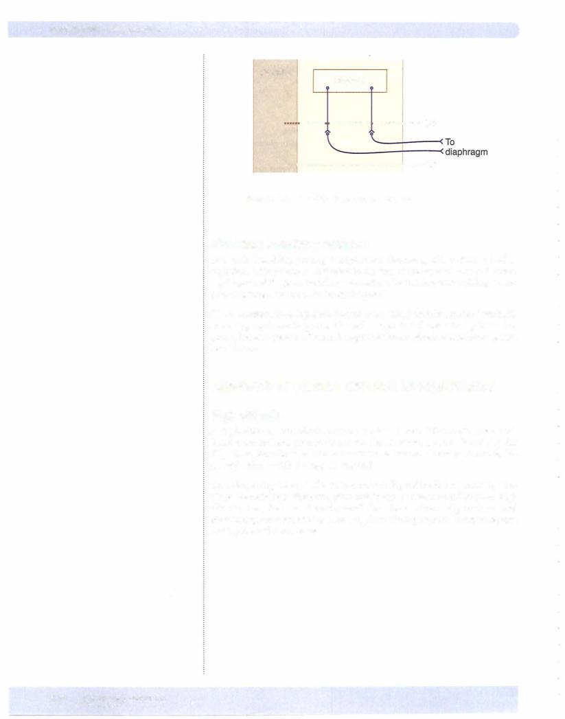
Central Respiratory Centers
Medullary centers
Site of the inherent rhythm for respiration.
Inspiratory center
Expiratory center
For spontaneous breathing, an intact medulla must be connected to the dia phragm (via the phrenic nerve). Thus a complete Cl or C2 lesion willprevent diaphragmatic breathing but not a complete C6 or lower lesion.
Figure VII-3-9 illustrates the main features involved in the central control of ventilation.
Bridge to PathologyI
Pharmacology
The normal C02 drive to breath is suppressed in COPD patients, and by narcotics a'nd general anesthetics.
Clinical Correlate
Although oxygen content is reduced in anemia, the Pa02 is normal; thus, anemia does not directly stimulate ventilation. However, the reduced
oxygen delivery can cause excess lactic acid production, which would in turn stimulate peripheral chemoreceptors.
MEDICAL 175

Chapter 3 • Transport of 02 and C02 and the Regulation ofVentilation
Table Vll-3-2.Acute Changes and Long-Term Adaptations (Acclimatization)
|
Acute Changes |
Acclimatization |
PA02 and Pa02 |
decreased |
remains decreased |
PAG02 and PaC02 |
decreased |
remains decreased |
Systemic arterial pH |
increased |
decreases to normal via |
|
|
renal compensation |
Fib coflcentral'ion |
no change |
increases (polycythemia) |
Hb % sat |
decreased |
remains decreased |
Systemic arterial 02 content |
decreased |
increases to normal |
At high altitude, hypoxia can develop, resulting in increased circulating levels of erythropoietin. Erythropoietin increases red blood cell production and eventu ally causes an adaptive polycythemia.
High-Pressure Environment
In a hyperbaric environment breathing room air (21% 02 and 79% N2), the par tial pressure of02 and N2 increase in the alveoli and systemic arterial blood. The pressure ofnitrogen also increases in other body compartments.
Oxygen
•Adverse effect is oxygen toxicity due to the production of oxygen radi cals.
•Clinical uses include carbon monoxide poisoning, compromised tissue graphs, and gas gangrene.
Nitrogen
•Rapture of the deep: a feeling of euphoria associated with high nitrogen levels
•Thebends (Caisson's disease): too-rapid decompression after exposure to high nitrogen pressures. It can result in nitrogen coming out of solution in joints (bends) or in the blood, resulting in air emboli in the vasculature.
Note
What principle explains the physiology of why nitrogen will be forced into
solution?
Answer: Henry's law. The amount of gas that will dissolve in a liquid varies directly with the pressure above that liquid. High pressures force gas into solution. However, solubilities and temperature also come into playwhen considering Henry's law. Even though a huge N2 gradient may exist between the air and plasma, nitrogen is barely soluble at all.
Bridge to Microbiology
Gas gangrene is caused by the bacteria
Clostridium perfringens. This bacteria thrives in an anaerobic environment,
explaining why hyperbaric oxygen can be helpful. Staphylococcus aureus and Vibrio vulnificus can cause similar
infections.
MEDICAL 1 77
Section VII • Respiration
Chapter Summary
•The only significant form in which oxygen is delivered to systemic tissues is
oxygen attached to hemoglobin. However, Pa02 created by dissolved oxygen is a force necessary to keep oxygen bound to hemoglobin.
•Normal hemoglobin in the systemic arterial system is almost completely saturated with oxygen when Pa02 is 100 mm Hg. Mixed venous hemoglobin in a resting individual is about 75°/o saturated.
•Increased W, C02, temperature, and 2,3-bisphosphoglycerate shift the
Hb-02 curve to the right. This assists in the unloading of oxygen to systemic tissues butdoes not prevent complete loading of oxygen in lung capillaries.
• The normal drive forventilation is C02, mainly on the central chemoreceptors.
•When the systemic arterial P02 dramatically decreases, the main drive for ventilation is the low P02 on the peripheral chemoreceptors.
•Spontaneous rhythmic breathing requires an intact medulla connected, via the phrenic nerve, to the diaphragm.
•At high altitude, there is a permanent depression in alveolar and systemic
arterial P02• The low P02 stimulates the peripheral chemoreceptors, inducing a hyperventilation and a decrease in alveolar and systemic arterial PC02• The loss of C02 produces a respiratory alkalosis. To compensate, the kidney loses bicarbonate to return arterial pH close to normal. Acutely, arterial oxygen content is depressed because of reduced hemoglobin saturation. Acclimatization returns oxygen content toward normal because of an increase in hemoglobin concentration.
178 MEDICAL


Section VII • Respiration
Regional Differences in Blood Flow
Even in a normal individual, there are regional differences in blood flow through the pulmonary circuit. These differences, for the most part, can be attributed to the effect of gravity.
•Moving toward the base (with gravity), pressure in the pulmonary arteries is higher compared to pressure in the pulmonary arteries of the apex (against gravity).
•Since the intravascular pressure in arteries is higher, there is more blood flow to the base of the lung compared to the apex.
Ventilation-Perfusion Relationships
The partial pressures of 02 and co2 in alveoli are determined by the combina tion ofventilation (adding 02, removing C02) and perfusion (removing 02 and adding C02). However, it is not the absolute amount of either that determines the composition of alveolar gases. Instead, it is the relative relationship between ventilation and perfusion that ultimately determines the alveolar gases. This is ventilation-perfusion matching.
In the normal situation, it would be "ideal" if ventilation and perfusion (blood flow) matched, i.e., the ventilation-perfusion ratio is one (Figure VII-4-3). If this were the case, then:
•PA02 = 100 mm Hg
•PAC02 = 40 mm Hg
•The blood draining the alveolus would have a pH = 7.40 (normal blood pH)
Although the above is "ideal;' it is not often encountered. Figure VII-4-3 illus trates ventilation, blood flow (Q) or perfusion, and the relative ventilation perfusion relationship for an upright individual. Toward the base of the lung:
•Alveolar ventilation is high relative to the apex (described above).
•Q is high relative to the apex (described above).
•However, relative to one another, Q is higher than alveolar ventilation, thus the ventilation-perfusion relationship is less than 1.0.
•In short, the alveoli are under-ventilated relative to the perfusion. If alveolar ventilation is inadequate, then it follows that P02 falls, PC02 rises, and blood pH falls (remember that C02 generates H+).
•Thus, the PA02 at the base is <100 mm Hg and the PAC02 is >40 mm Hg.
180 MEDICAL


Chapter It • Causes and Evaluation of Hypoxemia
Problem
If a person inhales a peanut that lodges in a peripheral airway, what changes would you expect for the following variables in the peanut-occluded unit?
PAC02 (increase)
PA02 (decrease)
pulmonary end capillary pH (decrease) blood flow in that lung unit (decrease)
All answers here are based on the fact that blocking the airway produces a shunt, as shown in Figure_YII-4-4. The blood flow decreases because of hypoxic vaso constriction. Low VA/Q ratios are associated with hypoxic vasoconstriction. If the pulmonary disease is severe and widespread, the alveolar hypoxia and subse quent arteriolar vasoconstriction increases pulmonary arterial pressure.
Problem
If a smallthrombus lodges in a pulmonary artery, what changes would you expect for the following variables in the thrombus-occluded unit?
PAC02 (decrease)
PA02 (increase)
pulmonary end capillary pH (increase)
Allanswers here are based on the fact that the thrombus increases the VA/Q ratio. This produces lung units that act as dead space, as shown in Figure VII-4-4.
Exercise
In exercise, there is increased ventilation and pulmonary blood flow. However, during exercise, ventilation increases more than cardiac output and VA/Q goes well above 1.0 as one approaches maximal oxygen consumption. Also, the base apexflows are more uniform.
REVIEW OF THE NORMAL LUNG
Before discussing the causes of hypoxernia let's review the normal state using standard values (Figure VII-4-5):
•The blood entering the alveolar-capillary unit is mixed venous blood.
-P02 = 40 and PC02 = 45 mm Hg
•PA02 = 100 mm Hg and PAC02 = 40 mm Hg
•Both gases are perfusion-limited and thus their partial pressures at the end of the capillary are the same as alveolar.
•Arterial blood gas (ABG) sample shows Pa02 = 95 mm Hg, and PaC02 = 40 mm Hg.
-The A-a gradient is 5 mm Hg (ranges 5-10 mm Hg but is influenced by age; see Clinical Correlate) and is primarily the result of anatomic shunts.
Clinical Correlate
As one ages, the A-a gradient increases because ventilation-perfusion matching becomes less and less "ideal." One formula fortakingthis into account is:
(age + 4) 4
MEDICAL 183

Section VII • Respiration
Normal
P02 = 40
Pa02 = 95
Figure Vll-4-5. Normal State
CAUSES OF HYPOXEMIA
Hypoventilation
Hypoventilation of the entire lung elevates alveolar PC02, and the increase in PC02 decreases P02. For example, ifalveolar ventilation decreases by 50%, alveo lar PC02 becomes 80 mm Hg (an increase of40 mm Hg). Assuming a respiratory ratio close to 1.0, alveolar P02 decreases by about 40 mm Hg to 60 mm Hg. Ifno other problem exists, pulmonary end capillary and systemic arterial P02 also de creases by 40 mm Hg. This is illustrated in Figure VII-4-6.
If PAC02 = 80, then:
Hypoventilation
Pa02 = 55
Figure Vll-4-6. Hypoventilation
184 |
MEDICAL |

Section VII • Respiration
Bridge to Pathology
Diffusion problems often occur in restrictive pulmonary diseases, such as pulmonary fibrosis, asbestosis, and sarcoidosis. In addition, pulmonary edema can cause a diffusion impairment.
Bridge to Pathology
Some conditions that often result in significantVA/Q mismatch include: severe obstruction (status asthmaticus, cystic fibrosis, anaphylaxis), infection (pneumonia), and partial occlusion of an airway (mucus plug, foreign object).
In summary
•There is an increase in A-a oxygen gradient.
•Supplemental oxygen can relieve the hypoxemia.
•End-tidal air does not reflect the arterial values.
•It is characterized by a decrease in DLCO.
•
Ventilation-Perfusion Mismatch: LowVA/Q Units
Ifventilation to a significant portion ofthe lungs is markedly compromised, then VA/Q is << 1 .0. As described earlier, low VA/Q creates alveolar and end-pulmo
nary capillary blood gases that ar approaching venous gases (low P02, and high C02). The blood from these low VA/Q units mixes in with blood draining normal alveolar-capillary units, resulting in systemic hypoxemia.
Because PA02 is normal in areas that don't have low VA/Q, the A-a gradient is elevated. Supplemental oxygen corrects the hypoxemia because the problem re gions stillhave some ventilation-it is just much lower than normal. Similar to diffusion impairment described above, the increased A-a gradient means end tidal P02 is not reflective of Pa02.
In summary:
•There is an increased A-a oxygen gradient.
•Supplemental oxygen corrects the hypoxemia.
•End-tidal air does not reflect the arterial values.
IntrapulmonaryShunt
By definition, systemic venous blood is delivered to the left side ofthe heart with out exchanging oxygen and carbon dioxide with the alveoli. A right-to-left shunt leads to hypoxemia.
Figure VII-4-8 illustrates the consequences of an intrapulmonary shunt. The sol id-line regions represent the normal areas ofthe lung. The dashed line represents the shunted blood, which is passing from the right heart to the left heart without a change in chemical composition.
186 MEDICAL


Section VII • Respiration
Chapter Summary
•As a consequence of gravity, there is more ventilation and blood flow near the base of the lung compared to the apex.
•The VA/Q relationship determines the alveolar gases and thus the gases of the blood draining the alveolus.
•The ideal VA/Q ratio at rest is close to 1.0. A ratio >1.0 is an overventilated lung unit and a ratio <1.0 is an underventilated lung unit.
•A low VA/Q ratio or any other decrease in alveolar P02 initiates a vasoconstriction ofthe pulmonaryvasculature.
•Hypoventilation is associated with equal decreases in the P02 ofthe alveolar, pulmonary end capillary, and systemic arterial compartments. There is no widening of the A-a gradient, and an increase in alveolar ventilation
returns arterial P02 to normal. This can also be achieved with supplemental oxygen. 
•Diffusion impairment is a structural problem ofthe lung. When it is severe, blood leaving a pulmonary capillary does not equilibrate with alveolar air. There is a widening ofthe A-a gradient, and supplemental oxygen returns arterial P02 toward normal.
•The development of regions of the lung with very low VA/Q causes hypoxemia with an elevated A-a gradient. The hypoxemia is corrected by providing supplemental oxygen.
•A pulmonary (right-to-left) shunt produces a widening ofthe A-a gradient and is the only cause of hypoxemia that typically does not respond significantly to supplemental oxygen.
•A left-to-right shunt can lead to pulmonary hypertension butdoes not produce hypoxemia.
190 MEDICAL

SECTION
Renal Physiology

Renal Structure and |
1 |
Glomerular Filtration |
OVERVIEW OF THE RENAL SYSTEM
Functions ofthe Kidney
•Excretes waste products: urea, uric acid, creatinine
•Water and electrolyte balance
•Acid/base balance
•Secretes the hormone erythropoietin and the enzyme, renin into the cir culation
•Hydroxylates 25-hydroxy-Vit D to form the active form ofVitamin D ( 1,25 dihydroxy-Vit D)
Functional Organization ofthe Kidney
Figure VI- 1- 1 illustrates the cortical versus the medullary organization of the kidney. Nephrons (the funcionting unit of the kidney) with glomeruli in the outer cortex have short loops of Henle (cortical nephrons). Those with glomeruli in the inner cortex have long loops of Henle that penetrate the medullary region (juxtamedullary nephrons).
•7/8 of allnephrons are cortical nephrons
•1/8 of allnephrons are juxtamedullary nephrons
Nephron structures in the medulla consist of the long loops of Henle and the terminal regions of the collecting ducts. Allother structures, including the first section of the collecting ducts, are in the cortex.
• In the cortex, the proximal and distal tubules, as well as the initial seg ment of the collecting duct, are surrounded by a capillary network, and the interstitium is close to an isotonic environment (300 mOsm/kg).
•The medullary region has capillary loops organized similar to the loops of Henle, known as the vasa recta.
•The slow flow through these capillary loops preserves the osmolar gradi ent of the interstitium.
•However, this slow flow also keeps the P02 of the medulla lower than that in the cortex and even though the metabolic rate of the medulla is lower than in the cortex, it is more susceptible to ischernic damage.
MEDICAL 193

Section VIII • Renal Physiology
Outer |
Collecting |
zone |
duct |
Figure Vlll-1 -1 . Nephron Structures
Function of the Nephron
There are 4 basic renal processes (figure VIII- 1 -2): filtration, reabsorption, secre tion, and excretion.
FiltraUon
•Blood is filtered by nephrons, the functional units of the kidney.
•Each nephron begins in a renal corpuscle (site of filtration), which is composed of a glomerulus enclosed in a Bowman's capsule.
•An ultrafiltrate resembling plasma enters Bowman's space.
•Filtration is driven by Starling forces.
1 94 MEDICAL

Chapter 1 • Renal Structure and Glomerular Filtration
•The ultrafiltrate is passed through, in turn, the proximal convoluted tubule, the loop of Henle, the dl.stal convoluted tubule, and a series of collecting ducts to form urine.
•Filtration rate or filtered load is the amount of a substance (in mg) that is filtered at the glomeruli in a min (mg/min; see chapter 2 for more details).
Reabsorption
•Tubular reabsorption is the process by which solutes and water are removed from the tubular fluid that was formed in Bowman's space and transported into the blood.
•Reabsorption rate is the amount (in mg) that is reabsorbed from the ultrafiltrate in a min (mg/min; see chapter 2 for more details) .
Secretion
•Tubular secretion is the transfer of materials from peritubular capillaries to the renal tubular lumen.
•Tubular secretion is primarily the result of active transport.
•Usually only a few substances are secreted.
•Many drugs are eliminated by tubular secretion.
•Secretion rate is the amount (in mg) that is secreted into the ultrafiltrate in a min (mg/min; see chapter 2 for more details).
Excretion
•Substances that are in the urine are excreted.
•A substance that is filtered and not completely reabsorbed is excreted in the urine.
•A substance that is filtered and then secreted is excreted in large amounts in the urine because it comes from 2 places in the nephron.
•Excretion rate is the amount (in mg) that is excreted in the urine in a min (mg/min; see chapter 2 for more details).
MEDICAL 195

Section VIII • Renal Physiology
afferent |
efferent |
afteriole |
Bowman's
space
1 : Filtration |
2 |
Peritubular |
2: Reabsorption |
3 |
capillaries |
3: Secretion |
4 |
|
4: Excretion |
|
Urine
Excretion = (filtration - reabsorption) + secretion
Figure Vlll-1-2. Renal Processes
The following equation is central to understanding renal physiology and will be addressed in detail in a later chapter.
Excretion rate (ER) = (filtration rate - reabsorption rate) + secretion rate
Blood flow throughout the kidney: renal artery arcuate artery afferent arte riole glomerular capillaries efferent arterioles peritubular capillaries vasa recta arcuate vein renal vein
The kidneys are very effective in autoregulating blood flow (figure VIII-1 -3). This is primarily due to changes in the resistance of the afferent arterioles, for which 2 mechanisms are involved:
•Myogenic responses
-The intrinsic property of smooth muscle is to contract when stretched (see also CV chapter)
•Tubuloglomerular feedback (TGF)
-Increased MAP leads to an increase in RBF and GFR
-High delivery of sodium ions to the macula densa (the part of the
nephron where the thick ascending loop of Henle connects with the beginning of the distal tubule) adenosine and ATP secretion vasoconstriction of the afferent arteriole decreases renal blood flow and GFR.
-Decreased delivery of sodium to the macula densa dilates the arteriole and leads to an increase in renal blood flow and GFR.
196 MEDICAL


Section VIII • Renal Physiology
Clinical Correlate
For a patient who has diarrhea, vomiting, or hemorrhaging, it is important to preserve extracellular volume; one way to do so is to increase reabsorption offluid and electrolytes at the proximal tubules.
Clinical Correlate
In nephrogenic diabetes insipidus (DI), ADH receptors are not functioning and it is not possible to increase reabsorption at the collecting duct. The patient loses free water and develops hypernatremia. Treatment is reduction of extracellular volume with a thiazide diuretic. This increases peritubular oncotic pressure, in turn increasing water reabsorption in the proximal tubule. The elevated water reabsorption, along with sodium loss in the urine (action of thiazide diuretics), corrects the hypernatremia.
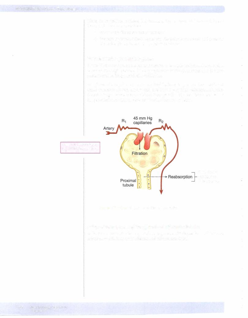
Hydrostatic pressure oftheglomerularcapillaries
PGC: The hydrostatic pressure of the glomerular capillaries is the only force that promotes filtration. Under normal conditions, this is the main factor that deter mines GFR.
Oncotic pressure oftheplasma
TIGC: The oncotic pressure ofthe plasma varies with the concentration ofplasma proteins. Because fluid is filtered but not protein, oncotic pressure, which opposes filtration, increases from the beginning to the end of the glomerular capillaries
Hydrostatic pressure in Bowman's space
PBS: The hydrostatic pressure in Bowman's capsule opposes filtration. Normally, it is low and fairly constant and does not affect the rate of filtration. However, it increases and reduces filtration whenever there is an obstruction downstream, such as a blocked ureter or urethra (postrenal failure).
Protein oroncoticpressure in Bowman's space
TIBS: This represents the protein or oncotic pressure in Bowman's space. Very little if any protein is present, and for all practical purposes this factor can be considered zero.
Normal Values PBS = 8 mm Hg PGC = 45 mm Hg
nBS = 0 mm Hg nGC = 24 mm Hg
Net filtration pressure = PGC - nGC - PBS = 45 - 24 - 8 = 13 mm Hg
To summarize, filtration at the glomeruli depends on starling forces:
•The glomerular capillaries have very high hydrostatic pressures this is why filtration occurs here
•Increasing the glomerular hydrostatic pressure increases GFR
•Decreasing the glomerular hydrostatic pressure decreases GFR
Oncotic pressure opposes GRF i plasma protein i oncotic pressure .J, GFR (no effect on RPF) .J, plasma protein .J, oncotic pressure i GRF (no effect on RPF)
The increased concentration ofprotein (increase oncotic pressure) is carried into the peritubular capillaries and promotes a greater net force ofreabsorption.
Important: If the main driving force for GFR is the hydrostatic pressure, what is the main driving force the reabsorption at the proximal tubule? The force that is driving reabsorption at the proximal tubule is the oncotic pressure in the peritu bular capillaries.
200 MEDICAL

Chapter 1 • Renal Structure and Glomerular Filtration
Filtering Membrane
The membrane ofthe glomerulus consists of3 main structures:
•Capillary endothelial wall with fenestrations that have a magnitude greater than proteins; in addition, the wall is covered with negatively charged compounds
•Glomerular basement membrane made up of a matrix ofextracellular negatively charged proteins and other compounds
•Epithelial cell layer of podocytes next to Bowman's space; the podocytes have foot processes bridged by filtration slit diaphragms
Around the capillaries is the mesangium, containing mesangial cells similar to monocytes.
The capillary wall with its fenestrated endothelium, the basement membrane with hydrated spaces, and the interdigitating foot processes of the podocytes combined with an overalllarge surface area, creates a high hydraulic conductivity (permeable to water and dissolved solutes). Passage oflarge proteins is restricted because ofnegative charge ofthe membrane system.
Inadditionto the net hydraulic force, GFR depends on both the permeability and the surface area ofthe filteringmembrane. The decrease in GFR in most diseased states is due to a reduction in the membrane surface area. This also includes a decrease in the number offunctioning nephrons.
Materials Filtered
The following are easily or freely filtered:
• Major electrolytes: sodium, chloride, potassium, bicarbonate
• Metabolic waste products: Urea Creatinine
• Metabolites: glucose, amino acids, organic acids (ketone bodies)
• Nonnatural substances: inulin, PAH (p-aminohippuric acid)
• Lower-weight proteins and peptides: insulin, myoglobin
The following are not freely filtered:
•Albumin and other plasma proteins
•Lipid-soluble substances transported in the plasma attached to proteins, such as lipid-soluble bilirubin, T4 (thyroxine), other lipid-soluble hor mones; unbound lipid-soluble substances such as free-cortisol are filtered and can appear in the urine
As blood flows though.theglomerular capillary, plasma is filtered, butalbumin is not, so the plasma albumin concentration and oncotic pressure increase.
Fluid Entering Bowman's Capsule
The fluid entering Bowman's space is an ultrafiltrate ofplasma; that is, the filtrate has the same concentration ofdissolved substances as plasma, except proteins.The osmolality ofthe filtrate is 300 mOsm/Kg. The criteria for effective osmolality are the same as those previously statedfor extracellular fluid (section I).
Bridge to Pathology
In nephrotic syndrome, there is marked disruption ofthe filtering membrane. As a result, plasma proteins now
pass through the membrane and are eliminated in the urine. This is typically associated with a non-inflammatory injury to the glomerular membrane system. The most common clinical signs are:
•Marked proteinuria >3.5 gm/day (because of disrupted glomerular membrane system)
•Edema (loss of plasma oncotic pressure)
•Hypoalbuminemia (albumin lost in urine)
•Lipiduria (disrupted membrane system and proteins in urine)
•Hyperlipidemia (increased lipid synthesis in liver)
In nephritic syndrome, there is an inflammatory disruption ofthe
glomerular membrane system. This disruption allows proteins and cells to cross the filtering membrane. The most common clinical signs are:
•Proteinuria <3.5 gm/day (evidence of disrupted membrane)
•Hematuria (disrupted membrane)
•Oliguria (inflammatory infiltrates reduce fluid movement across the membrane)
•Hypertension (inability of kidney to regulate the extracellularvolume)
•Azotemia (inability to filter and excrete urea)
MEDICAL 201
Section VIII • Renal Physiology
Ifa substance is freelyfilteredbythe kidney, the ratioofthe filtrate concentration to plasma concentration TF/P = LO. This means the concentrations in Bowman's space and the plasma is the same.
Filtration Fraction
The following formula for filtration fraction (FF) and the normal values given should be memorized.
FF = fraction ofmaterial entering the kidneythat is filtered and is normally0.20 or 20% for a freelyfiltered substance
FF= fraction ofthe material entering the kidneythatis filtered normally 0.20 or 20% for a freelyfiltered substance
FF = GFR |
GFR 120 mL/min |
RPF |
(renal plasma flow) = 600 mL/min |
RPF = |
120 mL/min = 0.20 or 20%
600mL/min
•FF affects oncotic pressure in the peritubular capillary (nPC). The great er the FF,the higher the oncotic pressure in the peritubular capillaries; that is because FF represents loss ofprotein-free fluid into Bowman's space, thereby increasing the concentration of protein in the plasma.
•IfFF decreases, then nPC decreases
•Only 20% ofthe renal plasma flow is filtered. Every minute 600 ml of plasma enters the kidneys. That is the renal plasma flow.
•20% or 120 mL ofplasma is filtered hence a GFR of 120 mL.
FactorsAffecting FF
Based on the precedingdiscussion,the following should be expected for afferent versus efferent constriction:
Afferent Constriction |
Efferent Constriction |
Glomerular filtration pressure |
i |
GFR |
i |
RPF |
-!. |
FF |
i |
Effects ofSympathetic Nervous System
Stimulation ofthe sympatheticneuronsto the kidneycausesvasoconstriction of the arterioles,buthas a greater effect onthe afferent arteriole. As a consequence:
•RPF decreases
•PGC decreases
•GFR decreases
•FF increases
•PPC decreases
202 MEDICAL

Chapter 1 • Renal Structure and Glomerular Filtration
•nPC increases
•i forces promoting reabsorption in the peritubular capillaries because of a lower peritubular capillary hydrostatic pressure and an increase in plasma oncotic pressure (FF increases)
Effects ofAngiotensin II
Angiotensin II (Ang II) is a vasoconstrictor. It constricts both the afferent and effer ent arterioles, but is has a bigger effect on the efferent arteriole. As a consequence:
•RPF decreases
•PGC increases
•GFR increases
•FF increases
•PPC decreases
•nPC increases
•i forces promoting reabsorption in the peritubular capillaries because of a lower peritubular capillary hydrostatic pressure and an increase in plasma oncotic pressure (FF increases)
During a stress response, there is an increase in both sympathetic input and very high levels of circulating angiotensin II. As a consequence:
•Increased sympathetic tone to the kidneys and very high levels of angio tensin II vasoconstrict both the afferent and the efferent arterioles. Because both arterioles constrict, there is a large drop in the RPF and only a small drop in the GFR.
•The net effect is an increase in FF.
•The increase in FF increase in oncotic pressure increase in the reabsorption in proximal tubules
•Overall, less fluid is filtered and a greater percentage of that fluid is reab sorbed in the proximal tubule, leading to preservation of volume in a volume depleted state
•There is also an increase in ADH due to the low volume state
•Activation of the sympathetic nervous system also directly increases renin release
The net effect of angiotensin II is to preserve GFR in volume-depleted state. In a volume-depleted state, a decrease in GFR is beneficial because less fluid filtered results in less fluid excretion (however, a very large decrease in GFR prevents removal of waste products like creatinine and urea). Angiotensin II prevents a large decrease in GFR.
MEDICAL 203

Sedion VIII • Renal Physiology
Clinical Correlate
A25-year-old man spends a week in the desert. Due to severe dehydration, his volume status is depleted high angiotensin II vasocontriciton of the efferent arterioles an increase in GFR and decrease in RPF an increase in FF more plasma filtered in the glomeruli higher albumin concentration (hence higher oncotic pressure) in the glomerular capillarieshigher oncotic pressure in the peritubular capillaries an increase in peritubular reabsorption. Therefore, an increase in the FF an increase is reabsorption at the proximal tubules.
Adehydrated patient needs to increase reabsorption offluid at the proximal tubules to preserve volume. Angiotension II helps preserve GRF and volume in a volume-depleted state.
Clinical Correlate
What would happen ifyou gave NSAIDs to the 75-year-old man who is hemorrhaging?
During a stress state the increase in sympathetic tone causes vasoconstriction ofthe afferent arterioles. The same stimuli activate
a local production of prostaglandins. Prostaglandins lead to vasodilation of the afferent arterioles, thus modulating the vasoconstriction. Unopposed, the vasoconstriction from the sympathetic nervous system and angiotensin II can lead to a profound reduction in RPF and GFR, which in turn, could cause renal failure. NSAIDs inhibit synthesis of prostaglandins and interfere with these protective effects.
Clinical Correlate
Administration ofAa inhibitors and ARBs
ACE inhibitors and ARBs are used for diabetic nephropathy because they lead to a reduction in glomerular capillary pressure and reduce damage and fibrosis ofthe glomuli (which will delay the need for hemodialysis). They treat hyperfiltration. In most cases, there is a small and transient drop in GFR.
Inhibition of angiotensin II leads to vasodilation ofthe efferent arteriole, which leads to decreased glomerular capillary pressure and decreased GFR. It also leads to increased RPF because of a decrease of resistance to flow. The pressure downstream from the efferent arteriole (peritubular capillary pressure) increases because there is a decreased resistance at the EA.
Use the following guidelines for using ACE inhibitors and ARBs:
•Give ACE inhibitors to patients with nephrotic syndrome and stable chronic renal failure.
•Avoid ACE inhibitors and ARBs in patients with severely compromised GFR (risk of hyperkalemia) and with acute renal failure.
•ACE inhibitors and ARBs may cause a type IV RTA because they block
aldosterone (leading to hyperkalemia); in this case they must both be held. If ACE inliibitors cause hyperkalemia, so will ARBs.
•Switch from ACE inhibitorto ARB in cases with ACE-inhibitor cough, not for hyperkalemia.
•ACE inhibitors and ARBs are contraindicated in bilateral renal artery stenosis, where both kidneys have such low perfusion that GFR is highly dependent on constriction of EE. When the effect of angiotensin II is removed, the result is a significant drop in GFR and acute renal failure.
There is no parasympathetic innervation ofthe kidney.
Although prostaglandins seem to play little role in the normal regulation ofrenal blood flow, they do become important in times of stress. For example, the va soconstriction produced by sympathetic activation is partially countered by the local release ofvasodilating prostaglandins (PGI2 and PGE2). This is thought to help prevent ischemic damage during times ofstress.
204 MEDICAL

Chapter 1 • Renal Structure and Glomerular Filtration
Chapter Summary
•Autoregulatory mechanisms maintain a fairly constant renal blood flow and GFR despite changes in arterial blood pressure.
•Individual nephrons are organized in parallel, but the vascular system of each nephron structure (afferent arteriole, glomerular capillaries, efferent arteriole, and peritubular capillaries) is connected in series.
•The major factor determining GFR is glomerular capillary hydrostatic pressure. The only other important factor in a normal kidney system is the oncotic pressure ofthe plasma proteins. The glomerular membrane system is permeable to all substances dissolved in plasma except proteins and provides a large surface area for filtration. A loss of surface area can be compensated for by an increase in glomerular capillary pressure.
•FF is the fraction of plasma that is filtered at the glomerular capillary. A decrease in flow tends to increase FF.
•Filtered load is the rate at which a substance is filtered into Bowman's space.
For a freely filtered substance it is determined by its plasma concentration and GFR.
•Nephrotic and nephritic syndromes are 2 classic patterns of glomerular disease.
-Nephrotic syndrome characteristically is a noninflammatory injury to the glomerular epithelium or basement membrane, resulting in a large proteinuria but only minimal changes in GFR.
-Nephritic syndrome is an inflammatory response to the glomerular injury, resulting in significantly decreased GFR and red blood cells appearing in the urine.
MEDICAL 205

Solute Transport: 2
Reabsorption and Secretion
SOLUTE TRANSPORT
Transport proteins in the cell membranes ofthe nephron mediate the reabsorp tion and secretion ofsolutes and water transport in the kidneys. Acquired defects in transport proteins are the cause of many kidney diseases.
In addition the transport proteins are important drug targets.
Transport Mechanisms
Simple diffusion
•Net movement represents molecules or ions moving down their electro chemical gradient.
•This doesn't require energy.
Facilitated diffus ion ffacilitated transport)
•A molecule or ion moving across a membrane down its concentration gradient attached to a specific membrane-bound protein.
•This doesn't require energy.
Active transport
•A protein-mediated transport that uses ATP as a source of energy to move a molecule or ion against its electrochemical gradient.
Dynamics of Protein-Mediated Transport
Uniport
•Transporter moves a single molecule or ion as in the uptake of glucose into skeletal muscle or adipose tissue. This is an example of facilitated diffusion.
Symport (cotransport)
•A coupled protein transport of 2 or more solutes in the same direction as in Na-glucose, Na-amino acid transporters.
MEDICAL 207

Section VIIIRenal• Physiology
Antiport (countertransport)
• A coupled protein transport of2 or more solutes in the opposite direction.
Saturation kinetics |
|
• As the concentration ofthe substance initially increases on one side of |
|
|
the membrane, the transport rate increases. |
• Once the transporters become saturated, transport rate is maximal |
|
|
(TM = transport maximum). Rate oftransport is dependent upon: |
|
Concentration ofsolute |
|
Number offunctioning transporters; the onlywayto increase TM is |
• |
to add more protein carriers to the membrane |
Once all the protein carriers are saturated, the solutes are transported |
|
• |
across the membrane at a constant rate. This constant rate is TM. |
There is no TM is simple diffusion. |
|
Chemical specificity
Tobe transported, the substance must have a certain chemical structure. Gener ally, onlythe natural isomer is transported (e.g., D-glucose but not L-glucose).
Competition for carrier
Substancesofsimilarchemicalstructuremaycompeteforthesametransporter.For example, glucoseandgalactose generallycompete forthesame transportprotein.
Primary andsecondary transport
•In primary active transport,ATP is consumed directly by the transport ing protein, (e.g., the Na/K-ATPase pump, or the calcium-dependent ATPase ofthe sarcoplasmic reticulum).
•Secondary active transport depends indirectly on ATP as a source of energy, as in the cotransport ofNa-glucose in the proximal tubule. This process depends on ATP utilized by the Na/K-ATPase pump.
•Glucose moves up a concentration gradient via secondaryactive trans port.
208 MEDICAL


Chapter 2 • Solute Transport: Reabsorption and Secretion
No Net Tubular Modification
•Filtered load = excretion rate
•The amount filtered and amount excreted per unit time are always the same, e.g., inulin, mannitol.
Net Reabsorption
•Filtered load > excretion
•Excretion is always less than filtered load, e.g., glucose, sodium, urea.
•If the substance is completely reabsorbed, the rate of filtration and the rate of reabsorption are equal.
•Excretion rate is 0
•If the substance is partially reabsorbed, excretion is less than filtration.
Net Secretion
•Filtered load < excretion
•Excretion is always greater than filtered load, e.g., PAH, creatinine.
•Creatinine is freely filtered, and a very small amount is secreted.
The following formula is sometimes used to calculate net transport. The sign of the calculated number will indicate the 3 basic categories:
0= no net transport
+ = net reabsorption
-= net secretion
net transport rate = filtered load - excretion rate
= (GFR x Px) - (Ux x V)
Question: Given the following information, calculate the reabsorption rate for glucose.
GFR = 120 mL/min
Plasma glucose = 300 mg/100 mL Urine flow = 2 mL/min
Urine glucose = 10 mg/mL
Answer: 340 mg/min
CLEARANCE
Clearance refers to a theoretical volume of plasma from which a substance is removed over a period oftime. Applying the principles of mass balance above, if a solute has an ER, then it is cleared by the kidney. In other words, ifit is filtered and not fully reabsorbed or is secreted, then it appears in the urine and is thus cleared from the body.
If, on the other hand, it is filtered and then allis reabsorbed, the ERand clearance is zero, and it is not cleared by the kidney.
For example, if the concentration of substance x is 4 molecules per liter and the excretion of x is 4 molecules per minute, the volume of plasma cleared of x is 1 L per minute. If the excretion of x decreases to 2 molecules per minute, the
GFR = glomerular filtration rate units = volume/time, e.g., ml/min
Px = free (not bound to protein) concentration of substance in the plasma
units = amount/volume, e.g., mg/ml
MEDICAL 211
Section VIII • Renal Physiology
volume cleared of x is only 0.5 L per minute. If the concentration ofx decreases to 2 molecules per liter ofplasma and excretion is maintained at 2 molecules per minute, the cleared volume is back to 1 L per minute. These numbers are sum marized in Table VIIl-2- 1 .
Table Vlll-2-1. Example Calculations ofClearance Values
Plasma Concentration |
Excretion Rate |
Volume Cleared |
(molecules/L) |
(molecules/minute) |
(L/minute) , |
4 |
4 |
1.0 |
4 |
2 |
0.5 |
2 |
2 |
1.0 |
Ux = urine concentration ofx
V = urine flow rate
Px = plasma concentration ofx
Thus, the 2 factors that determine clearance are the plasma concentration of the substance and its excretion rate.
Clearance ofx = |
Excretion rate ofx |
Ux X V |
|
Px |
|
Px |
|
The plasma concentration ofthe substance and its urine concentration must be in the same units, which then cancel.
Urine flow (V) is a volume per unit time, and the units of V become the units of clearance.
Clearance is a volume ofplasma cleared of a substance per unittime, mL/min or L/day.
Question: Using the following information, calculate the clearance of x, y, and z.
V = |
2 mL/min |
|
Ux = |
2 mg/mL |
Px = 2 mg/mL |
Uy = |
O mg/mL |
Py = 13.6 mg/mL |
Uz = |
0.5 mglmL |
Pz = 1 mg/mL |
Answer: x = 2 mL/min, y = 0, and z = 1 mL/min
TM TUBULAR REABSORPTION
Glucose
Figure VIII-2-4 graphically represents the dynamics of glucose filtration, reab sorption, and excretion. It is the application ofthe principle of mass balance and clearance discussed above. Manysubstances are reabsorbed via a TM system, and glucose serves as our prototypical example.
212 MEDICAL


Chapter z • Solute Transport: Reabsorption and Secretion
•When the PAH concentration is below the transport maximum, we can use the clearance of PAH to calculate the estimated RPF.
For example, a concentration of0.08 mg/ml means that out ofthe entire 480 mL ofplasma (80% ofthe RPF) thatwas not filtered, there is only38.4mg (0.08 x 480 mL) ofPAH because that is well below the transport maximum for PAH (80 mg/ min), it is all secreted.
Ifthe concentration ofPAH entering the kidneys in the RPF was high and it was past the TM for PAH, you would not be able to secrete it all because the carriers
are all saturated. Some would remain in the plasma ofthe renal vein; the clear ance ofPAH would decrease butyou willalways excrete the 20% ofthe RPF that
was filtered (because it is not reabsorbed). Therefore, as the plasma concentration ofPAH increases, the clearance ofPAH decreases.
For example, ifthe PAH concentration were 80 mg/mL, 20% ofthe RPF would be filtered (always) and excreted. That 20% is cleared (removed from the plasma).
Ofthe remaining 80% ofthe RPF, there willbe 80 mg/mL ofPAH, which means thatin 1 mL there willbe 80 mg ofPAH andyouwillonlybe able to secretethe 80
mg (because that is the TM),you willonly clear 1 mL ofplasma ofthe remaining
RPF. So 479 mL ofplasma out ofthe 600 ml that entered the kidney (the RPF) is not cleared and willbe present in the renal vein.
Therefore, you see a dramatic decrease in clearance (because there is more PAH in the renal vein, meaning it was not cleared).
Remember:
•Ifit is in the renalvein, it was not cleared.
•Ifit is in the urine, it was cleared.
Most ofwhatwas cleared in this casewas clearedbyfiltration, not secretion. Thus, calculating the clearance here is very similar to calculating the GRF, not the RPF.
Regardless ofthe plasma concentration ofPAH, the kidney will always clear the volume filtered (GFR), i.e., the volume of PAH that is filtered (GFR) is cleared from the plasma and excreted in the urine. At low PAH concentrations, as the plasma PAH concentration is increasing, both the secretion rate (SR) and filtered load (FL) are increasing. Thus, there is a very rapid increase in the excretion rate (ER) ofPAH and the slope ofthe line is very steep.
After transport maximum, the SR is constant because all the PAH protein carri ers are saturated. The slope ofthe ER decreases and parallels the FL because now, only the FL is increasing and the SR is constant. The ER still increases, just not as quickly.
The reason why the slope of the line decreases is because the PAH carriers are saturated and the SR has reached transport maximum.
ER = FL + SR
After transport maximum, the SR is constant, but as the plasma concentration increases, the FL increases, therefore, the ER increases and parallels the FL.
PAH clearance at low plasma concentrations is referred to as effective renal plasma flow (ERPF) because some plasma perfused the renal capsule. This flow (about 10%) is not cleared ofPAH. Thus, PAH clearance is only 90% ofthe true renal plasma flow.
MEDICAL 215

Section VIII • Renal Physiology
Renal blood flow = renal plasma flow/1 - HCT
If renal plasma flow is 600 mL/min and Hct is 50%, then renal blood flow is 1200 mL/min.
PAH is transported by a fairly nonspecific organic anion transporter (OAT). Many compounds compete for the carriers. In addition to PAH, some of those compounds include:
•Penicillin
•Furosernide
•Acetazolamide
•Salicylate
Because the organic anions allcompete for the same carriers, elevation of the plasmalevelofone ioninhibits the secretion and clearance ofthe others.
There is a similar transport secretorysystem formanyorganic cations. A slightly different transport mechanism isinvolvedbut, again, the system is fairlynonspe cific. Drugs using this pathway include:
•Atropine
•Morphine
•Procainamide
•Cimetidine
•Amiloride
Note that, because ofcompetitionforthe carrierproteins, the concurrent admin istration oforganic cations can increase the plasma concentration ofboth drugs to much higherlevelsthan when the drugs aregiven alone.
THE RENAL HANDLING OF SOME IMPORTANT SOLUTES
Theillustrations in figure VIII-2-6 representthenettransport ofspecific types of substances for a normal individual on atypicalWestern diet (contains red meat). The dashed lines represent the route followedbythe particular substance. Quan titative aspects are not shown. For example, in B, 20% ofthe substance entering the kidney is filtered and excreted, and the remaining 80% passes through the kidneys andback into the general circulationwithoutprocessing.
216 MEDICAL

Chapter 2 • Solute Transport: Reabsorption and Secretion
Vasculature
Note
These illustrations are meant to show
overall net transport only.
Entire nephron
A |
8 |
c |
A = protein CB = inulin
= potassium, sodium, urea D = glucose, bicarbonate
E = PAH
F = creatinine
D E F
Figure Vlll-2-6. Graphical Representation of Transport
MEDICAL 217


CLINICAL ESTIMATION OF GFR |
3 |
AND PATTERNS OF CLEARANCE |
CLEARANCE AS AN ESTIMATOR OF GFR
Estimates of GFR are used clinically as an index of renal function and to assess the severity and the course of renal disease. A fall in GFR means the disease is progressing, whereas an increase in GFR suggests a recovery. In many cases a fall in GFR may be the first and only clinical sign ofrenal dysfunction. Estimations of GFR rely on the concept of clearance.
Substances having the following characteristics can be used to estimate GFR.
•Stable plasma concentration that is easily measured
•Freely filtered into Bowman's space
•Not reabsorbed, secreted, synthetized, or metabolized by the kidney
Ideal substances include inulin, sucrose, and mannitol. Even though the clear ance of inulin is considered the gold standard for the measurement of GFR, it is not used clinically. Instead clinical estimates of GFR rely on creatinine.
Creatinine is released from skeletal muscle at a constant rate proportional to muscle mass. Muscle mass decreases with age but GFR also normally decreases with age. Creatinine is freely filtered and not reabsorbed by the kidney, though a very small amount is secreted into the proximal tubule.
Creatinine production = creatinine excretion = filtered load ofcreatinine = Per x GFR
Thus, if creatinine production remains constant, a decrease in GFR increases plasma creatinine concentration, while an increase in GFR decreases plasma cre atinine concentration.
Plasma creatinine, however, is not a very sensitive measure of reduced GFR. It only reveals large changes in GFR. As shown in Figure VIII-3-1, a significant re duction of GFR only produces a modest elevation of plasma or serum creatinine concentration.
MEDICAL 219


Chapter 3 • Clinical Estimation of GFR and Patterns of Clearance
lnulin
•The clearance of inulin is independent of the plasma concentration, thus plotting it on the graph produces a line parallel to the X-axis. This is because a rise in the plasma concentration produces a corresponding rise in filtered load and thus a corresponding rise in ER (recall that inu lin is neither secreted nor reabsorbed). In other words, the numerator and denominator of the clearance equation for inulin change in propor tion, leaving the quotient (clearance) unchanged.
•It is always parallel to the x axis, and the point of intersection with the y axis is always GFR.
•If GFR increases, the line shifts upward; likewise, if GFR decreases, the line shifts down.
Glucose
•At low plasma levels, the clearance of glucose is zero because all of the FL is reabsorbed.
•As the plasma levels rise, the FL exceeds the TM in some nephrons and as a result, glucose appears in the urine and thus has a clearance.
•The plasma level at which glucose first appears in the urine is called the plasma (or renal) threshold.
•As the plasma level rises further, the clearance increases and approaches that of inulin. The clearance never equals inulin because some glucose is always reabsorbed.
Creatinine
•Because there is some secretion of creatinine, the clearance is always greater than the clearance of inulin.
•However, because only a small amount is secreted, creatinine clearance parallels inulin clearance and is independent of production rate (excre tion rises as plasma concentration increases).
•Because it is endogenously produced, it is not necessary to infuse it to get a clearance measurement, as has to be done to measure inulin
clearance. Therefore, the clearance of creatinine is the preferred clinical method for determining GFR (see above).
PAH
•At low plasma concentrations, the clearance equals renal plasma flow.
•As the plasma concentration rises, the carriers in some nephrons hit
TM, resulting in some PAH appearing in the renal venous plasma.
•Plasma concentrations above TM reduce the clearance of PAH (described in chapter 2)
•As the plasma level rises further, the clearance approaches but never equals GFR because some PAH is always secreted.
Summary of the highest clearance to the lowest clearance:
PAH > creatinine > inulin > urea > sodium > glucose = albumin
MEDICAL 221
Section VIII • Renal Physiology
V= urine flow rate
uosm = urine osmolarity
Posm = plasma osmolarity
Remember, ifit is in the renal vein, it is not cleared. This could be because it was not filtered (like albumin) or it was filtered and all reabsorbed (like glucose).
FREE WATER CLEARANCE
Free water clearance is the best measure of the balance between solute and water excretion. Its use is to determine whether the kidneys are responding appropri ately to maintain normal plasma osmolality. Free water clearance is how much solute-free water is being excreted; it is as if urine consisted of plasma (with solutes) plus or minus pure water.
•If urine osmolality is 300 mOsm/kg (isotonic urine), free water clear ance is zero.
•If plasma osmolality is too low, urine osmolality should be lower still (positive free water clearance) in order to compensate.
•Positive-free water clearance tends to cause increased plasma osmolality; negative free water clearance causes reduced plasma osmolality.
•CH 0 (+) = hypotonic urine is formed (osmolality <300 mOsm/kg)
•220 (-) = hypertonic urine is formed (osmolality >300 mOsm/kg)
CHzO = V - |
Uosm V |
|
Posm |
Sample Calculation
V= 3.0 mL/min
Uosm = 800 mOsm/L
Posm = 400 mOsm/L
CHzO = -3 mL/min
Conclusion: The kidneys are conserving water; this is appropriate compensation for the excessive plasma osmolarity in this patient.
SODIUM AND UREA CLEARANCE
Sodium
•Sodium always appears in the urine, thus sodium always has a positive clearance.
•To clear a volume of plasma the substance in that volume must be excreted.
•Reabsorption of a filtered substance decreases the volume cleared.
•The higher the percentage of the filtered load reabsorbed, the smaller the volume cleared.
222 MEDICAL

Chapter 3 • Clinical Estimation of GFR and Patterns of Clearance
•Since almost the entire filtered load of sodium is reabsorbed its clear ance is just above zero. Aldosterone, by increasing the reabsorption of sodium, decreases its clearance. Atrial natriuretic factor increases the clearance of sodium by causing a sodium diuresis.
Urea
•Urea is freely filtered but partially reabsorbed.
•Because some urea is always present in the urine, you always clear a por
tion of the 120 mL/min filtered into Bowman's space.
•Since urea tends to follow the water and excretion is flow dependent, a diuresis increases urea clearance and an antidiuresis decreases urea clearance.
•ADH increases reabsorption of urea in the medullary collecting duct increase in BUN decrease in clearance if the plasma concentration is increasing in the renal venous plasma, less is cleared from the plasma
•With a small volume of concentrated urine, the concentration of urea is relatively high, but the excretion is less than in a diuresis that has a much lower concentration of urea. It is the large volume in the diuresis that increases the urea excretion and clearance.
Chapter Summary
• Substances that do not appear in the urine have a clearance of zero.
•Substances freely filtered and partially reabsorbed have a clearance less than the GFR.
•Substances freely filtered with no net transport like inulin have a clearance equal to GFR.
•Substances freely filtered and with net secretion like creatinine have a clearance greater than the GFR. Because only a very small amount of creatinine is secreted, it always has a clearance slightly greater than GFR. However, because the amount secreted is small, and it is a solute naturally produced in the body, estimating its clearance serves as an indicator of GFR.
•Substances freely filtered with the remainder entering the kidney completely secreted, have a clearance equal to renal plasma flow.
•Free water clearance is a volume excreted without solute. It is used to evaluate ifthe renal balance of solute and water excretion is appropriate.
•There is an obligatory solute loss from the kidney. The primary solutes lost are sodium and urea.
MEDICAL 223


Section VIII • Renal Physiology
ProximalTubule (Pl) Changes
Sodium
•Approximately two-thirds of the filtered sodium is reabsorbed in the proximal tubule (PT). The basolateral Na+__ K+ ATPase creates the gra dient for Na+ entry into the cell and its removal from the cell back into the bloodstream.
•Although it can be modified some, the PT captures two-thirds of the filtered sodium (referred to as glomerulotubular balance). Recapturing two-thirds of the sodium helps protect extracellular volume despite any changes that may occur in GFR.
•Catecholamine and angiotensin II stimulate the basolateral ATPase and thus enhance the fraction of sodium reabsorbed in the proximal tubule.
Bridge to Pharmacology
SGLT-2 blocker canagliflozin inhibits proximal tubule reabsorption of glucose and is used to treattype 2 DM.
Waterand electrolytes
•About two-thirds of the filtered H20, K+ and almost two-thirds of the filtered c1follow the sodium (leaky system to these substances), and the osmolality at the end of the proximal tubule remains close to 300 mOsm/kg (isosmotic reabsorption) . The chloride concentration rises slightly through the proximal tubule because of the large percentage of bicarbonate reabsorbed here.
•Therefore, at the end of the proximal tubule, osmolality and the concen trations of Na+ and K+ have not changed significantly from plasma, but only one-third of the amount originally filtered remains.
Metabolites
•Normally, all of the filtered glucose is reabsorbed in the PT via second ary active transport linked to sodium. This transporter is termed the sodium glucose-linked transporter (SGLT) and type 2 (SGLT-2) is the predominant form in the kidney.
•In addition, all proteins, peptides, amino acids, and ketone bodies are reabsorbed here via secondary active transport (requires luminal sodi um, linked to sodium reabsorption).
•Therefore, the concentration of the above should be zero in the tubular fluid leaving the proximal tubule (clearance is zero).
Bicarbonate
About 80% of the filtered bicarbonate is reabsorbed here. The mechanism for this reabsorption is:
•Bicarbonate combines with free H+ in lumen and is converted into C02 and H20, catalyzed by the luminal carbonic anhydrase enzyme (CA). H+ is pumped into the lumen in exchange with sodium (antiporter). Although not pictured, there is a H+--ATPase on the luminal membrane that contributes to pumping H+ into the lumen.
•C02, being very soluble, crosses the luminal membrane where it com bines with water to reform H+ and bicarbonate (note CA in the cell). The H+ is then pumped back into the lumen, while bicarbonate exits the baslolateral membrane to complete its reabsorption.
226 MEDICAL

•Because of this mechanism, bicarbonate reabsorption is dependent upon tt+ secretion and the activity of CA.
•The most important factor for tt+ secretion is the concentration of tt+ in the cell. Thus, tt+ secretion and bicarbonate reabsorption are increased during an acidosis and they decrease with an alkalosis.
•Angiotensin II stimulates the Na+--tt+ antiporter. Thus, in volume depleted states, the amount of bicarbonate reabsorption in the PT increases. This is thought to be the mechanism preventing bicarbonate loss when a patient develops a contraction alkalosis.
The small amount of bicarbonate that leaves the proximal tubule is normally re absorbed in subsequent segments.
Urate (uric acid)
The details of the renal handling of urate are too complex for the scope of this book. In short:
•Urate is formed by the breakdown of nucleotides
•Xanthine oxidase is the enzyme that catalyzes the final reaction to form urate.
•About 90% of the filtered urate is reabsorbed by the proximal tubule.
•If the FL of urate is high enough and the luminal pH is low, then more of the urate exists as uric acid, which can precipitate and form a kidney stone.
-This is not the most common type of kidney stone, but it can occur in patients with gout.
Secretion
The proximal tubule is where many organic anions and cations are secreted and cleared from the circulation including PAH, penicillin, atropine, and morphine.
Energyrequirements
Notice that all of the active processes illustrated in Figure VIIl-4-1 are powered by the Na/K- ATPase primary active pump. This pump is located in the proximal tubule basal and basolateral borders and is directly or indirectly responsible for most of the water and electrolyte reabsorption in the nephron. It thus represents the most energy-demanding process ofthe nephron.
Figure VIII-4-2 depicts the ratio of the concentration of the substance in the proximal tubular fluid (TF) to the concentration in the plasma (P), beginning in Bowman's space through the end ofthe PT.
Chapter If • RegionalTransport
Bridge to Pharmacology
The primary site of action for carbonic anhydrase inhibitors is the PT. Blocking CA reduces bicarbonate reabsorption and the activity ofthe Na+--w exchanger.
Clinical Correlate
An 85-year-old woman presents to the emergency room with confusion. She is on hydrochlorothiazide. Her bicarbonate is 34 mEq/L (normal = 24 mEq/L). What is the cause ofthe metabolic alkalosis?
This is what is called a contraction alkalosis. The thiazide diuretic is causing a decrease in the intravascular volume. You can see the same in sweating in the desert or vomiting (not diarrhea because you lose bicarbonate in the stools and get a metabolic acidosis). The low volume state increases renin secretion leading to high angiotensin II. Angiotensin II activates the sodium/ hydrogen exchanger . increasing the reabsorption of bicarbonate maintaining the metabolic alkalosis.
Clinical Correlate
Administration of a solute that is freely filtered but not reabsorbed (mannitol) and/or reducing/preventing the normal reabsorption of a solute results in the osmotic pull ofwater into the lumen and diuresis occurs. From the standpoint ofthe PT, glucose exceeding the TM is an important example.
MEDICAL 227


Section VIII • Renal Physiology
COLLECTING DUCT
The collecting duct (DC) is composed of principal cells and intercalated cells, both ofwhich are illustrated in figure VIIl-4-6.
Bridge to Pharmacology
Potassium sparing diuretics work by blocking ENaC, e.g., amiloride, or by blocking aldosterone receptors, e.g., spironolactone, or the production of aldosterone, e.g., blockers ofthe RAAS system. Because sodium reabsorption is reduced, potassium secretion is diminished.
Bridge to Pathology
Liddle's syndrome is a genetic disorder resulting in a gain offunction of
ENaC channels in the CD. This results in enhanced sodium reabsorption and potassium secretion. Patients are hypertensive, hypokalemic, and alkalotic.
Principal Cells
•The luminal membrane of principal cells contains sodium channels, commonly referred to as epithelial Na+ channels (ENaC). Because of these channels, sodium follows its electrochemical gradient (created by the basolateral Na+--K+ ATPase) into the cell.
•Some chloride does not follow the sodium, thus the reabsorption of sodium produces a negative luminal potential. This negative luminal potential causes potassium secretion.
•Thus, the reabsorption of sodium and secretion of potassium are linked.
•Mineralcorticoids such as aldosterone exert an important effect on these cells. Activation of the mineralcorticoid receptor increases the number of luminal ENaC channels, increases their open time, and induces syn thesis and trafficking of the basolateral Na+--K+ ATPase. The net effect is increased sodium reabsorption and potassium secretion.
•Although not illustrated on the slide, principal cells express aquaporins, which are regulated by anti-diuretic hormone (ADH), also known as arginine vasopressin (AVP). ADH acts on V2 (Gs-cAMP) receptors to cause insertion of aquaporins, which in turn, causes water (and urea) reabsorption.
Intercalated Cells
•Intercalated cells are intimately involved in acid-base regulation. The amount of fixed acid generated by an individual is mainly determined by diet. A high percentage of animal protein in the diet generates more fixed acid than a vegetarian-based diet.
•The luminal membrane contains a tt+--ATPase, which pumps tt+ into the lumen. Although free tt+ is pumped into the lumen, luminal pH can only go so low before it causes damage to cells, and thus most of the tt+ is eliminated from the body via buffers, phosphate and ammonia being the two most common.
•Monoprotonated phosphate is freely filtered at the glomerulus. About 80% is reabsorbed in the PT and another 10% is reabsorbed in the distal tubule. The remaining phosphate serves as a buffer for the secreted tt+. The tt+ pumped into the lumen binds to phosphate to form diproton ated phosphate, which is poorly reabsorbed, thus eliminating tt+ from the body. Phosphate is the primary titratable acid.
•In addition, the tt+ pumped into the lumen can combine with ammo nia to form ammonium, which is not reabsorbed and is thus excreted. Ammonia is produced by the catabolism of glutamine and this occurs in cells ofthe PT.
•For every tt+ excreted by the above buffers, bicarbonate is added to the body (new bicarbonate).
•Aldosterone stimulates the tt+--ATPase of intercalated cells. Thus, excess aldosterone results in a metabolic alkalosis.
232 MEDICAL


Section VIII • Renal Physiology
Causes:
•Autoimmune disorders
•Sjogren's syndrome
•Autoimmune hepatitis/primary biliary cirrhosis
•Systemic lupus erythematosus (may be hyperkalemic)
•Rheumatoid arthritis
•Ifosfamide
•Amphotericin B
•Lithium carbonate
•Toluene inhalation
•Hypercalciuric conditions (leading to precipitation of calcium stones in the renal pelvis or ureters)
•Hyperparathyroidism
•Vitamin D intoxication
•Sarcoidosis
•Idiopathic hypercalciuria
Renal TubularAcidosis Type IV: Hypoaldosterone States
•Inability to secrete potassium leading to hyperkalemia
•Decreased secretion of protons leading to metabolic acidosis
Causes:
•Diabetic nephropathy, due to low renin secretion with loss of kidney function
•Any drug that inhibits the RAAS system, such as ACE-inhibitors, ARBs, spironolactone, and aliskiren
•Trimethoprim
•Addison's disease due to loss of aldosterone secretion from the adrenal cortex
DISORDERS OF POTASSIUM HOMEOSTASIS
Potassium Balance
To keep the body amount constant, excretion of potassium must match dietary intake, and the kidneys regulate potassium excretion. A small percentage of in gested potassium is lost in the stool but this is not a major regulatory route under normal conditions.
234 MEDICAL


Section VIII • Renal Physiology
Intrarenal
In this condition, tubular damage occurs resulting in tubular dysfunction. Toxins, interstitial nephritis, ischemia, rhabdomyolysis, and sepsis are factors that could cause acute tubular necrosis and intrarenal failure. Characteristic signs are:
• Increased FeNa+: Tubules are damaged and thus unable to reabsorb Na+
• Casts/cells in the urine: Damages cells are sloughed offinto the tubule
• Low plasma BUN:Cr: Tubular damage prevents reabsorption of urea
Postrenal
This condition is caused obstruction of fluid outflow from the kidney, e.g., renal calculi, enlarged prostate.
•Early: Characteristics are similar to prerenal, i.e., reduced FeNa+, elevated plasma BUN:Cr
•Late: Build-up of pressure results in tubular damage, resulting in charac teristics of intrarenal failure, i.e., marked increase in FeNa+, low plasma BUN:Cr
Chronic Renal Failure
Althoughnephronsoftenrecoverfromthe sloughingofthe tubular epithelialcellsin acute renalfailure,inchronic renalfailurethere is an irreversibleloss ofnephrons. In order to compensate, the remaining nephrons have an increased glomerular capil lary pressure and hyperfiltration. One way oflooking at this is a "hypertension'' at the level ofthe nephron; the hyperfiltration combined with the increased workload promotes further injuryleadingto fibrosis, scarring, andloss ofadditionalnephrons.
Looking back at the function of the kidney and how it regulates a variety of physiologic variables, many of the consequences of chronic renal failure are pre dictable. These include:
•Inability to excrete waste products leads to a rise in plasma BUN and creatinine.
•Inability to regulate sodium and water: This can lead to hyponatremia, volume overload, and thus edema. In addition, patients are susceptible to rapid development of hypernatremia and volume depletion following vomiting and diarrhea.
•Inability to regulate potassium excretion leads to hyperkalernia.
•Inability to excrete fixed acids leads to metabolic acidosis (elevated anion gap-see next chapter)
•Inability to excrete phosphate leads to hyperphosphatemia. This hyperphosphaternia reduces plasma calcium, causing a rise in parathy roid hormone (PTH-secondary hyperparathyroidism) resulting in increased bone resorption (renal osteodystrophy).
•Inability to hydroxylate (1-alpha hydroxylase enzyme is in the kidney) 25,0H-cholecalciferol decreases circulating levels ofactive vitamin D, which contributes to the hypocalcemia.
•Inability to secrete erythropoietin results in anemia.
The most common cause ofchronic renal failure is the nephropathy produced by diabetes. The second most common cause is hypertension.
238 MEDICAL

Chapter 4 • Regional Transport
Chapter Summary
•In the proximal tubule, two-thirds of the water and electrolytes are reabsorbed, along with almost all the organic molecules filtered. An exception is urea. Because equal amounts of solutes and fluid are reabsorbed, the fluid remains isotonic.
•The loop of Henle, acting as a countercurrent multiplier, creates an
osmotic gradient in the medullary interstitium, with the tip reaching a maximum of 1200 mOsm/kg. This value determines the maximum osmolality ofthe urine.
•In the early distal tubule, additional electrolytes are reabsorbed and
the osmolality decreases further. In the collecting duct, aldosterone regulates the final electrolyte modifications and ADH the finalosmolality.
•Net acid lost in the urine is via phosphate and ammonium. Renal tubular acidosis is a metabolic acidosis caused by a failure of nephron cellular processes. Proximal failure is type II, distal failure (distal tubule/ collecting duct) is type I, and type IV is hypoaldosterone.
•Hyperkalemia can be caused either by a transcellular shift in potassium (ICF to ECF) or a failure ofthe kidney to excrete adequate amounts of potassium.
•Hypokalemia can be caused by a transcellular shift in potassium (ECFto
ICF) or to excess loss via the GI tract or kidney.
•Hyperand hypokalemia affect the excitability of nerves and cardiac muscle.
•Acute renal failure (disease) is a sudden loss of renal function (GFR) and is reversible.
•Chronic renal failure (disease) is a loss of functioning nephrons and is generally not reversible. Diabetes is the major cause of chronic renal failure followed by hypertension.
M EDICAL 239


SECTION
Acid-Base
Disturbances


Section IX • Acid-Base Disturbances
Question 2: What is the cause ofthe osis?
To answer this, one looks at bicarbonate next. In the section below, we will go into more detail.
Question 3: Was there compensation?
A calculation must be performed to answer this final question, and this will be covered in detail below. However, bear in mind the following:
•For respiratory disturbances, the kidneys alter total bicarbonate; whether or not compensation has occurred is based upon the patient's measured bicarbonate versus a calculated value of bicarbonate.
•The respiratory system responds quickly and it is important to determine if it has responded appropriately; respiratory compensation compares the patient's measured PC02 versus a calculated (predicted) value.
THE 4 PRIMARY DISTURBANCES
There are 4 primary acid-base disturbances, each of which results in an altered concentration of H+. The basic deviations from normal can be an acidosis (excess H+) or an alkalosis (deficiency of H+), either ofwhich may be caused by a respiratory or metabolic problem.
•Respiratory acidosis: too much C02
•Metabolic acidosis: addition of H+ (not of C02 origin) and/or loss of bicarbonate from the body
•Respiratory alkalosis: not enough C02
•Metabolic alkalosis: loss of H+ (not of C02 origin) and/or addition of base to the body
Normal systemic arterial values are as follows:
pH = 7.4 HC03- = 24 mEq/L PC02 = 40 mm Hg
Follow the Bicarbonate Trail
Question 2 asks for the cause of the osis. To answer this, look at the bicarbon ate concentration and remember the basic C02-bicarbonate reaction, applying mass action. The table below shows the 4 primary disturbances with the resultant bicarbonate changes.
244 MEDICAL


Chapter 1 • Acid-Base Disturbances
For example, a patient has a respiratoryacidosis (determined bysteps 1 and 2) with PaC02 60 mm Hg, which is 20 mm Hggreater than the normal of40 mm Hg.
If acute, then bicarbonate willbe -26 (20 x 0.1 = 2; 24 + 2 = 26). Ifchronic, then bicarbonate willbe -31 (20 x 0.35 = 24 + 7 =31).
MetabolicAcidosis
The patient's PaC02 should fall to a level that is ±2 mm Hg ofthe value computed by Winter's equation:
PaC02 = (1.5 x HC03-) + 8
•If the patient's PaC02 is within 2, then the patient has metabolic acido sis with respiratorycompensation.
•If it is higher than 2, then the respiratory response is inadequate and the patient has metabolic and respiratory acidosis.
•If the patient's PaC02 is too low, then the patient has a metabolic acido sis with arespiratoryalkalosis.
For example, a patient has a metabolic acidosis with a HC03of 10 mEq/L and a PaC02 of 23 mm Hg. Expected PaC02 is (1.5 x 10) + 8 = 23 mm Hg, which is what the patient has, thus respiratory compensation is adequate.
RespiratoryAlkalosis
Again, it is important to distinguish between acute (uncompensated) and chronic (compensated) respiratory disturbances.
• If acute,there is a 0.2 mEq/L fall in HC03for every 1 mm Hg decrease in PaC02 (1:0.2
• If chronic, there is a 0.5 mEq/L fall in HC03for every 1 mm Hg decrease in PaC02 (1:0.5 ratio).
For example, a patient has a respiratory alkalosis (determined by steps 1 and 2)
with a PaC02 of25 mm Hg, which is 15 mm Hgless than the normal of40 mm Hg. If acute, then bicarbonate willbe about 21 (15 x 0.2 = 3; 24 - 3 = 21), but if chronic it willbe around 16 (15 x 0.5 = 7.5; 24 - 7.5 = 16.5).
MetabolicAlkalosis
The patient's PaC02 should rise to a level that is ± 2 the value computed by the following equation (not to exceed 55 mm Hg):
Expected PaC02 = (0.7 x rise in HC03-) + 40
•Ifthe patient's PaC02 is within 2, the patient has metabolic alkalosis with respiratory compensation.
•If it is higher than 2, then the patient has metabolic alkalosis and respi ratoryacidosis.
•If the patient's PaC02 is too low, then the patient has a metabolic and respiratoryalkalosis.
MEDICAL 249


Chapter 1 • Acid-Base Disturbances
Answers
Example 1
What is the osis? pH is low, so acidosis.
Cause ofthe osis? HC03is low, so metabolic acidosis.
Compensation? Use Winter's to compute predicted PC02: (14 x 1.5) + 8 = 29. Patient's is 30, which is within 2, thus this is a metabolicacidosiswith respiratory compensation.
Example 2
What is the osis? pH is high, so alkalosis.
Cause ofthe osis? HC03- is low, so respiratory alkalosis.
Compensation? Must determine ifacute (uncompensated) or chronic (compen sated). PC02 is 20 below normal, thus acute: 20 x 0.2 = 4, so HC03- will be around 20 (24 - 4). If chronic, it would be 20 x 0.5 = 10, so HC03- would be
around 14 (24 - 10). The measured equals the predicted acute, thus this is an acute respiratory alkalosis.
Example 3
What is the osis? pH is low, so acidosis.
Cause ofthe osis? HC03- is high, so respiratory acidosis.
Compensation? Must determine ifacute (uncompensated) or chronic (compen sated). PC02 is 40 greater than normal, thus acute: 40 x 0.1 = 4, so HC03- will be around 28 (24 + 4). If chronic, it will be 40 x 0.35 = 14, so HC03- will be around
38 (24 + 14). The measured is much closer to the predicted acute, thus this is an acute respiratory acidosis.
Example 4
What is the osis? pH is high, so alkalosis.
Cause ofthe osis? HC03- is high, so metabolic alkalosis.
Compensation? The respiratory compensation is to reduce ventilation, thereby increasing PaC02. Thus, we need to compute what PaC02 should be in a patient with this acid-base disorder. Calculation: HC03- is 20 greater than normal of 24, 20 x 0.7 = 14, thus PaC02 should be 14 mm Hg greater than the normal of 40,
thus 40 + 14 = 54 (predicted PaC02). Patient's is 52, which is within 2, so this is a metabolic alkalosis with respiratory compensation.
Example 5
No pH is given, but we can still figure out the acid-base disorder. HC03- is low, so this must be a metabolic acidosis or respiratory alkalosis (review "follow the
bicarbonate trail" above). PaC02 is well above normal, ruling out a respiratory alkalosis, thus there is a mixed metabolic and respiratory acidosis. Note that
PaC02 and HC03- went in opposite directions, so we know there is a mixed disturbance. The low HCo3- indicates the metabolic acidosis, while the high PaC02 indicates respiratory acidosis. If this were simply a respiratory acidosis, HC03- would be elevated, not reduced.
MEDICAL 251
Section IX • Acid-Base Disturbances
I' 6' 0
GRAPHICAL REPRESENTATION (DAVENPORT PLOT)
The Davenport plot (figure IX-1 -4, panels A-D) provides a graphical representation ofthe preceding discussion. The pH is on the X-axis, and HC03- is on the Y-axis.
At pH 7.4 and HC03- 24, PaC02 is 40 (panel A). These represent the normal values indicated above. Knowing either of the 2 measurements allows one to cal culate the third, using the Henderson-Hasselbalch equation.
pH pK + log [HC03-J |
pK |
6.10 |
[HC03-J in mmol/L |
||
= |
aPC02 |
a |
|
0.0301 |
PC02 in mm Hg |
|
|
|
= |
|
|
Let's now walkthrough the 3 questions and see the correspondingvalues on the
Davenport plot.
Question 1: What is the osis?
Splitting the graph at pH 7.4, anything to the left represents an acidosis, while anything to the right represents an alkalosis (Figure X- 1 -4, panel B)
|
|
|
|
|
|
|
A |
_0J- |
|
Acidosis |
|
B |
|
|
|
|
|
|
|
|
|
|
|||||
30 |
|
|
|
|
|
|
|
|
..._ |
30 |
|
Alkalosis |
|
|
|
|
|
|
|
|
|
|
|||||
|
|
|
|
|
|
|
|
I' |
|
|
|
||
|
|
|
|
|
|
|
|
UJ |
25 |
|
|
|
|
|
|
|
|
|
|
|
|
0 |
|
|
|
||
25 |
- - - - - - - |
- .... |
|
|
|
|
|
E |
|
'I |
|
||
|
|
|
|
|
0"' |
|
|
||||||
'-. |
24 |
, ,PCO |
|
|
|
|
|
20 |
|
|
|
||
20 |
|
= 40 |
mmHg |
|
|
|
|||||||
|
I1 |
|
2 |
|
|
|
|
7.3 |
7.4 |
7.5 |
|||
|
|
|
|
|
|
|
|
|
|||||
|
|
|
|
|
|
|
|
|
|
|
|||
|
|
7.3 |
7.4 |
7.5 |
|
|
|
|
|
|
|||
|
|
|
|
|
|
|
|
|
pH |
|
|||
|
|
|
pH |
|
|
|
|
|
|
|
|
|
|
|
|
|
|
|
|
|
|
|
|
|
|
|
|
|
|
Figure IX-1 -4a |
|
|
|
|
|
|
Figure IX-1-4b |
||||
252 MEDICAL


SECTION
Endocrinology


|
|
|
|
Chapter 1 • GeneralAspects of the Endocrine System |
|
|
|
H H |
e.g., Cortisol |
|
|
|
Bound |
|
Free |
T4 (and T3) |
|
|
|
Estrogens |
|
||
|
hormone |
|
hormone |
testosterone |
|
|
Total HormoneActive Hormonein Blood = BoundFree Hormone+ Free |
|
|||
Figure X-1 -2. |
|
= |
|
||
|
|
|
Transport of Lipid-Soluble Hormones |
||
Role ofthe liver
If the liver changes its production and release of binding proteins, the circulating level of bound hormone will change. However, under most conditions the level of free hormone will remain constant.
Modulation
Liver dysfunction and androgens can decrease and estrogens can increase the circulating level of binding proteins. For example, a rise in circulating estrogen causes the release of more binding protein by the liver, which binds more free hormone. The transient decrease in free hormone reduces negative feedback to the hormone-secreting tissue. The increased secretion of free hormone quickly returns the plasma free hormone to normal.
This explains why during pregnancy and other states with a rise in estrogen levels:
•Total plasma lipid-soluble hormone increases.
•Free plasma hormone remains constant at a normal level; thus, the indi vidual does not show signs of hyperfunction.
Hormone Receptors
Hormone specificity
A hormone affects only cells that possess receptors specific to that particular hormone.
For example, both adrenocorticotropic hormone (ACTH) and luteinizing hor mone (LH) increase the secretion of steroid hormones. However, ACTH does so only in the adrenal cortex and LH only in gonadal tissue.
Hormone activity
Under normal conditions, receptors are not saturated; that is, extra receptors exist. Therefore:
• Normally, the number of hormone receptors is not rate-limiting for hormone action.
• The plasma concentration offree hormone is usually indicative of activity.
MEDICAL 261
Section X • Endocrinology
Resistance to hormone action
•Abnormalities in receptors or events distal to the ligand-receptor interac tion, often due to chronic elevation of circulating hormone (e.g., type II diabetes) or drug therapy.
•Under these conditions receptors are often saturated.
•Reduction of hormone levels often produces some recovery in sensitivity.
•The clinical presentation is often one of normal or elevated hormone levels but with reduced or absent peripheral manifestations of the hor mone and a failure of replacement therapy to correct the problem.
Permissive action
A phenomenon in which one type of hormone must be present before another hormone can act; for example, cortisol must be present for glucagon to carry out gluconeogenesis and prevent hypoglycemia.
Measurement ofHormone Levels
Plasma analysis
•Provides information at the time of sampling only and may not reflect the overall secretion rate
•When hormone secretion is episodic, single sampling may reflect peaks (erroneous hyperfunction) or nadirs (erroneous hypofunction). Pulsatile secretion, diurnal and cyclic variation, age, sleep entrainment, and hor mone antagonism must all be considered in evaluating circulating levels.
Growth hormone is secreted in pulses and mainly at night. This is not reflected in a fasting morning sample. However, growth hor mone stimulates the secretion of IGF-I which circulates attached to protein and has a long half-life (20 hours). Plasma IGF-I measured at any time during the day is usually a good index of overall growth hormone secretion.
Thyroid is a fairly constant system and T4 has a half-life of about 6-7 days. Thus, a random measurement of total T4 is usually a good estimate of daily plasma levels.
Urine analysis
•Restricted to the measurement of catecholamines, steroid hormones, and water-soluble hormones such as hCG and LH.
•A distinct advantage of urine analysis is that it provides an integrated sample.
A "24-hour urine free cortisol" is often necessary to pick up a low-level Cushing's syndrome and to eliminate the highs and lows of the normal circadian rhythm.
262 MEDICAL

Chapter 1 • GeneralAspects of the Endocrine System
DISORDERS OF THE ENDOCRINE SYSTEM
Hypofunction
•Can be caused by autoimmune disease (e.g., type I diabetes, hypothy roidism, primary adrenal insufficiency, gonadal failure), tumors, hemor rhage, infection, damage byneoplasms
•Evaluated by a stimulation test
-Hypothalamic hormones test anterior pituitary reserve
-Injection ofthe pituitary trophic hormone (e.g., ACTH) tests target gland reserve.
-Failure of growth hormone release after arginine injection
Hyperfunction
•Causedbyhormone-secreting tumors, hyperplasia, autoimmune stimu lation, ectopicallyproduced peptide hormones (e.g., ACTH, ADH)
•Evaluated by a suppression test
-Failure ofglucose to suppress growth hormone diagnostic for acro megaly
-Failure ofdexamethasone (low dose) to suppress cortisol diagnostic for hypercortisolism
-Multiple endocrine neoplasia (MEN) represents a group ofinher itable syndromes characterized by multiple benign or malignant tumors.
0MEN 1: hyperp,arathyroidism, endocrine pancreas, and pituitary edenomas
0MEN 2A: medullarycarcinoma ofthe thyroid, pheochromocytoma, hyperparathyroidism
0MEN 2B: medullary carcinoma ofthe thyroid, pheochromocytoma, hyperparathyroidism typically absent.
Gland Structure and Size
•When an endocrine gland does not receive its normal stimulus, it gener ally undergoes a reversible atrophy.
-Long-term high doses of glucocorticoids suppress the ACTH-adrenal axis. Withdrawal oftherapy can require up to a year for complete recovery.
•Overstimulation ofendocrine tissue can cause cell proliferation or hypertrophy in addition to hormone overproduction.
-In Graves' disease, overstimulation ofthe thyroid tissues causes cell proliferation and this polyclonal expansion creates a goiter in addi tion to hyperthyroidism.
•Tumors, which are generallymonoclonal expansions, may also cre ate a hyperfunction. Others produce little ifany hormone but are still disease-producing because of the compressive (mass) effect ofthe additional tissue.
MEDICAL 263
Section X • Endocrinology
Chapter Summary
•Lipid-soluble endocrine hormones: Receptors are inside cells. Because they must be synthesized as needed and must generate new proteins to carry out their actions, they represent slow-acting systems. The total plasma level
does not necessarily provide an index of activity because most is bound. It is the free hormone that determines activity. 
•Water-soluble hormones: Receptors are on the membrane surface. Second messengers carry out intracellular action. Because they are stored in vesicles and need only modify proteins to carry out their actions, they are fast-acting systems.
•Normally, receptors are not saturated. It is the plasma level offree hormone that determines activity.
•Tissue resistance elevates plasma hormone levels but reduces peripheral effects.
•Plasma hormonal analysis may not reflect overall secretion rate.
•Urine analysis provides an integrated sample for catecholamines and some steroid hormones.
•Hypofunction is evaluated by a stimulation test.
•Hyperfunction is evaluated by a suppression test.
•Lack of glandular stimulation causes a reversible atrophy.
•Overstimulation of a gland can cause hypertrophy or hyperplasia.
264 MEDICAL

Hypothalamic-Anterior 2
Pituitary System
HYPOTHALAMIC-ANTERIOR PITUITARY AXIS
•The hormones in this system are all water-soluble.
•The hypothalamic hormones are synthesized in the neuron cell body, packaged in vesicles, and transported down the axon to be stored and released from the nerve terminals.
•Pituitary is located in the bony sella turcica atthebase ofthe skull. It hangs from the hypothalamus by a stalk (the infundibulum) and is controlled by the hypothalamus. The dura membrane (diaphragm sellae) separates it from and prevents cerebrospinal fluid from entering the sella turcica.
•Optic chiasm is 5-10 mm above this diaphragm.
•In the hypothalamic-anterior pituitary system, hormonal release is mainly pulsatile. A possible exception is the thyroid system.
•The pulsatile release of GnRH prevents downregulation ofits receptors on the gonadotrophs of the anterior pituitary. A constant infusion of GnRH will cause a decrease in the release ofboth LH and FSH.
The hypothalamic-anterior pituitary system is summarized in Figure X-2-1.
Arcuate n.(dopamine, GHRH)
Paraventricular (CRH, TRH)
al
00
Figure X-2-1 . Hypothalamic-Anterior Pituitary Axis
MEDICAL |
265 |
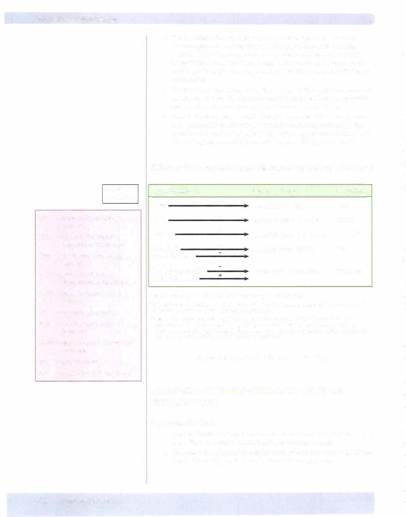

Chapter 2 • Hypothalamic-Anterior Pituitary System
• Isolated deficiency:
Growth hormone: sporadic or familial
Gonadotropins: Kallman syndrome - (tertiary) defective GnRH syn thesis; J, LH J, FSH J, sex steroids
ACTH, TSH, and prolactin extremely rare deficiencies, usually a sign ofpanhypopituitarism
•Craniopharyngioma is the most common tumor affecting the hypotha lamic-pituitary system in children (pituitary adenomas rare).
•Although one would predict the trophic hormones to be low, this is not often the case. Typically, they are in the normal range, but their level is inadequate to stimulate peripheral glands adequately.
•From an academic perspective, stimulation tests include: GnRH --7 LH, FSH; TRH --7 TSH, prolactin; insulin infusion --7 GH, ACTH
Sheehan syndrome
The pituitary in pregnancy is enlarged and therefore more vulnerable to infarc tion. Sometimes when delivery is associated with severe blood loss, the ensuing shockcauses arteriolar spasm in thepituitarywith subsequent ischernic necrosis. Some degree ofhypopituitarism has been reportedin 32% ofwomenwith severe postpartum hemorrhage. Symptoms vary, depending on the extent and location ofpituitary damage, but failure to resume normal menstrual cycles after weaning is a strong sign ofpotential pituitarydamage.
PituitaryAdenomas
•Most common cause of hypothalamic-pituitary dysfunction
•Microadenomas (< 1 cm. dia.): characterized by hormonal excess, no panhypopituitarism, treatable), e.g., ACTH (Cushing disease)
•Macroadenomas (> 1 cm. dia): mass effect, larger tumors with suprasel lar extension, associated with panhypopituitarism and visual loss
•Hypogonadism is the most common manifestation.
•Usually benign and can autonomously secrete hormone leading to hyper prolactinemia (60%), acromegaly (growth hormone 20%), and Cushing disease (ACTH 10%).
•Prolactinomas associated with hypogonadism and galactorrhea
•MEN 1 association
MEDICAL 267

Section X • Endocrinology
Chapter Summary
•Hypothalamic hormones affecting the pituitary are synthesized in the ventromedial, arcuate, and preoptic nuclei but are stored and released from the median eminence.
•Pulsatile system and the pulsatile release of GnRH prevents downregulation of gonadotroph receptors.
•Anterior pituitary hormones are regulated primarily by hypothalamic releasing hormones, except prolactin, which is mainly under the influence of PIF, an inhibiting hormone.
•Damage to the pituitary stalk causes a decrease in all anterior pituitary hormones except prolactin, which increases.
•Pituitary adenomas are the most common cause of anterior pituitary dysfunction.
•The mass effect causes sequential loss of GH and gonadotropin followed by TSH, ACTH, and finally prolactin.
•Prolactinomas are the most common tumor, and hypogonadism is the most common manifestation.
268 MEDICAL