


Adrenal Medulla |
5 |
HORMONES OF THE ADRENAL MEDULLA
•Secretion of the adrenal medulla is 20% norepinephrine and 80% epi nephrine.
•Phenylethanolamine-N-methyltransferase (PMNT) converts norepi nephrine into epinephrine.
•Half-life of the catecholamines is only about 2 minutes. Metabolic end products include metanephrines and vanillylmandelic acid (VMA) both of which can be measured in plasma and urine
•Removal of the adrenal medulla reduces plasma epinephrine to very low levels but does not alter plasma norepinephrine. Most circulating nor epinephrine arises from postganglionic sympathetic neurons.
•Because many of the actions of epinephrine are also mediated by nor epinephrine, the adrenal medulla is not essential for life.
•The vasoconstrictive action of norepinephrine is essential for the main tenance of normal blood pressure, especially when an individual is standing. Plasma norepinephrine levels double when one goes from a lying to a standing position. People with inadequate production of nor epinephrine suffer from orthostatic hypotension.
•Epinephrine is a stress hormone and rapidly increases in response to exercise, exposure to cold, emergencies, and hypoglycemia.
MAJOR METABOLIC ACTIONS OF EPINEPHRINE
•Liver: Epinephrine increases the activity of liver and muscle phosphory lase, promoting glycogenolysis. This increases glucose output by the liver.
•Skeletal muscle: Epinephrine promotes glycogenolysis but because muscle lacks glucose-6-phosphatase, glucose cannot be released by skeletal mus cle; instead, it must be metabolized at least to lactate before being released into the circulation.
•Adipose tissue: Epinephrine increases lipolysis in adipose tissue by increasing the activity of hormone-sensitive lipase. Glycerol from TG breakdown is a minor substrate for gluconeogenesis.
•Epinephrine increases the metabolic rate. This willnot occur without thy roid hormones or the adrenal cortex.
MEDICAL 305
Chapter 5 • Adrenal Medulla
PHEOCHROMOCYTOMAS
•Adrenal tumors that secrete epinephrine and norepinephrine in various ratios
•Usually unilateral benign tumors
•Characteristic ofMEN 2A and MEN 2B
•Paragangliomas are extrarenal pheochromocytomas ofsympathetic gan glia located primarilywithin the abdomen and that secrete norepineph rine.
•Most consistent feature is hypertension. Symptomsinclude headache, diaphoresis, palpitations, and anxiety. Increased metabolic rate and hyperglycemia also occur.
•Pheochromocytomas are highly vascular and encapsulated.
•Episodic release of hormone, particularlywhen it is mainly norepineph rine, can abruptly cause a hypertensive crisis. Can be induced by physi cal stimuli that displaces abdominal contents.
•Most reliable initial test is plasma metanephrines or 24-hour urine cat echolamines or metanephines.
•Usuallycurable but can be fatal if undiagnosed
•Treat with alphablocker followed by surgical removal.
Chapter Summary
•Circulating epinephrine originates mainly from the adrenals, whereas circulating norepinephrine originates mainly from sympathetic nerve endings.
•Epinephrine is a stress hormone secreted in response to exercise, exposure to cold, emergencies, and hypoglycemia.
•Epinephrine increases blood glucose via liver glycogenolysis. It also stimulates muscle glycogenolysis, but muscle does not release glucose.
•Epinephrine increases the release of fatty acids from adipose tissue by · increasing the activity of hormone-sensitive lipase.
•Pheochromocytomas secrete epinephrine and norepinephrine and are most consistently associated with hypertension.
•Episodic release, particularly of norepinephrine, can induce a hypertensive crisis.
•The most reliable test is urine metanephrines.
MEDICAL 307

Section X • l;ndocrinology
B
Figure X-6-2. Insulin
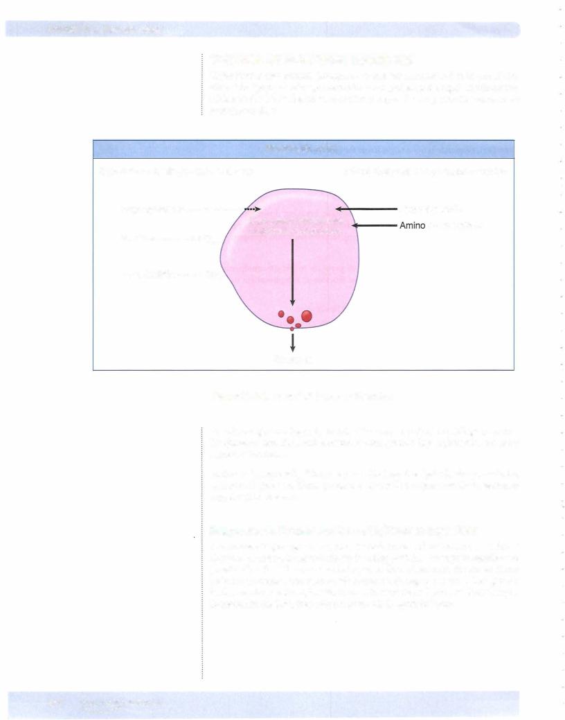
ACTIONS OF INSULIN
Insulin Receptor
•The portion of the insulin receptor that faces externally has the hormone-binding domain.
•The portion of the insulin receptor that faces the cytosol has tyrosine kinase activity.
•When occupied by insulin, the receptor phosphorylates itself and other proteins. (See Step 1 Biochemistry Lecture Notes for additional details.)
Peripheral Uptake ofGlucose
Glucose is taken up byperipheral tissues byfacilitated diffusion. Insulin facilitates this uptake in some tissues. Typically the insulin receptor causes the insertion of glucose transporters in the membrane.
Tissues that require insulin for effective uptake of glucose are:
•Adipose tissue
•Resting skeletal muscle (although glucose can enter working muscle without the aid of insulin)
•Liver because of glucokinase stimulation
Tissues in which glucose uptake is not affected by insulin are:
•Nervous tissue
•Kidney tubules
•Intestinal mucosa
•Red blood cells
•-cells ofpancreas
310 MEDICAL
Chapter 6 • Endocrine Pancreas
Metabolic Actions of Insulin
Insulin is a major anabolic hormone, which is secreted in response to a carbohy drateand/or protein-containing meal.
Anabolichormones tendtopromoteproteinsynthesis (increase leanbody mass).
Other anabolic hormones include:
•Thyroid hormones
•Growth hormone/IGF I
•Sex steroids (androgens)
Effects ofinsulin on carbohydrate metabolism
Insulin increases the uptake of glucose and its metabolism in muscle and fat. By increasing glucose uptake in muscle, its metabolism increases, i.e., its conversion to carbon dioxide and water is increased.
Insulin increases glycogen synthesis in liver and muscle. The activity ofenzymes that promote glycogen synthesis (glucokinase and glycogen synthetase) is increased. The activity of those enzymes that promote glycogen breakdown (phosphorylase and glucose-6-phosphatase) is decreased.
•Glucokinase and glucose-6-phosphatase are expressed by the liver but not by muscle.
Effects ofinsulin on protein metabolism
•Insulin increases amino acid uptake by muscle cells.
•Insulin increases protein synthesis.
•Insulin decreases protein breakdown (deficiency ofinsulin results in a breakdown ofprotein).
Effects ofinsulin onfatmetabolism
Insulin increases (Figure X-6-3):
•Glucose uptake byfat cells (increases membrane transporters). By increasing glucose uptake, insulin also makes triose phosphates available for triglyceride synthesis in adipose tissue.
•Triglyceride uptake by fat cells. It increases the activity oflipoprotein lipase (also called extracellular or clearing factorlipase). Lipoprotein lipase is located on the endothelium of capillaries and clears VLDL and chylomicrons from the blood.
•Triglyceride synthesis (lipogenesis) in adiposetissue and liver by stimu lating the rate-limiting step, namely the carboxylation ofacetyl CoA to malonyl CoA. In other words, insulin stimulates the conversion ofcar bohydrate into fat.
MEDICAL 311
Chapter 6 • Endocrine Pancreas
ACTIONS OF GLUCAGON
Glucagon is a peptide hormone. It is secreted by the a-cells ofthe pancreatic islets.
The primary target for glucagon action is the liver hepatocyte, where its action is mediated by an increase in the concentration of cAMP. The cAMP activates protein kinase A, which, by catalyzing phosphorylation, alters the activity of en zymes mediating the actions given below.
Note: Skeletal muscle is not a target tissue for glucagon.
SpecificActions ofGlucagon on the Liver
1. Increases liver glycogenolysis.
Glucagon activates glycogen phosphorylase, breaking down glycogen to glucose-I -phosphate. Glucagon inactivates glycogen synthetase, prevent ing the glucose- I-phosphate from being recycled back into glycogen.
2.Increases liver gluconeogenesis.
Glucagon promotes the conversion of pyruvate to phosphoenolpyruvate. Glucagon increases the conversion of fructose- I, 6-biphosphate to fruc tose-6-phosphate.
3.Increases liver ketogenesis and decreases lipogenesis.
Glucagon inhibits the activity of acetyl CoA carboxylase, decreasing the formation of malonyl CoA. When the concentration of malonyl CoA is low, ketogenesis is favored over lipogenesis.
4.Increases ureagenesis.
By increasing the production of glucose from pyruvate, glucagon indirectly stimulates the transamination of alanine to pyruvate. The amino group is eliminated as urea.
5.Increases insulin secretion.
The amino acid sequence of glucagon is similar to that of the duodenal hormone, secretin. Like secretin (and most other gut hormones), glucagon stimulates insulin secretion.
6.Increases lipolysis in the liver.
Glucagon activates hormone-sensitive lipase in the liver, but because the action is on the liver and not the adipocyte, glucagon is not considered a major fat-mobilizing hormone.
MEDICAL 315


Section X • Endocrinology
Type 2
•Accounts for about 90% of all the cases ofdiabetes
•Strong genetic component
•Body build is usually obese (particularly central or visceral).
•Usually, but not always, middle-aged or older
•The number ofyounger individuals in this category is increasing.
•Characterized by variable degrees of insulin resistance, impaired insulin secretion, and increased hepatic output of glucose. Insulin resistance precedes secretory defects and in the early stages hyperinsulinemia is able to overcome tissue resistance. Ultimately beta cell failure can occur.
•Insulin levels may be high, normal, or low.
•Resistance to insulin is not well understood. It is thought to be due to postreceptor defects in signaling, which ultimately lead to a decrease in the number of glucose transporters. Reducing plasma glucose and thus plasma insulin can increase receptor sensitivity toward normal.
•Plasma glucose good screening for type 2. Elevated glucose due to elevated hepatic output.
•With a controlled diet and exercise, the symptoms of type 2 diabetes often disappear without the necessity for pharmacologic therapy.
•Individuals tend to be ketosis resistant. The presence of some endog enous insulin secretion appears to protect from development of a ketoacidosis. If it does develop, it is usually the result ofsevere stress or infection (increased counterregulatory homones, suppressed insulin).
•In nonobese patients, a deficient insulin release by the pancreas is often the problem, but varying degrees ofinsulin resistance can also occur.
Metabolic Syndrome (Syndrome X)
A group of metabolic derangements that includes atherogenic dyslipidemia (low HLD) and high triglycerides, elevated blood glucose, hypertension, central obesity, prothromboticstate, andaproinflammatorystate. The clusteringoftheseriskfactors increases the probability ofdeveloping cardiovascular disease and type 2 diabetes.
Type l
•Genetic association less marked than in type 2
•Genetically predisposed individuals whose immune system destroys pan- creatic beta cells
•Symptoms do not become evident until 80% ofthe beta cells are destroyed.
•Body build usually lean
•Usually, but not always, early age of onset
•Due to an absence of insulin production
•Increased glucagon secretion also generally occurs
•Three target tissues for insulin-liver, skeletal muscle, and adipose tis sue-fail not only to take up absorbed nutrients but also to deliver inap propriate high levels to the bloodstream. This would include glucose, amino acids, and fatty acids.
318 M EDICAL
Section X • Endocrinology
Hyperosmolar Coma
•Severe hyperglycemia shifts fluid from the intracellular to the extracel lular space.
•Polyuria decreases volume ofthe extracellular space and leads to a decreased renal plasma flow and a reduced glucose excretion. Combined with the rise in counterregulatory hormones, the plasma glucose rises further.
•The severe loss of intracellular fluid from the brain causes the coma.
•Type 2 diabetics often present with the highest plasma glucose and greater states of dehydration. Thus these patients have a higher inci dence of coma.
Diabetic Ketoacidosis (DKA)
•Without any insulin, excessive lipolysis provides fatty acids to the liver, where they preferentially converted to ketone bodies because of the unopposed action of glucagon.
•Blood pH and bicarbonate decrease due to the metabolic acidosis.
•Increased hydrogen ion secretion (collecting duct) and the formation of an acid urine willoccur as a compensation for the ketoacidosis. The hydrogen ion secretion will tend to diminish potassium secretion, but the higher than normal tubular flow will promote potassium secretion.
•Increased alveolar ventilation partially compensates for the metabolic acidosis. When the arterial pH decreases to about 7.20, ventilation becomes deep and rapid (Kussmaul breathing).
•The severe acidosis is in addition to the dehydration and net decrease in total body sodium and potassium.
•Treatment is replacement of fluid and electrolytes and administration of insulin
•DKAtreatment is first 2-3 liters of normal saline and IV insulin. Subcutaneous insulin may not be fully absorbed because of decreased skin perfusion. Hyponatremia is common because of hyperglycemia. For each 100 point increase in glucose above normal, there is a 1.6 decrease in sodium.
100 mg ELEVATION glucose = 1.6 meqDECREASE sodium
When hyponatremia is present with hyperglycemia, management is cor rection of the elevated glucose level. When glucose comes to normal, the sodium corrects.
Hypokalemia in OKA
•Acidosis leads to hyperkalemia in blood
•Blood elevation in potassium leads to increased urinary loss of potassium
•Total body potassium is low
•Add potassium to IV fluids when potassium comes to normal
320 MEDICAL
Chapter 6 • Endocrine Pancreas
Hypoglycemia
•In the diabetic, overdosing with insulin causes hypoglycemia.
•Type 1 diabetics are particularly prone to hypoglycemia. In these indi viduals the glucagon response to hypoglycemia is absent.
•Initial symptoms due to catecholamine release followed by the direct effects of hypoglycemia include slowed mental processes and confusion.
PANCREATIC ENDOCRINE-SECRETING TUMORS
lnsulinomas
•Most common islet cell tumor
•Found almost exclusively within the pancreas and hypersecrete insulin
•Most common symptoms due to the hypoglycemia (confusion, disorienta tion, headache)
•Association with MEN 1
•Insulin measured to determine insulin-mediated versus noninsulinmediated hypoglycemia
•Insulin-secreting tumor: insulin and C-peptide both elevated
•Factitious hypoglycemia: C-peptide below normal
•Treat with removal
Other Endocrine-SecretingTumors
•Gastrinomas
•Glucagonomas
•Somostatinomas
•VIPomas
Management of all neuroendocrine tumors is localization with CT, then surgical resection.
Glucagonoma
•Alpha cell oversecretion
•Hyperglycemia/diabetes
•Localize with CT scan
•Surgically remove
MEDICAL 321
Chapter 1 • GeneralAspects of the Endocrine System
Chapter Summary
•Within the islets, beta cells are in the center, while the alpha and delta cells are in the periphery.
•(-peptide secreted in conjunction with insulin is an index of endogenous insulin secretion.
•The peripheral uptake ofglucose is via facilitated transport. In some tissues, such as adipose and resting skeletal muscle, the number of functioning transporters is regulated by insulin.
•Insulin facilitates the metabolism of glucose to carbon dioxide and water and also its conversion to glycogen in liver and muscle.
•Insulin promotes protein synthesis and decreases protein breakdown.
•Insulin promotes lipogenesis. It inhibits lipolysis by decreasing the activity of hormone-sensitive lipase. Hyperglycemia is the major promoter of insulin secretion.
•The majortarget tissue for glucagon is the liver, where its primary action is glycogenolysis and increased glucose output.
•Hypoglycemia is the main promoter and hyperglycemia the main inhibitor of glucagon secretion.
Type 2 Diabetes
•Strong genetic component and accounts for at least 90% of all cases
•Body build exhibits central obesity
•Insulin resistance, impaired insulin secretion, and increased hepatic output of glucose
•Fasting plasma glucose provides good screening.
•Individuals can have extremely high plasma glucose levels without ketoacidosis.
Type 1 Diabetes
•Generally autoimmune origin
•Absence of insulin secretion but elevated glucagon
•High circulating glucose (glycogen, gluconeogenesis), amino acids (protein), fatty acids (triglyceride)
•Polyuria, dehydration, and electrolyte depletion
•Ketoacidosis, Kussmaul breathing
•lnsulinomas: increased insulin, increased C-peptide, decreased plasma glucose
MEDICAL 323


Section X • Endocrinology
Plasma Calcium |
|
Protein-bound Ca2+ |
Free |
plus phosphateand |
|
citrate-bound Ca2+ |
2 |
Ca + |
Figure X-7-2. Relationship of Bound and Free Calcium
•Plasma calcium represents 50% ionized free, 40% attached to protein, 10% associated with anions such as phosphate and citrate.
•The free calcium is the physiologically active and precisely regulated form.
•Alkalosis (hyperventilation) decreases and acidosis increases free plasma calcium by varying the amount bound to protein.
•Alkalosis lowers free calcium by increasing protein-binding, while acidosis raises free calcium by decreasing protein-binding.
Relationship Between Calcium and Phosphate
Bone is a complex precipitate of calcium and phosphate to which hydroxide and bicarbonate ions are added to make up the mature hydroxyapatite crystals, which are laid down in a protein (osteoid) matrix. Whether calcium and phosphate are laid down in bone (precipitate from solution) or are resorbed from bone (go into solution) depends on the product oftheir concentrations rather than on their indi vidual concentrations.
When the product exceeds a certain number (solubility product or ion product), bone is laid down:
[Ca2+] x [P0-4] > solubility product = bone deposition
•Under normal conditions the ECF product of calcium times phosphate is close to the solubility product.
•Thus, an increase in the interstitial fluid concentration of either Ca2+ or phosphate increases bone mineralization.
•For example, an increase in plasma phosphate would increase the product of their concentrations, promote precipitation, and lower free calcium in the interstitial fluid.
•A malignant increase in the concentration of calcium or phosphate due to chronic renal disease or rhabdomyolysis can cause the precipitation of calcium phosphate within tissues.
When the product is below the solubility product, bone is resorbed:
[Ca2+] x [Po-4] < solubility product = bone resorption
•Thus, a decrease in the interstitial concentration of either Ca2+ or phos phate promotes the resorption of these salts from bone (demineraliza tion).
•For example, a decrease in plasma phosphate alone would promote bone demineralization. Increasing renal excretion of phosphate would promote bone demineralization and a rise in interstitial free calcium.
326 MEDICAL
Chapter 7 • Hormonal Control of Calcium and Phosphate
It is the free Ca2+, not the phosphate, that is regulated so precisely. Hormonal control of free Ca2+ levels is via a dual hormonal system; parathyroid hormone and vitamin D.
Vitamin D should be considered more of a prohormone than a vitamin.
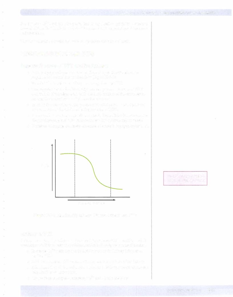
PARATHYROID HORMONE (PTH)
General Features of PTH and Its Actions
•PTH is a peptide hormone released from the parathyroid glands in response to lowered plasma free Ca2+ (Figure X-7-3).
•Free Ca2+ in the plasma is the primary regulator of PTH.
•The negative feedback relationship between plasma calcium and PTH secretion is highly sigmoidal, with the steep portion of the curve repre senting the normal range of plasma free calcium.
•To sense the free calcium, the parathyroid cell depends upon high levels of expression of the calcium-sensing receptor (CaSR).
•In most cells exocytosis depends on a rise in intracellular free calcium. In the parathyroid gland, a fall in intracellular free calcium causes release.
•Depletion of magnesium stores can create a reversible hypoparathyroidism.
PTH
Normal range region between dashed= lines
Ionized Calcium
Figure X-7-3. Relationship between Plasma Calcium and PTH
Actions of PTH
A decrease in the free calcium is the signal to increase PTH secretion and the function of PTH is to raise free calcium, which it does by several mechanisms.
•Increases Ca2+ reabsorption in distal tubule of the kidney (chapter 4, section VIII)
•Inhibits phosphate (Pi) reabsorption in proximal tubule of the kidney.
•Stimulates the 1-alpha-hydroxylase enzyme in kidney, converting inactive vitamin D to its active form.
•Causes bone resorption, releasing Ca2+ and Pi into the blood.
MEDICAL 327

Chapter 7 • Hormonal Control of Calcium and Phosphate
CALCITONIN
•Calcitonin (CT) is a peptide hormone secreted by the parafollicular cells (C cells) of the thyroid gland. It is released in response to elevated free
calcium.
• Calcitonin lowers plasma calcium by decreasing the activity of osteoclasts, thus decreasing bone resorption. Calcitonin is useful in the treatment of Paget's disease, severe hypercalcemia, and osteoporosis.
• Calcitonin is not a major controller of Ca2+ in humans. Removing the thyroid (with the C cells) or excess of calcitonin via a C cell tumor (med ullary carcinoma ofthe thyroid) has little impact on plasma calcium.
• No deficiency or excess disease has been described.
BONE REMODELING
•Bone is undergoing continual remodeling throughout life, although the turnover is faster in younger individuals. As many as 300,000 bone remodeling sites are active in a normal person.
•Peak bone mass is generally obtained in early adulthood. It then tends to decline (at least in sedentary individuals) and accelerates in women in the perimenopausal period.
•Remodeling occurs more in cancellous (trabecular or low-density) bone than in cortical (compact or high-density) bone.
•Remodeling occurs along force lines generated by mechanical stresses. The osteocytes are mechano-receptors that pick up vibrations and relay this information to the bone surface and osteoblasts, which then initiate the remodeling process. For example, a bowing deformity initiates new bone deposition on the concave side of the stressed bone, thus increas ing overall strength.
Weight-BearingStress
Weight-bearing mechanical stress increases the mineralization ofbone.
The absence ofweight-bearing stress (being sedentary, bedridden, or weightless) pro motes the demineralization ofbone. Under these conditions, the following occurs:
•Plasma Ca2+ tends to be in the upper region of normal.
•Plasma PTH decreases.
•Urinary calcium increases.
Indices
Indices can be utilized to detect excess bone demineralization and remodeling:
•Increased serum osteocalcin and alkaline phosphatase are associated with osteoblastic activity.
•Increased urinary excretion of hydroxyproline is a breakdown product of collagen
MEDICAL 329


Section X • Endocrinology
DISORDERS IN CALCIUM AND PHOSPHATE
Hypercalcemia
Hypercalcemia ofprimaryhyperparathyroidism
•Initiating factor is primary hypersecretion of PTH
•Consequences include increased plasma calcium, decreased plasma phos phate, polyuria, hypercalciuria, and decreased bone mass
•80% due to a single parathyroid adenoma
•High calcium inactivates ADH, leading to nephrogenic diabetes insipi dus. This is why there is massive volume deficit in hypercalcemia.
•High calcium makes it harder to depolarize neural tissue. This is why hypercalcemia causes lethargy, confusion, and constipation.
•15% due to primary hyperplasia as in MEN 1 or MEN 2A
•Parathyroid carcinoma rare
•Ectopic hormonal hypercalcemia usually PTHrP.
•Most patients asymptomatic
•Symptoms include lethargy, fatigue, depression, neuromuscular weakness, and difficulty in concentrating
•Increased plasma alkaline phosphatase, osteocalcin and increased excretion of cAMP (second messenger for PTH in the kidney), and hydroxyproline
•Severe dehydration
•Bone manifestation is osteitis fibrosa cystica in which there are increased osteoclasts in scalloped areas of the surface bone and replacement of marrow elements with fibrous tissue. Increased alkaline phosphatase is due to high bone turnover.
•Hypercalcemia decreases QT interval and in some cases causes cardiac arrhythmias.
Related causes ofhypercalcemia
•Lithium shifts the sigmoid Ca/PTH curve to the right. Higher calcium levels are thus needed to suppress PTH. A rare familial defect which reduces the Ca receptor sensitivity in a similar way results in hypercalce mia. Lithium causes hypercalcemia.
•Sarcidosis and other granulomatous disorders ( 10%) due to increased activity of 1 -alpha hydroxylase activity in granulomas.
•Thyrotoxicosis, milk-alkali syndrome
•Thiazide diuretics increase renal calcium absorption.
332 MEDICAL
Chapter 7 • Hormonal Control of Calcium and Phosphate
Differential diagnosis and treatment
•Elevated plasma calcium and PTH normal or elevated; conclusion is primary hyperparathyroidism
•Elevated plasma calcium and decreased PTH; conclusion is something other than primary hyperparathyroidism
•Treatment is usually surgery; i.e., removing the adenoma or with hyper- plasia removing most of the parathyroid tissue
•Treat with high volume fluid replacement
•Bisphosphonates need 2-3 days to be fully effective
•Calcitonin rapidly inhibits osteoclastic activity
Hypocalcemia
Hypocalcemia ofprimary hypoparathyroidism
•Can be hereditary or autoimmune
•Caused by thyroid surgery or surgery to correct hyperparathyroidism
•The initiating factor is inadequate secretion of PTH by the parathyroid glands.
•The decrease in plasma calcium is accompanied by an increased plasma phosphate. Even though less phosphate is resorbed from bone, plasma phosphate increases because the normal action of PTH is to inhibit phosphate reabsorption and increase excretion by the kidney. Therefore, without PTH, more of the filtered load is reabsorbed.
•Symptoms focus on the hypocalcemic induced increased excitability of motor neurons creating muscular spasms and tetany
•Chvostek's sign is induced by tapping the facial nerve just anterior to the ear lobe.
•Trousseau's sign is elicited by inflating a pressure cuff on the upper arm. A positive response is carpal spasm.
•Hypomagnesemia prevents PTH secretion and induces hypocalcemia. This condition responds immediately to an infusion of magnesium.
Pseudohypoparathyroidism
•This is a rare familial disorder characterized by target tissue resistance to parathyroid hormone:
•Exhibits same signs and symptoms as primary hypoparathyroidism except PTH elevated
•It is usually accompanied by developmental defects: mental retardation, short and stocky stature, one or more metacarpal or metatarsal bones missing (short 4th or 5th finger) .
MEDICAL 333
Section X • Endocrinology
Additional causes ofhypocalcemia
•Acute hypocalcemia can occur even with intact homeostatic mechanisms. Included would be alkalosis via hyperventilation, transfusions of citrated blood, rhabdomyolysis or tumor lysis, and the subsequent hyperphos phatemia
•Hyperphosphatemia of chronic renal failure
•Failures with vitamin D system
•Congenital absence of parathyroids rare (DiGeorge's syndrome)
•Damaged muscle binds calcium. Rhabdomyolysis binds free calcium.
Predictive indicesfora primarydisorder
When plasma calcium and phosphate levels are changing in opposite directions, the cause is usually a primary disorder. An exception may be chronic renal failure. This state is not a primary disorder but is usually associated with hypocalcemia and hyperphosphatemia (hypocalcemic-induced secondary hyperparathyroidism).
Renal Failure and Secondary Hyperparathyroidism
•Most common cause of secondary hyperparathyroidism
•Loss of nephrons prevents kidneys from excreting phosphate (Pi)
•Elevated Pi lowers free Ca2+, which in turn increases PTH
Vitamin D Deficiency and Secondary Hyperparathyroidism
•Causes include a diet deficient in vitamin D, inadequate sunlight expo sure, malabsorption of vitamin D, enzyme deficiencies in the pathway to activation of vitamin D
•In all cases there is a decrease in plasma calcium, which elicits an increase in PTH secretion and a secondary hyperparathyroidism.
•A similar consequence is the increased demand for calcium as in pregnancy.
•Characterized by increased PTH, decreased plasma calcium, and decreased plasma phosphate. Even though the elevated PTH increases phosphate resorption from bone, PTH also inhibits phosphate reabsorp tion by the kidney, thereby promoting phosphate excretion and a drop in plasma phosphate.
•Bone mass is lost to maintain plasma calcium.
•Diagnostic test is a low plasma 25(0H) vitamin D.
ExcessVitamin D and Secondary Hypoparathyroidism
• An excessive intake of vitamin D raises plasma calcium, which will elicit a decrease in PTH
•Characterized by decreased PTH, increased plasma calcium, and increased plasma phosphate but normal or decreased phosphate excre tion. Because PTH increases the excretion of phosphate by inhibiting reabsorption in the proximal tubule, decreased PTH will cause increased reabsorption of phosphate and drive plasma levels higher.
•Excessive vitamin D promotes bone resorption and bone mass decreases.
334 MEDICAL
Chapter 7 • Hormonal Control of calcium and Phosphate
Predictive indicesfora secondary disorder
When the plasma calcium and phosphate are changing in the same direction, the origin is usually a secondary disorder.
•Secondary hyperparathyroidism: both decrease
•Secondary hypoparathyroidism: both increase
Note also that in either a deficiency or an excess ofvitamin D, there is a decrease in bone mass but for completely different reasons.
METABOLIC BONE DISORDERS
Osteoporosis
•Osteoporosis is a loss ofbone mass (both mineralization and matrix) with fractures, due to normal age-related changes in bone remodeling as well as additional factors that exaggerate this process.
•If bone mineral density is 2.5 standard deviations below the average, then this equals osteoporosis.
•Ifbone mineral density is 1 to 2.5 standard deviations below the aver age, then this equals osteopenia.
•Bone mass reaches a peak subsequent to puberty. Heredity accounts for most ofthe variation but physical activity, nutrition, and reproductive hormones play a significant role, especially estrogens even in men.
•Secondary osteoporosis can occur in thyrotoxicosis and particularly with elevations in glucocorticoids.
•A mainstay oftreatment involves the use ofbisphosphonates that are rapidly incorporated into bone and reduce the activity of osteoclasts.
•Calcitonin inhibits bone resorption.
Osteoporosis can also be treated with:
•Denosumab: inhibitor of RANKL. RANKL is a TNF family of cytokine that activates osteoclasts; denosumab therefore, inhibits osteoclasts.
•Teriparitide: synthetic PTH. When used intermittently, teriparitide has a stimulatory effect on osteoblastic bone formation.
•Calcitonin
•Raloxifene: selective estrogen receptor modifier
Rickets and Osteomalacia
•Origin is the abnormal mineralization ofbone and cartilage
•Rickets is before plate closure, osteomalacia is after plate closure.
•In rickets there is expansion ofthe epiphysealplates and the most striking abnormalities are the bowing ofthe legs and protuberant abdomen.
•In osteomalacia, symptoms are more subtle.
•Most common cause in adults is a malabsorption disorder, e.g., celiac disease; a vitamin D deficiency can also cause
•Rarely, it can be caused by enzyme deficiencies when substrate availability is normal.
MEDICAL 335
Chapter 8 • Thyroid Hormones
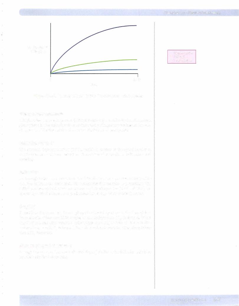
% Uptake of
1231odine
0 Time 24 hr Figure X-8-3. Relationship of Thyroid Function and Iodine Uptake
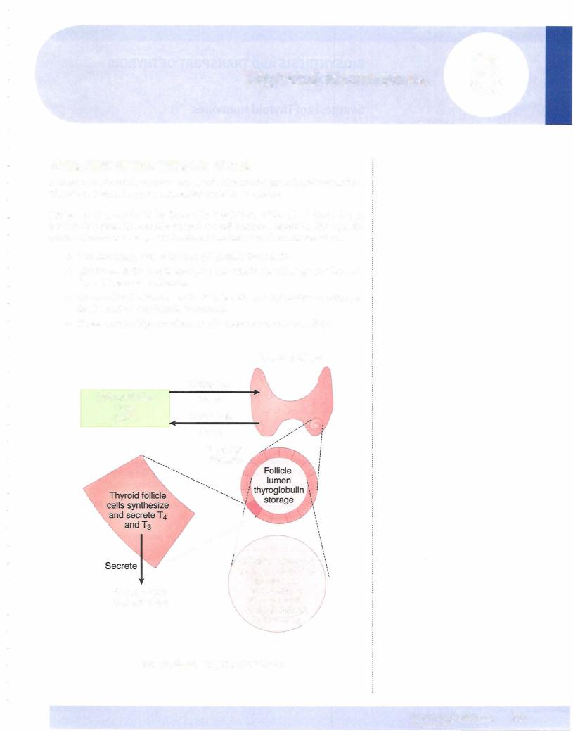
Thyroglobulin synthesis
A high molecular weight protein (>300,000 daltons) is synthesized in ribosomes, glycosylated in the endoplasmic reticulum, and packagedinto vesicles in the Gol gi apparatus. The thyroglobin then enters the lumen via exocytosis.
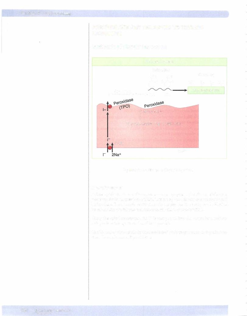
Oxidation ofr to 1°
The enzyme, thyroperoxidase (TPO), which is located at the apical border of the follicle cell, catalyzes oxidation. Peroxidase also catalyzes iodination and coupling.
Iodination
As thyroglobulin is extruded into the follicular lumen, a portion (<20%) of its tyrosine residues are iodinated. The catalyst for this reaction is peroxidase. The initial products of iodination are monoand diiodotyrosine (MIT and DIT), re spectively, with the latter form predominating, except when iodine is scarce.
Coupling
Peroxidase also promotes the coupling of iodinated tyrosine in the thyroglobu lin molecule. When two DITs couple, tetraiodothyronine (T4) is formed. When one DIT and one MIT combine, triiodothyronine (T3) is formed. When iodine is abundant, mainly T4 is formed. But when iodine becomes scarce, the produc tion of T3 increases.
Storage ofthyroid hormones
Enough hormone is stored as iodinated thyroglobulin in the follicular colloid to last the body for 2-3 months.
-Hyperthyroid
-Normal
-Hypothyroid
MEDICAL 339
Section X • Endocrinology
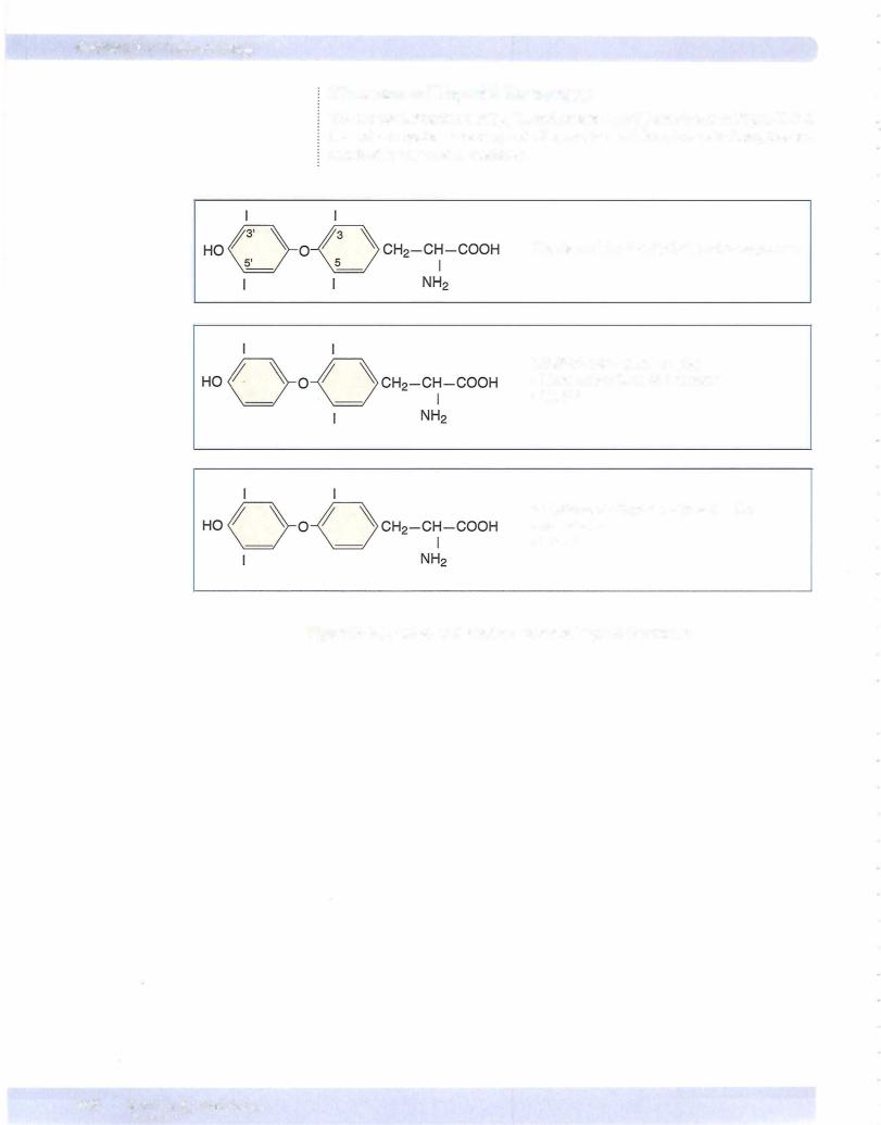
Structure ofThyroid Hormones
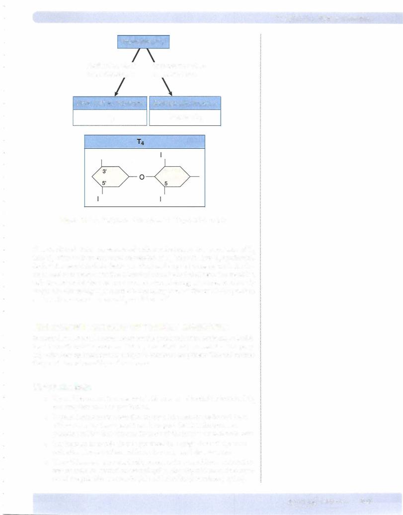
The chemical structures ofT4, T3, and reverse T3 (rT3) are shown in Figure X-8-4. Do not memorize structure; note the number and location of iodines, instead, attached to the tyrosine residues.
Thyroxine (T4) 3,5,3',5',-tetra-iodothyronine
3,5,3'-tri-iodothyronine (T3) More active form of hormone ·• No 5' I
3,3',5'-tri-iodothyronine (reverse T3)
No activity
·• No 5 1
Figure X-8-4. Active and Inactive Forms of Thyroid Hormones
340 MEDICAL

Section X • Endocrinology
Growth and Maturation (T4 and T3 Anabolic Hormones)
Fetal growth rates appear normal in the absence of thyroid hormone production (i.e., if the fetus is hypothyroid). However, without adequate thyroid hormones during the perinatal period, abnormalities rapidly develop in nervous system maturation.
•Synapses develop abnormally and there is decreased dendritic branching and myelination. These abnormalities lead to mental retardation.
•These neural changes are irreversible and lead to cretinism unless replacement therapy is started soon after birth.
Lipid Metabolism
•Thyroid hormone accelerates cholesterol clearance fromtheplasma.
•Thyroid hormones are required for conversion ofcarotene to vitamin A, and, as a consequence, hypothyroid individuals can suffer from night blindness and yellowing ofthe skin.
CHO Metabolism
•Thyroid hormone increases the rate ofglucose absorption fromthe small intestine.
Cardiovascular Effects
•Thyroid hormones have positive inotropic and chronotropic effects on the heart.
•The increased contractility is partly direct and partly indirect: they increase the number and affinity of -adrenergic receptors in the heart, therebyincreasing the sensitivity to catecholamines.
•Acting on the SA node, they directly increase heart rate.
•Cardiacoutput is increased, and both heart rate and strokevolume are elevated.
•Systolic pressure increases are due to increased stroke volume, and dia stolic pressure decreases are due to decreased peripheral resistance.
•Thyroid hormones in the normal range are required for optimum cardiac performance.
Additional Effects
•Thyroid hormones maintain the ventilatory response to hypoxia, increase erythropoietin, and increase gut motility and bone turnover.
•Hypothyroidism is associated with an increased prolactin. TRH in excess amounts will stimulate prolactin.
344 MEDICAL
Chapter 8 • Thyroid Hormones
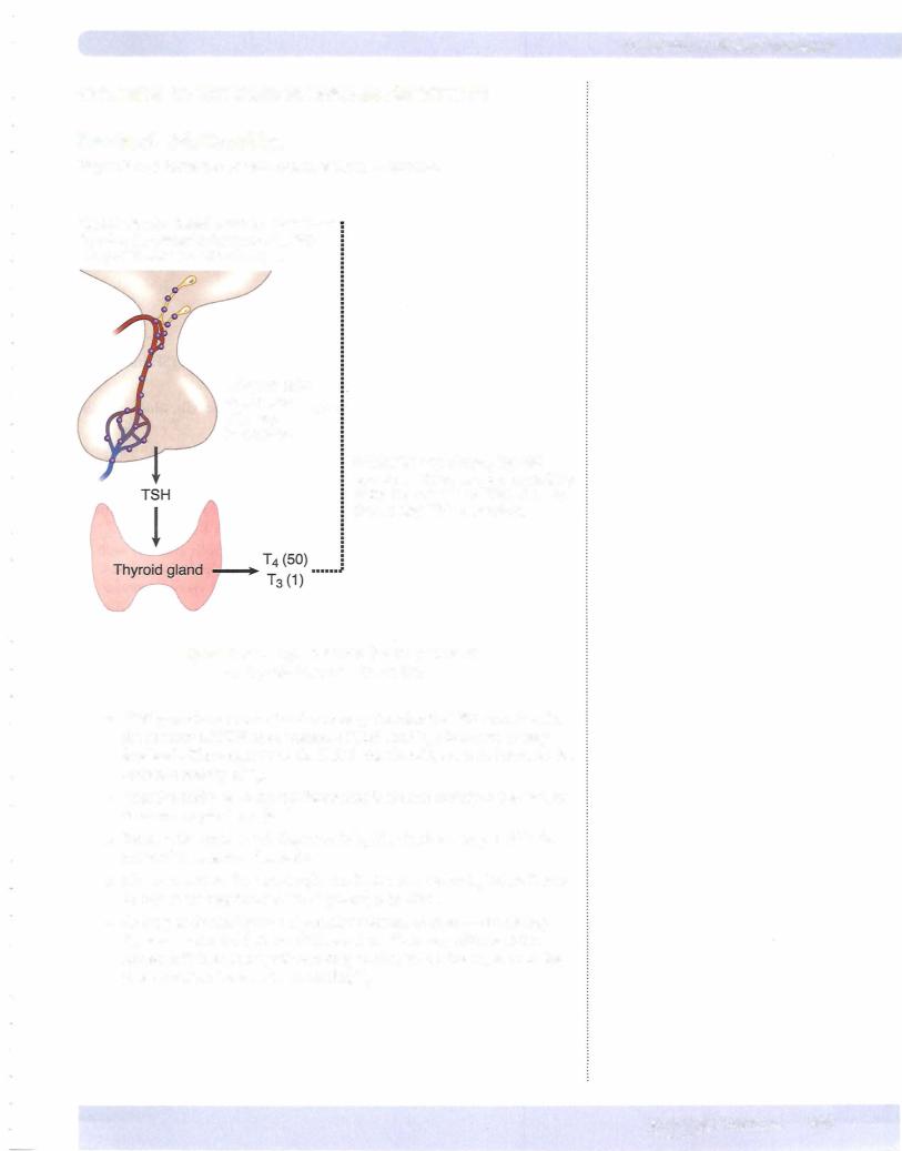
CONTROL OF THYROID HORMONE SECRETION
Feedback Relationships
Figure X-8-8 shows the overall control ofthyroid function.
Hypothalamic nuclei secrete thyrotropin-releasinghormone.....(TRH):············· into portal vessels stimulating ...
...thyrotrophs
of anterior
pituitary ......
to secrete...
Within the thyrotroph, thyroid hormones decrease the sensitivity of the thyrotroph to TRH, thereby decreasing TSH secretion.
Hypothalamic-Pituitary Control
Figure ofX-Thyroid8-8. -Hormone Secretion
•TRH provides a constant and necessary stimulus for TSH secretion. In the absence ofTRH, the secretion ofTSH (and T4) decreases to very low levels. The target tissue for TSH is the thyroid, where it increases the secretion mainly ofT4.
•Negative feedback ofthyroid hormones is exerted mainly at the level of the anterior pituitary gland.
•Because the main circulating form is T4, it is T4 that is responsible for most ofthe negative feedback.
•However,within the thyrotrophs the T4 is converted to T3 before it acts
•to reduce the sensitivity ofthe thyrotroph to TRH.
MEDICAL 345

Section X • Endocrinology
Overall Effects ofThyrotropin {TSH) on the Thyroid
Rapidlyinduced TSH effects
TSH tends to rapidly increase (within minutes or an hour) all steps in the syn thesis and degradation ofthyroid hormones, including:
•Iodide trapping
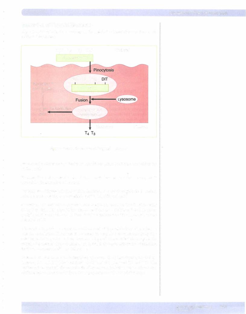
•Thyroglobulin synthesis and exocytosis into the follicular lumen
•Pinocytotic reuptake ofiodinated thyroglobulin back into the thyroid follicular cell
•Secretion ofT4 into the blood
Slowlyinduced TSH effects
Changes that occur more slowly (hours or days) in response to TSH include:
•Increased blood flow to the thyroid gland
•Increased hypertrophy or hyperplasia ofthe thyroid cells,which initially leads to increased size ofthe gland or goiter
Tests ofThyroid Function
• Determining the serum TSH is the first step in evaluating thyroid func tion.
• Secondly, free T4 (FT4) measurements are now readily available and would confirm an initial conclusion based on the TSH measurement. An alternative test would be an index ofthe free T4 via resin uptake.
• Autoimmune thyroid disease is sometimes detected by measuring circu lating antibodies. Most notably are the TPO antibodies, which are ele vated in Hashimoto's thyroiditis (hypothyroidism) and Graves' disease (hyperthyroidism).
•Additional antibodies are those against thyroglobulin and the TSI anti bodies that stimulate the TSH receptor in Graves' disease.
•Uptake of iodine isotopes by the thyroid allows thyroid imaging and quantitation oftracer uptake.
•Subacute thyroiditis: overall a below-normal uptake ofisotope
•Graves' disease: increasedtracer uptake that is distributed evenly throughout the enlarged gland
•Toxic adenomas: local areas ofincreased uptake with below-normal uptake in the remainder ofthe gland
•Toxic multinodular goiter: enlarged gland that often has an abnormal architecture and with multiple areas ofhigh and low uptake.
346 MEDICAL

Section X • Endocrinology
Primary Hypothyroidism
Primary changes and clinical presentation
•Most common cause is Hashimoto's thyroiditis, an autoimmune destruc tion ofthe thyroidwith lymphocytic infiltration; i TPO antibodies; early stages have a diffusely enlarged thyroid progressing in the later stages to a smaller atrophic andfibrotic gland.
•i TSH, 1 FT4; in subclinical hypothyroidism the TSH is on the high side ofnormal and the FT4 is on the low side ofnormal.
•Decreased basal metabolicrate and oxygen consumption
•Plasma cholesterol and other blood lipids tend to be elevated.
•Increased TRH drives a hyperprolactinemia. In women it may result in amenorrhea with galactorrhea; more often anovulatory cycles with men orrhagia. In men infertility and gynecomastia.
•Decreased GFR and an inabilityto excrete a water load, which may lead to hyponatremia.
•Inability to convert carotene to vitaminA may cause yellowing ofthe skin and night blindness.
•Slow thinking and lethargy; some patientshave severe mental symp- toms, dementia, or psychosis ("myxedema madness")
•Decreased food intake but individuals tend to be overweight
•Deep tendon reflexes with slow relaxation phase
•In the earlystages a decreased cardiac performance due to decreased loading conditions. In the later stages cardiac features suggestive of cardiomyopathy
•Anemia, constipation, hoarseness in speech, and the skin is dry and cool
•A decreasedventilatory drive to hypercapnia and hypoxia
•Accumulation ofsubcutaneous mucopolysaccharides that give rise to a nonpitting edema (myxedema)
•Myxedema coma is the end stage ofuntreated hypothyroidism. The major features are hypoventilation, fluid and electrolyte imbalances, and hypothermia and ultimately shock and death.
Cretinism
•Untreated postnatal hypothyroidism results in cretinism, a form of dwarfism with mental retardation.
•Individuals often appear normal following delivery but may display some respiratory difficulty, jaundice, feeding problems, and hypotonia.
•Abnormalities rapidlydevelop in nervous system maturation,which are irreversible and result in mental retardation.
•Prepubertal growth, including bone ossification, is retarded in the absence ofthyroid hormones. A stippled epiphysis is a sign ofhypothy roidism in children.
•There is no evidence thatthyroid hormones act directly on growth or bone formation. Rather, thyroid hormone appears to be permissive or act synergistically with growth hormone or growth factors acting
directly on bone. Thyroid hormone is· required for normal synthesis and secretion ofgrowth hormone.
348 MEDICAL
Chapter 8 • Thyroid Hormones
•Acquired hypothyroidism during childhood results in dwarfism but there is no mental retardation.
•At puberty, increased androgen secretion drives an increased growth hormone secretion. This will not occur with depressed levels ofthyroid hormones.
Additional causes ofhypothyroidism
•Secondary generally associated with panhypopituitarism
•Secondary or tertiarycharacterizedby J. FT4 and inappropriately normal TSH.
•Severe iodine deficiency (not in the United States)
•Drug induced, e.g. lithium
•Failure to escape from the Wolff-Chaikoff effect following excessive iodine intake
•Rarely there can be resistance to thyroid hormone
Treatment
•Replacement doses of T4. The goal is to give enough T4 to normalize serum TSH.
•Because metabolism ofT4 decreases and the plasma half-life increases with age, higher doses ofT4 are required in younger individuals.
•Overalllevels ofTSH must be checked on occasion to make sure ofthe proper dosage of T4.
•In women beyond menopause, overprescribing T4 can contribute to the development of osteoporosis.
Primary Hyperthyroidism (Graves' Disease)
•Thyrotoxicosis by definition is the clinical syndrome whereby tissues are exposed to high levels ofthyroid hormone (= hyperthyroidism)
•The most commoncause ofthyrotoxicosis is Graves' disease, a primary hyperthyroidism.
•Graves' disease is an autoimmune problem in which one antibody is directed againstthe thyroid receptor. It is referred to as the thyroid stimu lating antibody (TSI or TSH-R).
•In addition TPO antibodies andthose against thyroglobulin are also found in Graves' disease.
•i FT4, J. TSH; it is the TSI stimulating the TSH receptor on the thyroid that is driving the hyperthyroidism.
•In Graves' disease the thyroid is symmetrically enlarged.
•Increased radioiodine uptake by the thyroid and decreased serum cholesterol.
•Only Graves' disease has thyroid-stimulating antibodies. The onlytypes ofhyperthyroidism with increased radioactive iodine uptake are Graves' disease and toxic nodular goiter.
Subacute thyroiditis and "silent" or "painless" thyroiditis do not have increased radioactive iodine uptake; they are "leaking" ofthyroid hormone out of a gland damaged by antibodies.
MEDICAL 349

Section X • Endocrinology
•Increased metabolic rate and heat production. Individuals tend to seek a cool environment.
•Cardiac output, contractility, and heart rate are increasedwith possibly palpitations and arrhythmias (increased 13-adrenergic stimulation)
•Many symptoms suggest a state of excess catecholamines but circulating catecholamines are usually normal.
•Weight loss with increased food intake, protein wasting, and muscle weakness.
•Tremor, nervousness, and excessive sweating.
•The wide-eyed stare (exophthalmos) in patients with Graves' is caused by an infiltration oforbital soft tissues and extraocular muscles and the resulting edema, and this process is caused bythe antibodies.
•Untreated hyperthyroidism may decompensate into a condition called "thyroid storm."
•The end-stage of Graves' disease is often a hypothyroidism.
Acute Treatment
•Beta blockers are the most rapid in effect
•Methimazole or propylthiouracil stops the production ofhormone
•Iodine in high dose stops incorporation ofiodine into the gland
•Steroids such as dexamethasone stop conversion ofT4 to T3
•Long-term permanent cure is ablation ofthe gland with radioactive iodine
Additional origins ofhyperthyroidism (thyrotoxicosis)
•Autonomously functioning thyroid adenoma
•Toxic multinodular goiter
•Subacute and silent thyroiditis
•TSH-secreting pituitary adenoma (secondary hyperthyroidism) (very rare)
Goiter
•A goiter is simply an enlarged thyroid and does not designate functional status. A goiter can be present in hypo-, hyper-, and euthyroid states. There is no correlation between thyroid size and function.
•A generalized enlargement ofthe thyroid is considered a "diffuse goiter:'
•Diffuse enlargement often results from prolonged stimulation by TSH or TSH-like factor; e.g., Hashimoto's thyroiditis, Graves' disease, diet deficient in iodine
•An irregular or lumpy enlargement ofthe thyroid is considered a "nod ular goiter."
•With time, excessive stimulation by TSH can result in a multinodular goiter e.g. iodine deficiency initially produces a diffuse nontoxic goiter. Long term however, focal hyperplasia with necrosis and hemorrhage results in the formation ofnodules. Nodules vary from "hot nodules" that can trap iodine to "cold nodules " that cannot trap iodine.
350 MEDICAL

Chapter 8 • Thyroid Hormones
Chapter Summary
•Thyroid hormones are anabolic and are required fornormal growth and maturation.
•Thyroid follicles store several months supply ofthyroid hormone.
•The thyroid synthesizes and releases mainly T4 but on an iodine-deficient diet, the production and release ofT3, a more active form ofthe hormone, increases.
•Most of the activity ofT4 is due to the peripheral conversion to T3.
•Thyroid hormones increase metabolic rate, conversion of carotene to vitamin A and increase cardiac performance.
•Thyroid hormone is required for postnatal brain maturation. Cretinism is a form of dwarfism with mental retardation.
•CirculatingT4 creates most ofthe negative feedback to the anterior pituitary but the T4 entering the thyrotropes must be converted into T3 before it creates negative feedback.
•Tests of thyroid function focus on the circulating TSH and FT4•
•Primary hypothyroidism is mainly due to Hashimoto's thyroiditis and results in decreased FT4 and increased TSH.
•Primary hyperthyroidism is mainly due to Graves' disease. The autoimmune factor stimulating the thyroid increases FT4 and decreases TSH.
•A goiter, which can be described as diffuse or nodular, is simply an enlarged thyroid and does not designate functional status.
•Individuals on an iodine-deficient diet develop large goiters but they are usually euthyroid.
MEDICAL 351

Growth, Growth Hormone, |
9 |
and Puberty |
IN-UTERO AND PREPUBERTAL GROWTH
Intrauterine Growth
•Important roles for growth hormone, IGF-11 (early in gestation), IGF-I (later in gestation) and insulin
•Infants of diabetic mothers have increased insulin levels and are large.
•Smoking decreases vascularity ofthe placenta and decreases birth weight.
•Poor maternal nutrition leading cause oflow birth weight worldwide.
Postnatal Growth
•Although fetal hypothyroidism does not decreasebirth weight, hypothy roidism following delivery causes irreversible abnormalities in nervous system maturation, which in turn lead to mental retardation (cretinism).
•Growth hormone, insulin, and thyroid hormone play major roles. Acquired hypothyroidism later in childhood will slow growth and reduce bone advancement more than growth hormone deficiency, but will not cause mental retardation.
•Replacement ofhormone deficiencies creates a period ofcatch-up growth, but it is soon replaced with a normal growth rate.
•There is no major role for gonadal sex steroids on prepubertal growth or for glucocorticoids but glucocorticoid excess will slow growth.
•Hypersecretion ofgrowth hormone pre-puberty (pituitary adenoma) results in giantism. It also delays pubertal changes, and the subsequent hypogonadism contributes to the giantism.
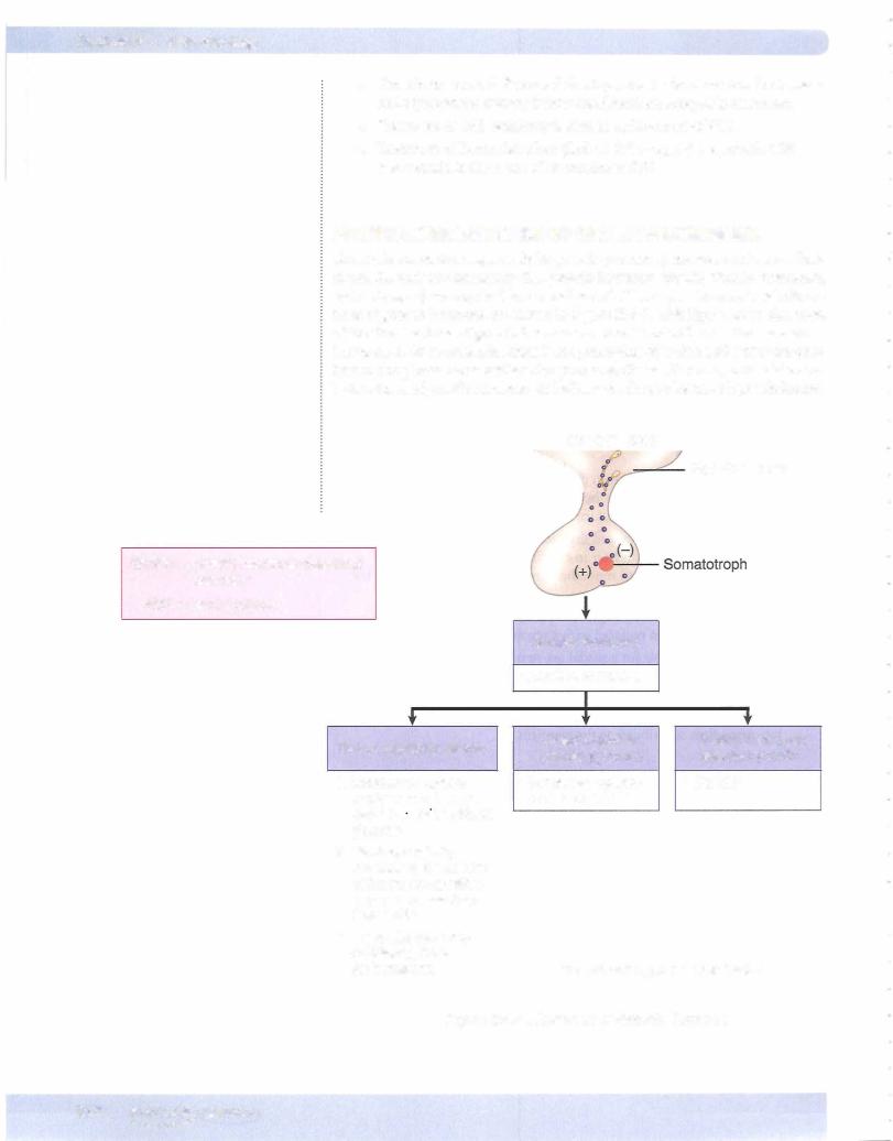
Prepubertal Growth Hormone Deficiency
•Congenital deficiency is associated with decreased birth length.
•Idiopathic deficiency is due to decreased growth hormone-releasing hor mone (GHRH).
•Classic congenital deficiency creates short stature, delayed skeletal matu ration, proneness to episodes ofhypoglycemia, and a chubby, immature facial appearance.
•Acquired deficiency maybe due to a hypothalamic-pituitary tumor.
•Growth hormone deficiencyleads to dwarfism.
•Tissue resistance to growth hormone (i growth hormone, .j, IGF-I) results in Laron syndrome (Laron dwarfism).
•Stimulation test is with an arginine infusion.
MEDICAL 353

Chapter 9 • Growth, Growth Hormone, and Puberty
•Near the end ofpuberty, androgens promote the mineralization (fusion or closure) ofthe epiphyseal plates oflong bones. Estrogen can also cause plate closure, even in men.
•In females, the growth spurt begins early in puberty and is near comple tion by menarche. In males, the growth spurt develops near the end of puberty.
ACROMEGALY
•It is caused by a post pubertal excessive secretion ofgrowth hormone.
•It is almost always due to macroadenoma (> 1 cm dia) ofthe anterior pituitary and second in frequency to prolactinomas.
•There is a slow onset of symptoms, and the disease is usually present for 5 to 10 years before diagnosis.
•Ectopic GHRH secretion has been documented but is rare.
•Some tumors contain lactotrophs, and elevated prolactin can cause hypogonadism and galactorrhea.
•Increased IGF-I causes most ofthe deleterious effects ofacromegaly but
growth hormone excess directly causes the hyperglycemia and insulin resis
tance.
•There is characteristic proliferation of cartilage, bone and soft tissue, visceral, and cardiomegaly.
•Observable changes include enlargement of the hands and feet (acral parts) and coarsening ofthe facial features, including downward and forward growth ofthe mandible. Also, increased hat size.
•Measurement of IGF-I is a useful screening measure and confirms diag nosis with the lack ofgrowth hormone suppression by oral glucose.
•Diagnosis: Treatment should only be done when 3 steps are completed: Elevated IGF level
Failure of suppression of GH/IGF after giving glucose
MRI shows lesion in brain in pituitary
Never start with a scan in endocrinology. Benign pituitary "inciden taloma" is common in 2-10% of the population. Always confirm the presence of an overproduction ofa hormone before doing a scan. This is true for adrenal lesions as well.
•Treatment:
-Surgical removal by trans-sphenoidal approach is first. Removal of an over-producing adenoma is the first treatment in most of endocrinol ogy with the exception ofprolactinoma.
-Ifsurgical removal fails, use the growth hormone receptor antagonist, pegvisomant, or octreotide. Octreotide is synthetic somatostatin. Cabergoline is a dopamine agonist used when other medications have failed.
 Radiation is used last, only after surgery, pegvisomant, octreotide and cabergoline have failed.
MEDICAL 357

Section X • Endocrinology
Chapter Summary
•Anabolic hormones except thyroid hormone are required for intrauterine growth.
•Thyroid hormone is required in the perinatal period to prevent mental retardation.
•Sex steroids have no major role prepuberty.
•Excess glucocorticoids slow growth.
•Excess growth hormone accelerates growth, delays pubertal changes, and creates gigantism.
•Prepubertal growth hormone deficiency or tissue resistance to growth hormone (Laron syndrome) leads to dwarfism.
•Most of the direct actions of growth hormone are consistent with its actions as a stress hormone; i.e., it decreases the peripheral uptake of glucose and promotes lipolysis.
•Most ofthe anabolic actions ofgrowth hormone are indirect via growth factors.
•The most important growth factor is IGF-1, which increases the synthesis of cartilage in the epiphyseal plates of long bones.
•Growth hormone secretion is pulsatile and a great deal is released during the night.
• Plasma IGF-1 is usually a good index of overall growth hormone secretion.
•The first noted female change at puberty is breast development; in the male it is testes enlargement.
•The increased secretion of growth hormone during puberty is driven by a concurrent increase in androgen secretion.
•The acute factors regulating growth hormone secretion are similarto those regulating glucagon and are consistentwith their role as stress hormones.
•Acromegaly is almost always due to pituitary adenoma and most ofthe deleterious effects due to IGF-1. Noted changes include acral enlargement and coarse facial features. Measurement of IGF-1 is useful screening, lack of growth hormone suppression by oral glucose diagnostic.
358 MEDICAL


Section X • Endocrinology
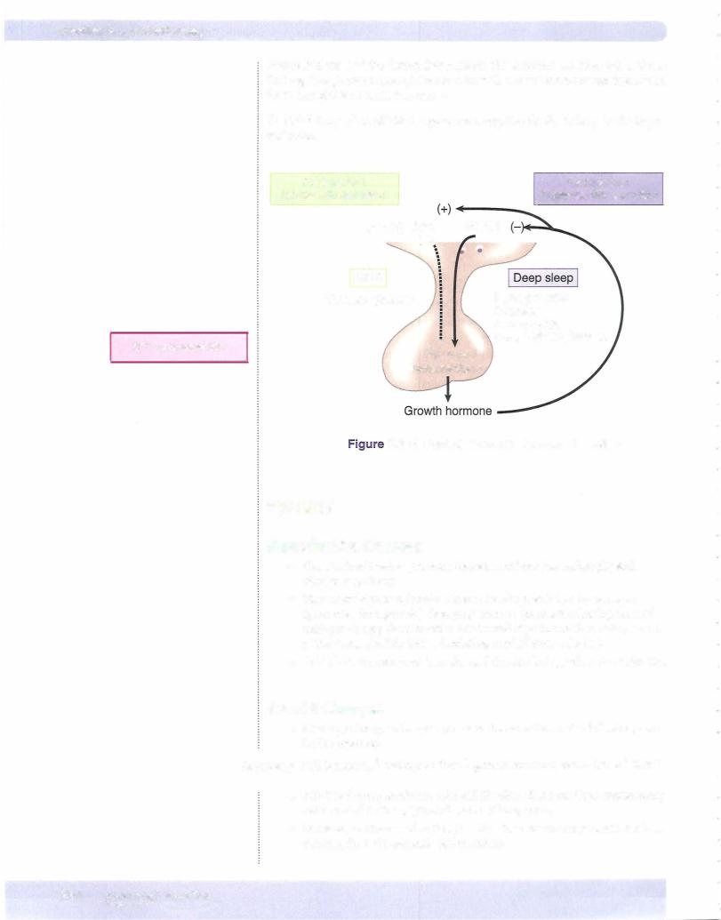
GnRH is synthesized in the hypothalamus and secreted in pulses into the hypophyseal portal vessels. In the anterior pituitary it stimulates the gonado trophs, which produce a pulsatile release ofLH and FSH.
LH and FSH are glycoproteins. TSH and human chorionic gonadotropin (hCG) are also glycoproteins. All four hormones are composed ofan ex and asubunit. The ex subunits all have the same structure. It is the subunit that provides specificity.
LH/Leydig Cells
•Leydig cells express receptors for LH
•LH is a peptide hormone that activates Gs--cAMP, which in turn initi ates testosterone production by activating steroidogenic acute regulatory protein (StAR).
•Testosterone diffuses into Sertoli cells (high concentration) and into the blood.
•Circulating testosterone provides negative feedback to regulate LH secretion at the level ofthe hypothalamus and anterior pituitary.
Sa-reductase
Some target tissue express this enzyme, which converts testosterone into the more potent dihydrotestosterone. Some important physiologic effects primarily mediated by dihydrotestosterone are as follows:
•Sexual differentiation: differentiation to form male external genitalia
•Growth ofthe prostate
•Male-pattern baldness
•Increased activity of sebaceous glands
•Synthesis of NO synthase in penile tissue
FSH/Sertoli Cells
•FSH binds to Sertoli cells and activates a Gs--cAMP pathway.
•Sertoli cells release inhibin B, which has negative feedback on FSH secretion.
360 MEDICAL
Chapter 10 • Male Reproductive System
1. Fetal life
The development ofmale and female internal and external structures depends on the fetal hormonal environment. The Wolffian and Miillerian ducts are initially present in both male and female fetuses. Ifthere is no hormonal input (the situ ation in the normal female fetus), female internal and female external structures develop (Milllerian ducts develop, Wolffian ducts regress).
Normal male development requires the presence of 3 hormones: testosterone, dihydrotestosterone, and the Miillerian inhibiting factor (MIF).
1 . (hCG) + LH -7 Leydig cells -7 testosterone -7 Wolffian ducts
5-a-reductase
2. testosterone -7 dihydrotestosterone -7 urogenital sinus & genital organs
3.Sertoli cells -7 MIF -7 absence offemale internal structures
- In the absence oftestosterone,the Wolffian ducts regress.
•Dihydrotestosterone induces the urogenital sinus and genitaltubercle to differentiate into the external scrotum, penis, and prostate gland.
2. Childhood
Within a few months afterbirth, LH and testosterone drop to low levels and re main low until puberty. The cause ofthis prolonged quiescence ofreproductive hormone secretion during childhood is not known. Interestingly, LH secretion remains low in spite oflow testosterone.
3. Puberty
Near the onset ofpuberty, the amplitude ofthe LH pulses becomes greater, driv ing the mean level of LH higher. Early in puberty, this potentiation of the LH pulses is especially pronounced during sleep. This increased LH stimulates the Leydig cells to again secrete testosterone.
4. Adult
During adulthood, LH secretion drives testosterone secretion. Thus, it is not sur prising that the relativelevels ofthe two hormones parallel one another.
5. Aging adult
Testosterone and inhibin secretions decrease with age. Men in their seventies generally secrete only 60-70% as much testosterone as do men in their twenties. Nevertheless, there is no abrupt decrease in testosterone secretion in men that parallels the relatively abrupt decrease in estrogen secretion that women experi ence at menopause. The loss of feedback will cause an increase in LH and FSH secretion.
MEDICAL 363
Section X • Endocrinology
Effect on Muscle Mass
The capacity of androgens to stimulate protein synthesis and decrease protein breakdown, especially in muscle, is responsible for the larger muscle mass in men as compared with women. Exogenous androgens (anabolic steroids) are some times taken by men and women in an attempt to increase muscle mass.
Spermatogenesis is Temperature Dependent
Effectonfertility
For unknown reasons, spermatogenesis ceases at temperatures typical of the ab dominal cavity. Thus, when the testes fail to descend before or shortly after birth, and the condition (cryptorchidism) is not surgically corrected, infertility results.
Cooling mechanisms
Normally, the scrotum provides an environment that is 4°C cooler than the ab dominal cavity. The cooling is accomplished by a countercurrent heat exchanger located in the spermatic cord. Also, the temperature of the scrotum and the tes tes is regulated by relative degree of contraction or relaxation of the cremasteric muscles and scrotal skin rugae that surround and suspend the testes.
Effe ct on FSH and LH
Sertoli cells, and therefore germ cell maturation, are adversely affected by the elevated temperatures of cryptorchid testes. In adults with bilaterally unde scended testes, FSH secretion is elevated, probably as a result of decreased Sertoli cell production of inhibins. Testosterone secretion by the Leydig cells of cryptorchid testes also tends to be low, and as a result, LH secretion ofadults with bilateral cryptorchidism is elevated.
ERECTION, EMISSION, AND EJACULATION
Erection
•Erection is caused by dilation of the blood vessels (a parasympathetic response) in the erectile tissue of the penis (the corporaand ischiocav ernous sinuses).
•This dilation increases the inflow of blood so much that the penile veins get compressed between the engorged cavernous spaces and the Buck's and dartos fasciae.
•Nitric oxide (NO), working through cGMP, mediates the vasodilation.
Emission
•Emission is the movement of semen from the epididymis, vas deferens, seminal vesicles, and prostate to the ejaculatory ducts.
•The movement is mediated by sympathetic (thoracolumbar) adrenergic transmitters.
364 MEDICAL
Chapter 10 • Male Reproductive System
•Simultaneouslywith emission, there is also a sympathetic adrenergic mediated contraction ofthe internal sphincter ofthe bladder, which prevents retrograde ejaculation ofsemen into the bladder. Destruction of this sphincter by prostatectomy often results in retrograde ejaculation.
•Emission normally precedes ejaculation but also continues during ejaculation.
Ejaculation
•Ejaculation is causedbythe rhythmic contraction ofthe bulbospongiosus and the ischiocavernous muscles,which surround the base ofthe penis.
•Contraction ofthese striated muscles that are innervated by somatic motor nerves causes the semen to exitrapidly in the direction ofleast resistance, i.e., outwardly through the urethra.
GONADAL DYSFUNCTION IN THE MALE
The consequences of deficient testosterone production depend upon the age of onset:
•Testosterone deficiencyin the second to third month of gestation results in varying degrees of ambiguity in the male genitalia and male pseudo hermaphrodism.
•Testosterone deficiency in the third trimester leads to problems in tes ticular descent (cryptorchidism) alongwith micropenis.
•Pubertaltestosteronedeficiencyleads to poor secondary sexual develop ment and overall eunuchoid features.
•Postpubertal testosterone deficiency leads to decreased libido, erectile dysfunction, decrease in facial and body hair growth, low energy, and infertility.
Causes of Hypogonadism
•Noonan syndrome
•Klinefelter's syndrome
•Hypothalamic-pituitary disorders (Kallman's syndrome, panhypopitu itarism)
•Gonadal failure/sex steroid synthesis failure
Definitions
•Pseudohermaphrodite: an individual with the genetic constitution and gonads of one sex and the genitalia ofthe other.
•Female pseudohermaphroditism: female fetus exposed to androgens during the 8th to 13th week of development, e.g., congenital virilizing adrenal hyperplasia.
•Male pseudohermaphroditism: lack ofandrogen activity in male fetus, e.g., defective testes, androgen resistance
•When the loss ofreceptor function is complete, testicular feminizing syn
drome results. Here MIF is present and testosterone is secreted, usually at elevated levels. The external structures are female, but the vagina ends blindlybecause there are no female internal structures.
MEDICAL 365

Female Reproductive System 1 1
MENSTRUAL CYCLE
The Phases
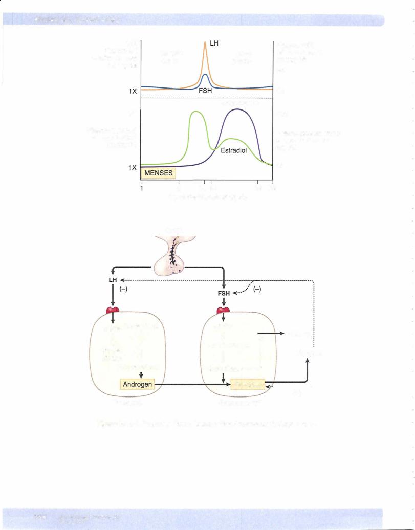
The menstrual cycle (approximately 28 days) can be divided into the following phases or events. By convention, the first day of bleeding (menses) is called day 1 of the menstrual cycle.
•Follicular phase (first 2 weeks) is also called the proliferative or preovula tory phase. This phase is dominated by the peripheral effects of estrogen, which include the replacement ofthe endometrial cells lost during menses.
•Ovulation (approximately day 14) is preceded by the LH surge, which induces ovulation.
•Luteal phase (approximately 2 weeks) is dominated by the elevated plasma levels of progesterone, and along with lower levels of secreted estrogen, creates a secretory quiescent endometrium which prepares the uterus for implantation.
•Menses. Withdrawal ofthe hormonal support of the endometrium at this time causes necrosis and menstruation.
Follicularphase (approximately days 1 to 14)
•By convention, the first day ofbleeding (menses) is called day 1 of the menstrual cycle.
•During the follicular phase, FSH secretion is slightly elevated, causing proliferation of granulosa cells and increased estrogen secretion within a cohort of follicles.
•One follicle, possibly the one with the best blood supply, secretes more estradiol than the others. Because estradiol acts locally within the follicle to increase the granulosa cell's sensitivity to FSH, this follicle becomes the dominant follicle, i.e., the one destined to grow and rupture. The remaining follicles, lacking sufficient FSH, synthesize only androgen and become atretic (die).
Figures X- 1 1 - 1 through X- 1 1-4 illustrate the hormonal regulation ofthe men strual cycle. The graphs represent the plasma hormonal levels throughout the cycle. The length ofthe menstrual cycle varies, but an average length is 28 days. Each ofthe plasma hormone concentrations is plotted relativeto the dayonwhich its concentration is lowest, i.e., just prior to menses (day 28). The accompanying diagram illustrates specific aspects ofthe phase under consideration.
MEDICAL 367

Chapter 11 • Female Reproductive System
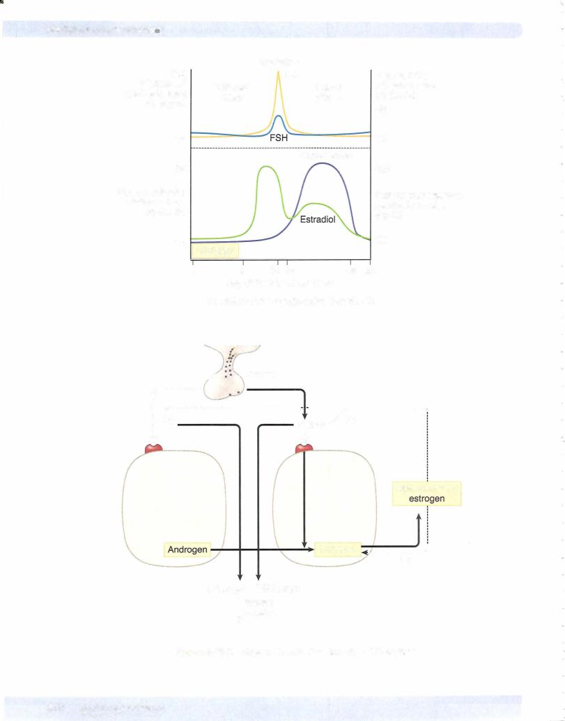
ThecaCells: Under LH stimulation, whichacts intracellularlyvia cAMP, cholesterol is transported into the mitochondria (StAR is activated). The pathway continues through intermediates to androgens. Little androgen is secreted into theblood; most ofthe androgen enters the adjacent granulosa cells.
Granulosa Cells: Possess the follicle's onlyFSH receptors. When coupled to FSH, these act via cAMP to increase the activity ofaromatase; aromatase converts the androgens to estrogens (mainly estradiol).
Estrogen: Some of the estrogen produced by the granulosa cells is released into the blood and inhibits the release of LH and FSH from the anterior pituitary. However, another fraction ofthe estrogen acts locally on granulosa cells, increas ing their proliferation and sensitivity to FSH.
•This local positive effect of estrogens causes a rising level of circulat ing estrogens during the follicular phase, but at the same time FSH is decreasing because of the inhibitory effect of estrogen on FSH release.
•Granulosa cells also release inhibin B.
•Inhibin B inhibits the secretion of FSH by the pituitary but their role in the menstral cycle is poorly understood.
Peripheral effects of estrogen produced by the granulosa cells during the fol licular phase include:
•Circulating estrogens stimulate the female sex accessory organs and sec ondary sex characteristics.
•Rising levels of estrogens cause the endometrial cells of the uterine mucosal layers to increase their rate ofmitotic division (proliferate).
•Circulating estrogens cause the cervical mucus to be thin and watery, making the cervix easy for sperm to traverse.
Ovulation
Ovulation takes place approximately on day 14. This is an approximation. Since ovulation is always 14 days before the end ofthe cycle, you can subtract 14 from the cycle length to find the day ofovulation.
Cycle length - 14 = ovulation day
MEDICAL 369
Chapter u • Female Reproductive System
Estrogen Levels
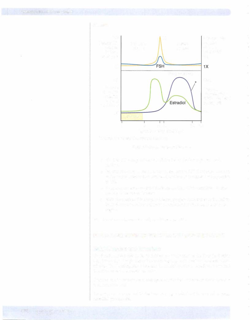
As shown in Figure X-11-2, near the end ofthe follicular phase, there is a dra matic rise in circulating estrogen. When estrogens rise above a certain level, they no longer inhibit the release ofLH and FSH. Instead, theystimulate the release of LH and FSH (negative feedbackloop to positive feedback loop).
This causes a surge in the release ofLH and FSH. Only the LH surge is essential for the induction ofovulation and formation of the corpus luteum. Notice from the figure that the LH surge and ovulation occur after estrogen peaks. Therefore, if estrogens are still rising, ovulation has not occurred.
Follicularrupture occurs 24-36 time interval, LH removes the in prophase for years. The first body is extruded.
hours afterthe onset ofthe LH surge. Duringthis restraint upon meiosis, which has been arrested meiotic division is completed, and the first polar
Positive feedback loops are rare in the body. Only ovulation with estrogen and parturition with oxytocin represent positive feedbackloops.
MEDICAL 371
Chapter u • Female Reproductive System
Preovulatory Follicle
In the latter stages ofthe follicular phase, intracellular changes within the granu losa and theca cells occur in preparation for their conversion into luteal cells.
•Estradiol, in conjunction with FSH, causes the granulosa cells to pro duce LH receptors.
•The metabolic pathways are then altered to favor the production of pro gesterone.
•This would include a decrease in the activity ofaromatase and a drop in estrogen production.
LH Surge
Induced by the elevated estrogens, it causes the granulosa cells and theca cells to be transformed intoluteal cells and increases the secretion ofprogesterone.
Corpus Luteum
The process of luteinization occurs following the exit of the oocyte from the follicle. The corpus luteum is made up ofthe remaining granulosa cells, thecal cells, and supportive tissue. Once formed, theluteal cells are stimulatedbyLH to secrete considerable progesterone and some estrogen. Progesterone inhibits LH secretion (negative feedback).
The increased plasma level ofprogesterone has several actions:
•It causesthe uterine endometrium to become secretory, providing a source ofnutrients for the blastocyst.
•It causes the cervical mucus to become thick, sealing offthe uterus from further entry of sperm or bacteria.
•It has thermogenic properties, causing the basal body temperature to increase by 0.5-1.0° F.
MEDICAL 373

Chapter 11 • Female Reproductive System
Monitoringthe Menstrual Cycle
The amount of sex steroids excreted in the urine can be used to monitor the menstrual cycle. For example:
•Lowprogesterone metabolites and lowbut slowly rising estrogen metabo lites characterizethe early follicular phase.
•Low progesterone metabolites and rapidly rising estrogen metabolites characterize the latter part ofthe follicular phase just before ovulation.
•Elevated levels ofprogesterone metabolites characterize the luteal phase and pregnancy. In the early luteal phase progesterone is rising, in the lat ter halfit is falling.
Estrogens and Androgen Formation
•Estrogen: Generic term for any estrus-producing hormone, natural or synthetic
•17 -Estradiol: Major hormone secreted by the ovarian follicle
•Estrone: Some is secreted from the ovarybut much is formed in periph eral tissues such as adipose tissue from androgens. These androgens originate from both the ovaryand the adrenal glands. This is the main circulating estrogen following menopause. Fat cells have aromatase. Adipose tissue creates modest levels of estrogen.
•Estriol: Major estrogen synthesized from circulating androgens by the placenta
•Potency: Estradiol > estrone > estriol
•Androgens: The follicles also secrete androgen; DHEA, androstenedione, and testosterone.Additional testosterone production is from the peripheral conversion ofadrenal and ovarian androgen. Some testosterone is also con vertedvia 5 a-reductase to dihydrotestosterone in the skin.
New Cycle
Duringthe 3 days prior to and during menses, plasmalevels ofprogesterone and estradiol are at their low point; negative feedback restraint for gonadotropin se cretion is removed. FSH secretion rises slightly and initiates the next cycle of follicular growth.
The length ofthe follicular phase ofthe menstrual cycle is more variable than the length of the luteal phase. Long cycles are usually due to a prolonged follicular phase and short cycles to a short follicular phase. Once ovulation has occurred, menses generally follows in about 14 days. The length ofthe menstrual cycle in days minus 14 gives the most likely dayofovulation.
MENSTRUAL IRREGULARITIES
Amenorrhea
•By definition, amenorrhea means the lack of menstral bleeding.
•Though in itselfit does not cause harm, it may be a sign ofgenetic, endocrine, or anatomic abnormalities.
MEDICAL 375

Section X • Endocrinology
•In the absence of anatomic abnormalities (and pregnancy), it usually indicates a disruption of the hypothalamic-pituitary axis or an ovarian problem.
•A hypothalamic-pituitary origin would include Kallrnan's syndrome, functional hypothalamic amenorrhea, amenorrhea in female athletes, eating disorders, hypothyroidism, and pituitary tumors such as prolacti nomas.
•Ovarian causes could be premature ovarian failure (premature meno pause), repetitive ovulation failure, or anovulation (intermittent bleeding), or a polycystic ovary.
•Hypothyroidism increases prolactin levels. TRH stimulates prolactin.
Polycystic Ovarian Syndrome
•Characterized by infertility, hirsutism, obesity, insulin resistance, and amenorrhea or oligomenorrhea
•The enlarged polycystic ovaries are known to be associated with increased androgen levels (DHEA).
•It originates in obese girls. The high extraglandular estrogens (mainly estrone) selectively suppress FSH. Ovarianfollicles do have a suppressed aromatase activity and thus a diminished capacityto convert androgen into estrogen, but the adrenals mayalso contribute to the excess androgens as well.
•High androgens promote atresia in developing follicles and disrupt feedback relationships. Look for high LH and DHEA levels.
•The overall result is anovulation-induced amenorrhea with an estrogen induced endometrial hyperplasia and breakthrough bleeding.
•Although poorlyunderstood the hyperinsulinernia is believed to be a key etiologic factor.
•Treat amenorrhea in PCOS with metformin.
•Treat androgenization with spironolactone.
Hirsutism
•Defined as an excessive generally male pattern of hair growth.
•Virilization refers to accompanying additional alterations, such as deepening of the voice, clitoromegaly, increased muscle bulk, and breast atrophy.
•It is often associated with conditions of androgen excess such as con genital adrenal hyperplasia and polycystic ovarian syndrome.
•Axillary and pubic hair are sensitive to low levels of androgen.
•Hair on the upper chest, face (scalp region not involved), and back requires more androgen and represents the pattern seen in males.
•Circulating androgens involved are testosterone, DHEA, DHEAS, and androstenedione in response to LH and ACTH.
•Measurements of DHEAS as well as a dexamethasone suppression test helps in separating an adrenal from an ovarian source.
•Polycystic ovarian syndrome is the most common cause of ovarian androgen excess.
376 MEDICAL

Chapter u • Female Reproductive System
PREGNANCY
Ovum Pickup and Fertilization
In women, the ovum is released from the rupturing follicle into the abdominal cavity, where it is "pickedup"bythe fimbria ofthe oviduct. Failure ofovumpick up may result in ectopic pregnancy, i.e., the implantation ofthe blastocyst at any site other than the interior ofthe uterus.
Fertilization occurs in the upper end ofthe oviductwithin 8-25 hours after ovu lation. After this, the ovum loses its ability to be fertilized. Sperm retain their capacity to fertilize an ovum for as long as 72 hours after ejaculation. For about 48 hours around the time ofovulation the cervical mucus is copious and slightly alkaline. This environment represent a good conduit forthe sperm.
Weeks ofgestation (gestational age) to estimate the delivery date are commonly taken from the first day ofthe last menstrual period.
Sperm are transported from the vagina to the upper ends ofthe oviduct by con traction ofthe female reproductive tract. The swimming motions ofthe sperm are important forpenetration ofthe granulosa celllayer (cumulus oophorus) and membranes surrounding the ovum.
Low sperm counts (<20 million/mL ofejaculate) are associated with reduced fer tility because sperm from ejaculates with low counts often contain many sperm withpoor motility and an abnormal morphology. Thefirst step in infertility eval uation is semen analysis.
Implantation
At the time ofimplantation, which occurs about 5-7 days after fertilization, the developmentis atthe blastocyst stage. The trophoblasticcells ofthe fetus nowbe gin to secrete a peptide hormone, human chorionic gonadotropin (hCG). HCG starts 10 days after fertilization.
Fetal hCG possesses a subunit similar tothatofLH, and therefore ithas consid erable LH activity. However, a subunit alone will not function as FSH.
The presence ofthebeta subunit ofhCG inthe urine canbe detectedbya variety oftest kits forthe detection ofpregnancy.
Hormonal Maintenance ofthe Uterine Endometrium
Figure X- 1 1 -5 illustrates the production of estrogen and progesterone during pregnancy. The figure is divided into 3 phases:
•Part ofthe luteal phase before implantation
•Early pregnancy
•Late pregnancy
MEDICAL 377

Chapter u. • Female Reproductive System
Third month to term
Placenta secretes enough progesterone and estrogen to maintain the uterus. This is not controlled by hCG. At this time, the ovaries (corpus luteum) can be re moved andpregnancycontinues.
Progesterone secretion ofthe placenta is limited onlybythe amount ofprecursor (cholesterol) deliveredbylow-densitylipoproteins (LDL) to theplacenta. Proges teronemaintainsuterine quiescence duringpregnancy.
Estrogen secretion during pregnancy involves a transfer ofsteroids from the fetal adrenal cortex and fetal liver to the placenta and then to the maternal circulation.
During midpregnancy, the fetal adrenal cortex, which is as large as the fetal kid ney, secretes considerable dehydroepiandrosterone (DHEA) and dehydroepi androsterone sulfate (DHEAS) (weak androgens that do not create problems in a female fetus).
Sequential enzymatic action bythe fetal liver andthe placenta convert these an drogens into estrogens, which enter the maternal circulation. The main estrogen produced is estriol. Rising serum or urinary estriol is considered an excellent index ofboth placental function and fetal well-being.
Peripheral Effects of Hormonal Changes
The large amount of estrogen and progesterone secreted by the placenta during pregnancy stimulates the following important changes within the mother:
•Massive growth ofthe uterus, especially the myometrium
•Increased growth of all components (glands, stroma, and fat) ofthe breasts
Additional hormonal changes
Increased prolactin secretion by the pituitary in response to elevated estrogens
Secretion of human chorionic somatomammotropin (hCS), also referred to as human placental lactogen (hPL), bythe placenta. This markedly increases during the latter halfofthe pregnancy.
•hCS (hPL) has considerable amino acid sequence homologywith growth hormone but has verylittle growth-stimulating activity.
•hCS (hPL) has metabolic actions similar to growth hormone; that is, it increases maternal lipolysis and ketogenesis and decreases maternal glu cose utilization, thereby making maternal energy stores more available for the fetus.
•During the second trimester pregnancy becomes a hyperinsulinemic state with peripheral resistance to the metabolic effects ofinsulin. This reserves glucose for fetal needs and the mother depends more heavily on fatty acids as a source of energy. Under these conditions even modest fasting can cause ketosis.
MEDICAL 379
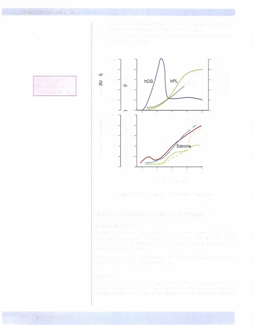

Chapter 11 • Female Reproductive System
spared. Failure to lactate is the most common clinical sign. Other manifestations would include the consequences ofhypothyroidism and hypocortisolism.
Estrogen increases the circulating steroid-bindingglobulins and bound hormone increases but FT4 is normal. Hyperthyroidism increases the risk ofpreterm deliv ery. Hypothyroidism is unusual in pregnancy.
Estrogen increases renin secretion, and overall increased activity of the renin angiotensin-aldosterone system causes fluid retention and hemodilution.
Changes inducednearthe endofpregnancy
The pubic symphysis, cervix, and vagina become more distensible. These changes make passage of the fetus through the birth canal easier. The peptide hormone relaxin, which is secreted by the ovary, also promotes these changes. Its action is not essential. Parturition in humans is normal in the absence ofovaries.
In response to elevated plasma estrogens, oxytocin receptors increase in the myo metrium. Thus, the sensitivity ofthe uterine myometrium to the excitatory action ofoxytocin is increased.
Parturition
The factors that initiate parturition are not well understood, but the following facts are known:
•Although oxytocin can be administered to induce uterine contractions, during normal parturition plasma oxytocin is not elevated until the fetus enters the birth canal (a few minutes before birth).
•Thus, increased oxytocin secretion does not initiate the rhythmic uter ine contractions characteristic of the onset of labor.
•Oxytocin does, however, cause the uterus to contract immediately after the fetus is expelled, thus limiting blood flow and blood loss.
•Acting locally on the myometrium, prostaglandins increase contractions. Oxytocin increases uterine synthesis ofprostaglandins.
•When a fetus dies, toxic products originating from the fetus increase prostaglandin release in the uterus, thus initiating contractions and a spontaneous abortion (miscarriage). Similarly, administration ofprosta glandins induces abortion.
LACTATION
Mammary Gland Growth and Secretion
Growth ofmammary tissue is stimulated bythe female sex steroids estrogen and progesterone. However, for these steroids to stimulate maximum growth, prolac tin, growth hormone, and cortisol also must be present.
During pregnancy, the high levels ofplasma estrogen greatly increase prolactin secretion, but milk synthesis does not occur because the high level of estrogen (and progesterone) blocks milk synthesis. At parturition, plasma estrogen drops, withdrawing the block on milk synthesis. As a result, the number of prolactin receptors in mammary tissue increases several-fold, and milk synthesis begins.
MEDICAL 381
Section X • Endocrinology
Maintaining Lactation
Suckling is required to maintain lactation.
Magnocellular nuclei of hypothalamus (synthesizes oxytocin)
Posterior pituitary (stores and releases oxytocin)
neurons
·Hypothalamusdecrease in release of PIF = increase in prolactin
· decrease in release of GnRH = decrease in FSH and LH
Anterior pituitary
Oxytocin |
Prolactin |
"" |
|
\ |
stimulatesSuckling ofneuralthe baby |
|
receptors in the nipple |
Figure X-11-7. Lactation and the Suckling Reflex
The suckling of the baby at the mother's breast stimulates receptors in the mother's nipples. Signals from these receptors are transmitted to the hypo thalamus and have the following effects:
•Oxytocin synthesis and secretion are increased. Oxytocin causes the myo epithelial basket cells that surround the alveoli to contract. Preformed
milk is ejected into the ducts and out the openings ofthe nipple; that is, milk ejection is initiated.
•The release ofprolactin-inhibiting factor (PIF = dopamine) bythe hypo thalamus into the hypophyseal portal vessels is inhibited. This removes a chronic restraint on prolactin secretion. Prolactin secretion increases, and milk secretion is stimulated each time the baby suckles.
•The secretion of GnRH into the hypophyseal portal vessels is inhibited; secretion of FSH and LH decreases. Thus, follicular growth, estrogen secretion, ovulation, and menses cease. High prolactin levels also con tribute to the amenorrhea.
382 MEDICAL

Chapter u • Female Reproductive System
For the suckling stimulus to inhibit GnRH secretion completely, the stimulus must be prolonged and frequent. Supplementation ofthe mother's milkwith other fluids or sources ofenergyreducesthebaby's sucklingandallows gonadotropin secretion, follicular growth, and ovulation to occur.
Womenwho do not wish tobreastfeed their children are sometimes administered large doses of estrogen. The estrogen inhibits lactation (by its inhibitory action ofmilk synthesis), even though estrogen promotes increased prolactin secretion.
Breast feeding as a form ofcontraceptive is about 95% protective, i.e., breastfeed ing willstop about 95% ofconceptions. This isbecause breastfeeding should stop ovulation.
MEDICAL 383
Section X • Endocrinology
Chapter Summary
•In the early stages ofthe follicular phase, estrogen is slowly rising. This is followed by a more rapid rise as ovulation approaches. This latter rise occurs because estrogen acts locally to enhance its own production.
•Once estrogen rises above a certain level, it no longer suppresses LH and FSH secretion but instead enhances their secretion. This induces a surge in the secretion of both LH and FSH. However, only the LH surge is required for ovulation.
•In the luteal phase, LH stimulates the luteal cells to secrete considerable progesterone as well as estrogen. Progesterone in this phase inhibits LH secretion.
•It is the drop in progesterone (and estrogen) that withdraws the hormonal support ofthe endometrium and that causes menstruation.
•Variations in the length ofthe menstrual cycle are due to the follicular phase. Once ovulation has occurred, menstruation begins almost exactly 14 days later.
•Fertilization and biological pregnancy occur at the beginning of the luteal
phase. Implantation occurs at about the middle ofthe luteal phase.
• In the first 2 to 3 months of pregnancy, fetal production of hCG is required for continuation ofthe secretion of progesterone and estrogen by the ovary.
•In pregnancy cardiac output increases, TPR decreases and blood pressure is usually below prepregnancy levels.
•Pregnancy increases GFR and decreases renal threshold.
•Pregnancy is a euthyroid state but there is activation ofthe renin angiotensin-aldosterone system, which increases ECFvolume.
•The ovaries are not required for the last 6 months of pregnancy because the placenta takes over the secretion ofboth progesterone and estrogen.
•hCG is an index offetal well-being in early pregnancy. hPL is an index of placental function later in pregnancy. Estriol in an index offetal well-being and placental function.
•Postpartum hemorrhaging can cause pituitary infarction (Sheehan's syndrome)
•Near the end of pregnancy, estrogen induces the appearance of oxytocin receptors in the myometrium. Once this occurs, oxytocin can be administered to induce labor. However, it is unlikely that a rise in oxytocin is the natural signal that begins delivery.
•During pregnancy, the rising estrogens are driving an increase in prolactin secretion, but the estrogen also blocks milk synthesis.
•At delivery, it is the drop in estrogen that initiates milk synthesis, but suckling is required to maintain lactation.
384 MEDICAL

SECTION
Gastrointestinal
Physiology

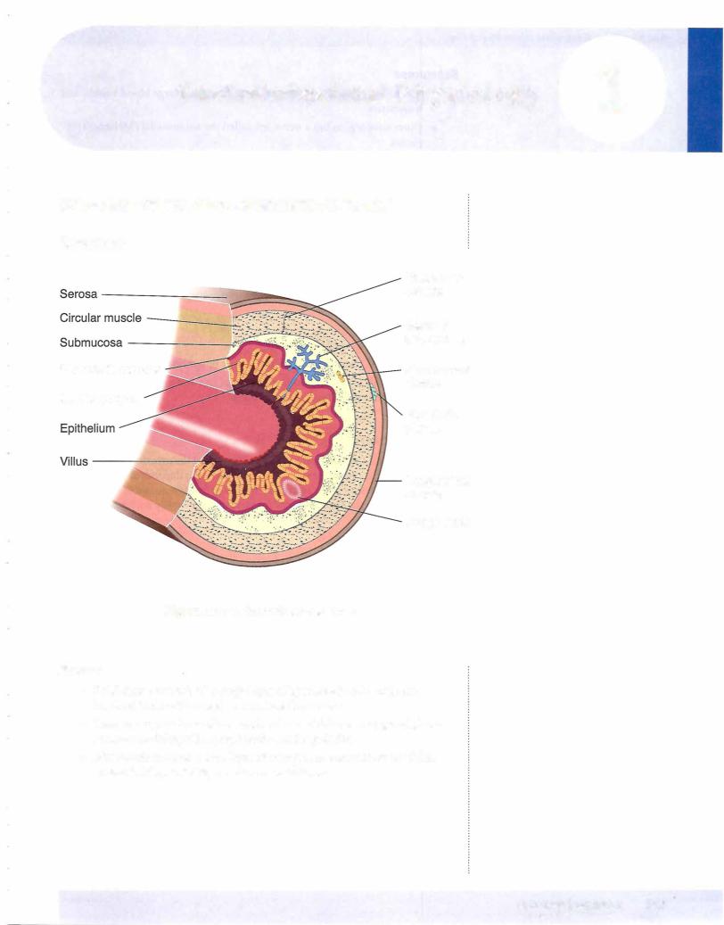

Chapter 1 • Gastrointestinal Physiology
Endocrine Control
Table Xl-1-1.The Endocrine Control of the GI System
Hormone** |
Source |
Stimulus |
Stomach Motility |
Pancreas |
Gallbladder |
|
|
|
and Secretion |
|
|
Secretin |
S cells |
Acid entering |
Inhibits |
Stimulates |
|
|
lining |
duodenum |
|
fluid secretion |
|
|
duodenum |
|
|
(HC03-) |
|
CCK |
Cells lining |
Fat and amino |
Inhibits emptying |
Stimulates |
1. Contraction |
|
duodenum |
acids entering |
|
enzyme |
2. Relaxation |
|
|
duodenum |
|
secretion |
sphincter (Oddi) |
Gastrin |
G cells of |
Stomach |
Stimulates |
|
|
|
stomach |
distension |
|
|
|
|
Antrum |
Parasym (GRP) |
|
|
|
|
|
Peptides |
|
|
|
|
Duodenum |
Stomach acid |
|
|
|
|
|
inhibits* |
|
|
|
GIP |
Duodenum |
Fat, CHO, |
Inhibits |
|
Increases insulin |
GLP |
|
amino acids |
|
|
Decreases glucagon |
CCK cholecystokinin; GIP = gastric inhibitory peptide (glucose insulinotropic peptide), GLP = glucagon·like peptide |
|||||
=
*Note: In a non-acid·producingstomach (e.g., chronic gastritis), the reduced negative feedback increases circulating gastrin. **All four hormones stimulate insulin release.
MOTILITY
Characteristics ofSmooth Muscle
Electrical activity
•Resting membrane potential -40 to -65 mV. Close to depolarization.
•Oscillation ofmembrane potential is generated by interstitial cells (inter stitial cells ofCajal) that act as pacemakers. This is referred to as slow waves or basic electrical rhythm, and ifthreshold is reached it generates action potentials.
•Action potentials are generated by the opening of slow channels that allow the entry ofboth sodium and calcium.
•The duodenum contracts the most often.
Motoractivity
•Stretch produces a contractile response.
•Gap junctions create an electrical syncytium within the smooth muscle.
•Slowwaves createlowlevel contractions, and action potentials strengthen the contractions.
•Pacemaker activity from the interstitial cells creates the intrinsic motor activity.
•Tonic contraction at sphincters act as valves.
Note
Anticholinergic medications such as atropine or tricyclic antidepressants slow GI motility.
Note
Nerve gas increases GI and bronchial secretions.
MEDICAL 389
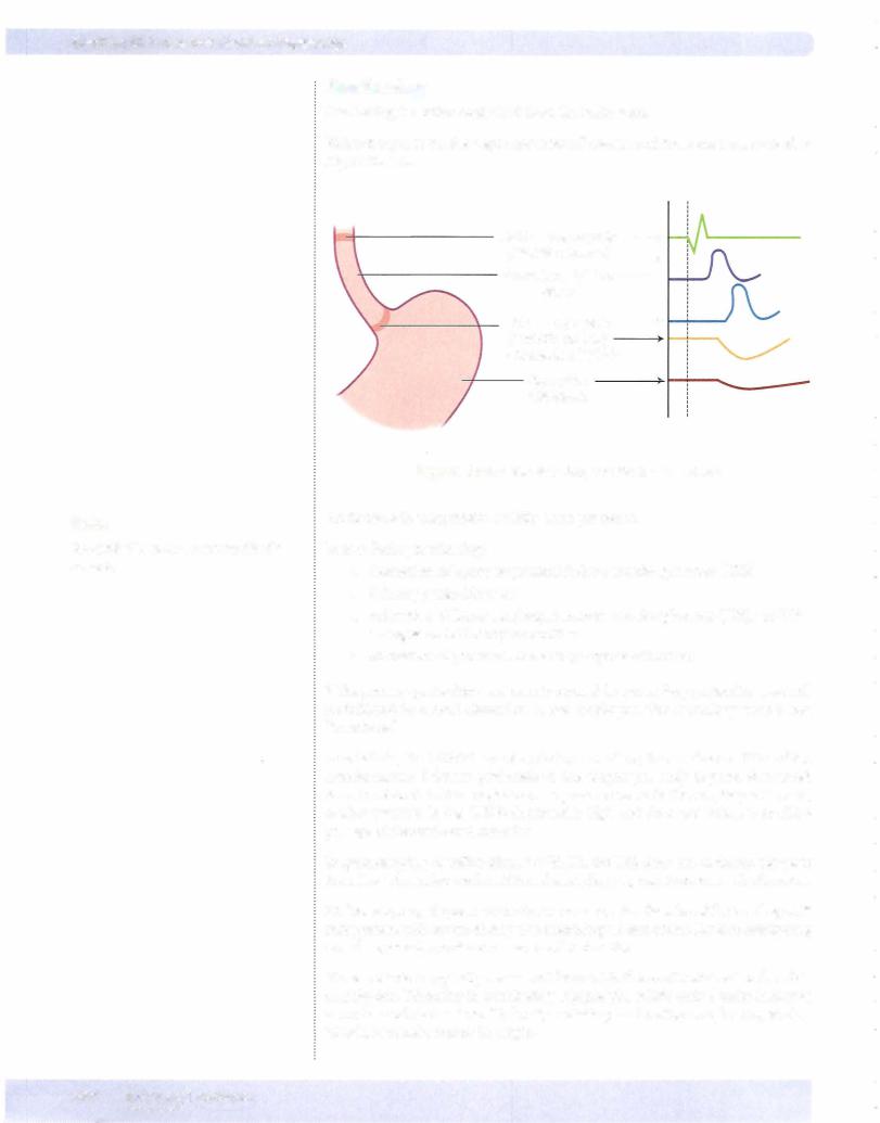

Section XI • Gastrointestinal Physiology
Colon Motility
•Segmentation contractions createbulges (haustrations) along the colon.
•Mass movements, which are propulsive, are more prolonged than the peristaltic movements ofthe small intestine.
Migrating Myoelectric Complex (MMC)
•A propulsive movement initiated during fasting that begins in the stomach and moves undigested material from the stomach and small intestine into the colon.
•Repeats every 90-120 minutes during fasting.
•When one movement reaches the distal ileum, a new one starts in the stomach.
•Correlated with high circulating levels ofmotilin, a hormone ofthe small intestine
•This movement prevents the backflow ofbacteria from the colon into the ileum and its subsequent overgrowth in the distal ileum.
Defecation
•Defecation is a reflex involving the central nervous system.
•A mass movement in the terminal colon fills the rectum and causes a reflex relaxation ofthe internal anal sphincter and a reflex contraction ofthe external anal sphincter.
•Voluntary relaxation ofthe external sphincter accompanied with pro pulsive contraction ofthe distal colon complete defecation.
•Lack of a functionalinnervation ofthe external sphincter causes invol untary defecation when the rectum fills.
SECRETIONS
Salivary Secretions
•Parotid gland secretions are entirely serous (lack mucin).
•Submandibular and sublingual gland secretions are mixed mucus and serous.
•They are almost entirely under the control ofthe parasympathetic system, which promotes secretion.
•The initial fluid formation in the acinus is via an indirect chloride pump (secondary active transportpoweredby the Na/K ATPase pump), and the electrolyte composition is isotonic and similar to interstitial fluid.
Duct cells modify the initialacinarsecretion (Figure XI-1-4).
392 MEDICAL

Chapter 1 • Gastrointestinal Physiology
Sympathetic (stimulates)
Figure Xl-1 -4. Salivary Secretion
Composition ofsalivary secretions
• Low in Na+, Clbecause of reabsorption
• High in K+, HC03 because ofsecretion (pH = 8)
•Low tonicity: Salivary fluidis hypotonic because ofreabsorption of NaCl and impermeability ofducts to water.
•a-Amylase (ptyalin): secreted in the active form and begins the digestion ofcarbohydrates
•Mucus, glycoprotein
•Immunoglobulins and lysozymes
Gastric Secretions
•The epithelial cells that cover the gastric mucosa secrete a highly viscous alkaline fluid (mucin plus bicarbonate) that protects the stomach lining from the caustic action ofHCL
•Fluid needs both mucin andbicarbonate to be protective.
•Nonsteroidal anti-inflammatory drugs such as aspirin decrease the secretion ofthe mucin and bicarbonate.
•Surface ofthe mucosa studded with the openings ofthe gastric glands
•Except for the upper cardiac region and lower pyloric region whose glands secrete mainly a mucoid fluid, gastric glands secrete a fluid whose pH can be initiallyas low as 1.0.
MEDICAL 393
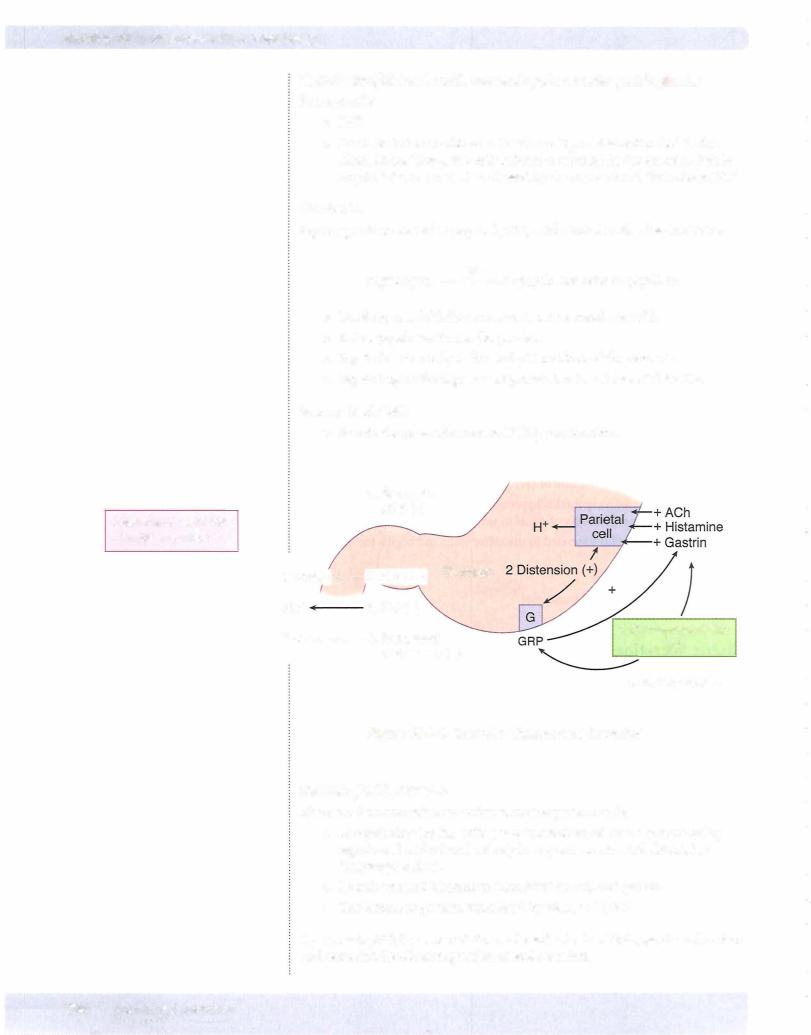

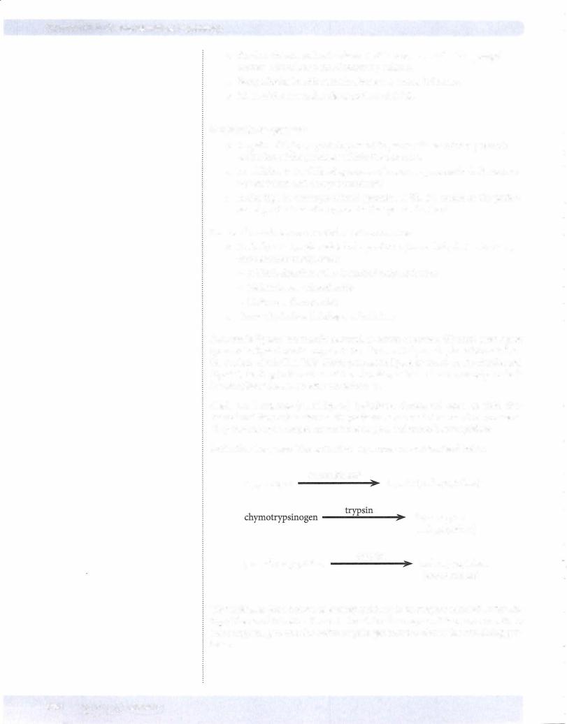

Chapter 1 • Gastrointestinal Physiology
Bilesalts and micelles
•Primary bile acids known as cholic acid and chenodeoxycholic acid are synthesized by the liver from cholesterol.
•The lipid-soluble bile acids are then conjugated primarily with glycine.
•The conjugated forms are water-soluble but contain a lipid-soluble segment.
•Because they are ionized at neutral pH, conjugated bile acids exist as salts of cations (Na+) and are, therefore, called bile salts.
•Bile salts are actively secreted by the liver.
•Secondary bile acids are formed by deconjugation and dehydroxylation of the primary bile salts by intestinal bacteria, forming deoxycholic acid (from cholic acid) and lithocholic acid (from chenodeoxycholic acid) .
•Lithocholic acid has hepatotoxic activity and is excreted.
•When bile salts become concentrated, they form micelles. These are water-soluble spheres with a lipid-soluble interior.
•As such, they provide a vehicle to transport lipid-soluble materials in the aqueous medium of the bile fluid and the small intestine.
•Micelles are vital in the digestion, transport, and absorption of lipid soluble substances from the duodenum to the distal ileum.
•In the distal il um, and only in the distal ileum, can the bile salts be actively reabsorbed and recycled (enterohepatic circulation).
•Lack of active reabsorbing mechanisms (or a distal ileal resection) causes loss in the stool and a general deficiency in bile salts, as the liver has a limited capacity to manufacture them.
•This deficiency can lead to fat malabsorption and cholesterol gallstones.
Bile pigments
A major bile pigment, bilirubin is a lipid-soluble metabolite of hemoglobin. Transported to the liver attached to protein, it is then conjugated and excreted as water-soluble glucuronides. These give a golden yellow color to bile.
Stercobilin is produced from metabolism of bilirubin by intestinal bacteria. It gives a brown color to the stool.
Salts and water
The HC03- component is increased by the action of secretin on the liver.
The active pumping of sodium in the gallbladder causes electrolyte and water reabsorption, which concentrates the bile.
Bile pigments and bile salts are not reabsorbed from the gallbladder.
Phospholipids (mainly lecithin)
Insoluble in water but are solubilized by bile salt micelles
Cholesterol
Present in small amounts. It is insoluble in water and must be solubilized by bile salt micelles before it can be secreted in the bile.
Bridge to Pathology
Increased levels of plasma bilirubin produce jaundice. If severe, bilirubin can accumulate in the brain, producing profound neurological disturbances (kernicterus).
MEDICAL 399

Chapter 1 • Gastrointestinal Physiology
•Luminal Cl then pulls water, Na, and other ions into the lumen, creating the isotonic secretion. This is the general scheme ofthe chloride pump.
•Neurotransmitter secretagogues include VIP andACh.
•The CIchannels are opened by increases in cytosolic Ca2+ and/or cAMP. The cAMP-dependent Ca2+ channels are CFTR channels.
DIGESTION
Figure XI-1-10 summarizes the regional entry ofthe major digestive enzymes proceeding from the mouth, stomach, andthroughthe small intestine.
Triglyceride |
CHO |
*Lipase Colipase
*Bile micelles
2'-Monoglycerides |
Glucose, Galactose, |
Fatty acids |
Fructose |
Protein
a-Amylase
---Pepsin
*a-Amylase ---*Trypsin, etc.
*Sucrase ---*Peptidases *Lactase
(lactose
intolerant)
Amino acids (40%)
Di-, Tripeptides (60%)
Mouth
Stomach
Pancreas
Gallbladder
Small intestine (brush border)
*Required for digestion
Figure Xl-1-10. Summary of Digestive Processes
MEDICAL 401
Section XI • Gastrointestinal Physiology
Digestive Enzymes and End Products
Triglycerides
Stomach: Fatty materials are pulverized to decrease particle size and increase surface area.
Small intestine: Bile micelles emulsify the fat, and pancreatic lipases digest it. Micelles and pancreatic lipase are required for triglyceride digestion. The major end products are 2-monoglycerides andfatty acids.
Carbohydrates
Mouth: Salivary a-amylase begins the digestion, and its activity continues in the stomach until acid penetrates the bolus; however, it is not a required enzyme.
Small intestine: Pancreatic a-amylase, a required enzyme for CHO digestion, continues the process. Hydrolysis of starch by a-amylase goes on in solution in the lumen of the small intestine, mostly in the duodenum. Further processing or splitting of these trisaccharides, disaccharides, and oligosaccharides is neces sary but does not take place in solution; rather, it occurs on the brush border. The enzymes-a-dextrinase (or a-glucoamylase), isomaltase, and maltase-are all bound to the brush border (apical membrane of enterocytes). Brush border enzymes have their highest activity in the jejunum (upper). These brush border enzymes are required for digestion mainly because disaccharides-e.g., sucrose, lactose-are not absorbed from the gut.
•The a-dextrinase cleaves terminal a-1,4 bonds, producing free glucose.
•Lactase hydrolyzes lactose into glucose and galactose. Lactase deficiency (lactose intolerance) leads to osmotic diarrhea.
•Sucrase splits sucrose into glucose and fructose.
•Maltase (also a brush border enzyme) breaks down the maltose and maltotriose to form 2 and 3 glucose units, respectively.
•The monosaccharide end products-glucose, galactose, and fructose are readily absorbed from the small intestine, also mainly in the jejunum.
Proteins
Stomach: Pepsin begins the digestion of protein in the acid medium of the stomach; however, it is not an essential enzyme.
Small intestine: Digestion continues with the pancreatic proteases (trypsin, chymotrypsin, elastase, and carboxypeptidases A and B), which are essential enzymes.
Protein digestion is completed by the small intestinal brush border enzymes, dipeptidases, and an aminopeptidase. The main end products are amino acids (40%) and dipeptides and tripeptides (60%).
Pancreatic enzymes are required for triglyceride, CHO, and protein digestion. Circulating CCK is almosttotallyresponsible for their secretionfollowinga meal.
402 MEDICAL
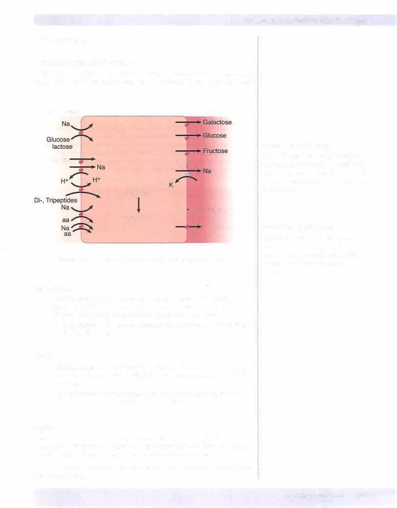
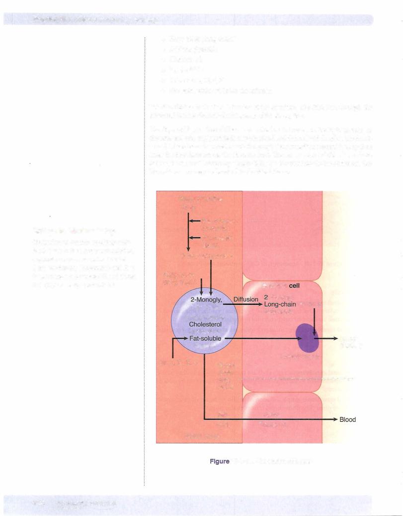

Chapter 1 • Gastrointestinal Physiology
Electrolytes
The net transport of electrolytes along the length of the small and large intestine is summarized in Figure XI- 1-13.
Duodenum
•Hypertonic fluid enters this region, and following the movement of some water into the lumen, the fluid becomes and remains isotonic (see crypt secretion above).
•The absorption of most divalent ions and water-soluble vitamins begins here and continues through the small intestine.
•Injested iron and calcium tend to form insoluble salts. The acid envi ronment of the stomach redissolves these salts, which facilitates their absorption in the small intestine. Iron and calcium absorption is dimin ished in individuals with a deficient stomach acid secretion.
•Calcium absorption is enhanced by the presence of calbindin in intestinal cells, and calcitriol (active vitamin D) induces the synthesis of this protein.
•Intestinal cells express the protein ferritin, which facilitates iron absorption.
Jejunum
•Overall, there is a net reabsorption of water and electrolytes.
•The cellular processes involved are almost identical to those described in the renal physiology section for the cells lining the nephron proximal tubule.
Ileum
•Net reabsorption of water, sodium, chloride, and potassium continues, but there begins a net secretion of bicarbonate.
•It is in the distal ileum, and only in the distal ileum, where the reabsorp tion of bile salts and intrinsic factor with vitamin B12 takes place.
Colon
•The colon does not have digestive enzymes or the protein transporters to absorb the products of carbohydrate and protein digestion.
•Also, because bile salts are reabsorbed in the distal ileum, very few lipid soluble substances are absorbed in the colon.
•There is a net reabsorption of water and sodium chloride, but there are limitations.
•Most of the water and electrolytes must be reabsorbed in the small intestine, or the colon becomes overwhelmed.
•Most of the water and electrolytes are absorbed in the ascending and transverse colon; thereafter, the colon has mainly a storage function.
•The colon is a target for aldosterone, where it increases sodium and water reabsorption and potassium secretion.
•Because there is a net secretion of bicarbonate and potassium, diarrhea usually produces a metabolic acidosis and hypokalernia. It commonly presents as hyperchlorernic, nonanion gap metabolic acidosis, as described in the acid-base section.
MEDICAL 405
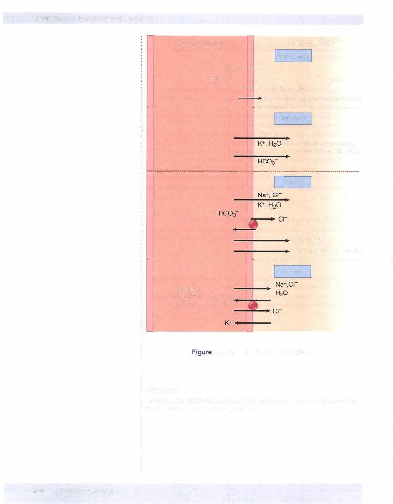

Chapter 2. • Gastrointestinal Physiology
Chapter Summary
•Sympathetic activity slows processes in the GI tract, whereas parasympathetic does the opposite.
•Secretin is required for releasing pancreatic bicarbonate, which neutralizes stomach acid entering the duodenum.
•CCK is required to release pancreatic enzymes and for the release of bile into the duodenum.
•Gastric stomach activity and its secretion is inhibited by stomach acid.
•Because of regional pacemaker activity, there is always some basal motor activity in the GI tract.
•Swallowing and defecation are reflexes requiring the central nervous system. Relaxation ofthe lower esophageal sphincter is due to the release ofthe inhibitory transmitter VIP.
•Following a meal, local distension and parasympathetic activity increase stomach motility. An overload of the duodenum decreases stomach motility.
•There are mixing and propulsive movements in the small intestine and colon.
•Salivary secretions are regulated via parasympathetic input. The reabsorption of sodium chloride produces a fluid that is hypotonic.
•Gastric acid secretion via a hydrogen/potassium-ATPase pump is stimulated by acetylcholine, histamine, and gastrin. A low stomach pH and a duodenal overload inhibit acid secretion.
•Pancreatic enzymes are required forthe digestion of carbohydrate, protein, and lipids.
•Pancreatic fluid high in bicarbonate is required to neutralize acid entering the duodenum.
•Bile salts form micelles that are required for the digestion and absorption of lipids. Only in the distal ileum can they be actively reabsorbed. They must be recycled because the liver has a limited ability fortheir synthesis.
•Bile pigments are water-soluble compounds excreted via the bile and intestine.
•Electrolytes and water, but not bile pigments or bile salts, can be absorbed from the gall bladder lumen.
•Digestion of lipid requires bile micelles and pancreatic lipases. End products absorbed include 2-monoglycerides and fatty acids.
(Continued)
MEDICAL 407
