7.3 ● Oligosaccharides |
221 |
R O H
C +
R' OH
Hemiacetal
Hemiketal
|
R |
O |
H |
|
R'' |
OH |
|
C |
|
|
|
R' |
O |
R'' |
|
|
|
Acetal |
|
|
R |
O |
R''' |
|
R'' |
OH |
|
C |
|
|
|
R' |
O |
R'' |
|
|
|
Ketal |
|
FIGURE 7.16 ● Acetals and ketals can be formed from hemiacetals and hemiketals, respectively.
Acetals, Ketals, and Glycosides
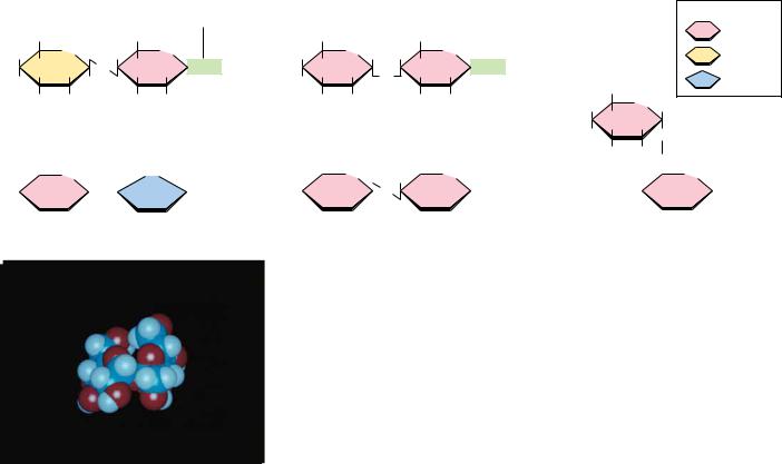
Hemiacetals and hemiketals can react with alcohols in the presence of acid to form acetals and ketals, as shown in Figure 7.16. This reaction is another example of a dehydration synthesis and is similar in this respect to the reactions undergone by amino acids to form peptides and nucleotides to form nucleic acids. The pyranose and furanose forms of monosaccharides react with alcohols in this way to form glycosides with retention of the - or -configuration at the C-1 carbon. The new bond between the anomeric carbon atom and the oxygen atom of the alcohol is called a glycosidic bond. Glycosides are named according to the parent monosaccharide. For example, methyl- -D-glucoside (Figure 7.17) can be considered a derivative of -D-glucose.
7.3 ● Oligosaccharides
|
|
|
CH2OH |
|
|
|
|
H |
O |
H |
|
|
|
H |
|
|
|
|
|
|
|
|
|
|
|
|
|
OH |
H |
|
|
|
|
HO |
|
O |
CH3 |
|
|
|
H |
OH |
|
|
|
|
Methyl-α -D-glucoside |
|
|
|
CH2OH |
|
|
|
|
H |
O |
|
|
|
|
O |
CH3 |
|
|
|
|
|
H |
|
|
|
|
|
|
|
OH |
H |
|
|
|
|
|
|
H |
|
HO |
|
|
|
|
H |
OH |
|
|
|
Methyl-β -D-glucoside
FIGURE 7.17 ● The anomeric forms of methyl-D-glucoside.
Given the relative complexity of oligosaccharides and polysaccharides in higher organisms, it is perhaps surprising that these molecules are formed from relatively few different monosaccharide units. (In this respect, the oligoand polysaccharides are similar to proteins; both form complicated structures based on a small number of different building blocks.) Monosaccharide units include the hexoses glucose, fructose, mannose, and galactose and the pentoses ribose and xylose.
Disaccharides
The simplest oligosaccharides are the disaccharides, which consist of two monosaccharide units linked by a glycosidic bond. As in proteins and nucleic acids, each individual unit in an oligosaccharide is termed a residue. The disaccharides shown in Figure 7.18 are all commonly found in nature, with sucrose, maltose, and lactose being the most common. Each is a mixed acetal, with one hydroxyl group provided intramolecularly and one hydroxyl from the other monosaccharide. Except for sucrose, each of these structures possesses one free unsubstituted anomeric carbon atom, and thus each of these disaccharides is a reducing sugar. The end of the molecule containing the free anomeric carbon is called the reducing end, and the other end is called the nonreducing end. In the case of sucrose, both of the anomeric carbon atoms are substituted, that is, neither has a free OOH group. The substituted anomeric carbons cannot be converted to the aldehyde configuration and thus cannot participate in
FIGURE 7.18
222 |
Chapter 7 ● |
Carbohydrates |
|
|
|
|
|
|
|
|
|
Free anomeric carbon |
|
|
|
|
Simple sugars |
|
|
|
|
(reducing end) |
|
|
|
|
Glucose |
|
CH2OH |
|
CH2OH |
|
CH2OH |
|
CH2OH |
|
|
|
|
|
|
|
HO |
O |
O |
O |
HOH |
O |
|
O |
HOH |
Galactose |
|
OH |
OH |
OH |
O |
OH |
Fructose |
|
|
|
|
HO |
|
|
|
|
|
|
|
|
|
|
|
CH2OH |
|
OH |
|
OH |
|
OH |
|
OH |
|
|
|
|
|
|
O |
|
Lactose (galactose-β -1,4-glucose) |
Maltose (glucose-α -1,4-glucose) |
OH |
|
|
|
|
|
|
|
|
|
|
|
|
|
|
|
|
|
|
HO |
|
|
|
|
|
|
|
|
|
|
|
|
|
CH2OH |
|
|
CH2OH |
|
|
O |
|
|
|
|
O H |
|
OH |
|
O |
|
|
HO |
|
HO |
|
|
|
|
|
|
CH2OH |
|
|
|
|
|
|
|
|
|
OH |
|
|
|
OH |
Sucrose (glucose-α -1,2-fructose)
|
|
CH2OH |
|
CH2OH |
|
OH |
|
CH2 |
O |
|
|
|
|
|
|
|
O |
|
O |
|
|
|
|
|
|
OH |
O OH |
HOH |
|
OH |
|
|
HOH |
|
HO |
|
|
|
|
|
|
HO |
|
|
|
|
|
|
|
|
|
OH |
|
OH |
|
|
|
|
|
OH |
|
Cellobiose (glucose-β -1,4-glucose) |
Isomaltose (glucose-α -1,6-glucose) |
● The structures of several important disaccharides. Note that the notation OHOH means that the configuration can be either or . If the OOH group is above the ring, the configuration is termed . The configuration is if the OOH group is below the ring as shown. Also note that sucrose has no free anomeric carbon atoms.
Sucrose
the oxidation–reduction reactions characteristic of reducing sugars. Thus, sucrose is not a reducing sugar.
Maltose, isomaltose, and cellobiose are all homodisaccharides because they each contain only one kind of monosaccharide, namely, glucose. Maltose is produced from starch (a polymer of -D-glucose produced by plants) by the action of amylase enzymes and is a component of malt, a substance obtained by allowing grain (particularly barley) to soften in water and germinate. The enzyme diastase, produced during the germination process, catalyzes the hydrolysis of starch to maltose. Maltose is used in beverages (malted milk, for example), and because it is fermented readily by yeast, it is important in the brewing of beer. In both maltose and cellobiose, the glucose units are 1n4 linked, meaning that the C-1 of one glucose is linked by a glycosidic bond to the C-4 oxygen of the other glucose. The only difference between them is in the configuration at the glycosidic bond. Maltose exists in the -configuration, whereas cellobiose is . Isomaltose is obtained in the hydrolysis of some polysaccharides (such as dextran), and cellobiose is obtained from the acid hydrolysis of cellulose. Isomaltose also consists of two glucose units in a glycosidic bond, but in this case, C-1 of one glucose is linked to C-6 of the other, and the configuration is .
The complete structures of these disaccharides can be specified in shorthand notation by using abbreviations for each monosaccharide, or , to denote configuration, and appropriate numbers to indicate the nature of the linkage. Thus, cellobiose is Glc 1–4Glc, whereas isomaltose is Glc 1–6Glc. Often the glycosidic linkage is written with an arrow so that cellobiose and isomaltose would be Glc 1n4Glc and Glc 1n6Glc, respectively. Because the linkage carbon on the first sugar is always C-1, a newer trend is to drop the 1- or
7.3 ● Oligosaccharides |
223 |
A D E E P E R L O O K
Trehalose—A Natural Protectant for Bugs
Insects use an open circulatory system to circulate hemolymph (insect blood). The “blood sugar” is not glucose but rather trehalose, an unusual, nonreducing disaccharide (see Figure). Trehalose is found typically in organisms that are naturally subject to temperature variations and other environmental stresses— bacterial spores, fungi, yeast, and many insects. (Interestingly, honeybees do not have trehalose in their hemolymph, perhaps because they practice a colonial, rather than solitary, lifestyle. Bee colonies maintain a rather constant temperature of 18°C, protecting the residents from large temperature changes.)
What might explain this correlation between trehalose utilization and environmentally stressful lifestyles? Konrad Bloch* suggests that trehalose may act as a natural cryoprotectant. Freezing and thawing of biological tissues frequently causes irreversible structural changes, destroying biological activity. High concentrations of polyhydroxy compounds, such as sucrose and glycerol, can protect biological materials from such damage.
Trehalose is particularly well-suited for this purpose and has been shown to be superior to other polyhydroxy compounds, especially at low concentrations. Support for this novel idea comes from studies by P. A. Attfield,† which show that trehalose levels in the yeast Saccharomyces cerevisiae increase significantly during exposure to high salt and high growth temperatures—the same conditions that elicit the production of heat-shock proteins!
H |
H |
H |
H |
CH OH |
|
OH |
HO |
|
|
OH |
|
|
|
|
H |
H |
2OH |
|
OH |
|
H |
|
H |
O |
|
H |
|
|
H |
* Bloch, K., 1994. Blondes in Venetian Paintings, the Nine-Banded Armadillo, and Other Essays in Biochemistry. New Haven: Yale University Press.
†Attfield, P. A., 1987. Trehalose accumulates in Saccharomyces cerevisiae during exposure to agents that induce heat shock responses. FEBS Letters 225:259.
1n and describe these simply as Glc 4Glc and Glc 6Glc, respectively. More complete names can also be used, however, so that maltose would be O- -D- glucopyranosyl-(1n4)-D-glucopyranose. Cellobiose, because of its -glycosidic linkage, is formally O- -D-glucopyranosyl-(1n4)-D-glucopyranose.
-D-lactose (O- -D-Galactopyranosyl-(1n4)-D-glucopyranose) (Figure 7.18) is the principal carbohydrate in milk and is of critical nutritional importance to mammals in the early stages of their lives. It is formed from D-galactose and D-glucose via a (1n4) link, and because it has a free anomeric carbon, it is capable of mutarotation and is a reducing sugar. It is an interesting quirk of nature that lactose cannot be absorbed directly into the bloodstream. It must first be broken down into galactose and glucose by lactase, an intestinal enzyme that exists in young, nursing mammals but is not produced in significant quantities in the mature mammal. Most humans, with the exception of certain groups in Africa and northern Europe, produce only low levels of lactase. For most individuals, this is not a problem, but some cannot tolerate lactose and experience intestinal pain and diarrhea upon consumption of milk.
Sucrose, in contrast, is a disaccharide of almost universal appeal and tolerance. Produced by many higher plants and commonly known as table sugar, it is one of the products of photosynthesis and is composed of fructose and glucose. Sucrose has a specific optical rotation, [ ]D20, of 66.5°, but an equimolar mixture of its component monosaccharides has a net negative rotation ([ ]D20 of glucose is 52.5° and of fructose is 92°). Sucrose is hydrolyzed by the enzyme invertase, so named for the inversion of optical rotation accompanying this reaction. Sucrose is also easily hydrolyzed by dilute acid, apparently because the fructose in sucrose is in the relatively unstable furanose form. Although sucrose and maltose are important to the human diet, they are not taken up directly in the body. In a manner similar to lactose, they are first hydrolyzed by sucrase and maltase, respectively, in the human intestine.
224 Chapter 7 ● Carbohydrates
A D E E P E R L O O K
Honey—An Ancestral Carbohydrate Treat
Honey, the first sweet known to humankind, is the only sweetening agent that can be stored and used exactly as produced in nature. Bees process the nectar of flowers so that their final product is able to survive long-term storage at ambient temperature. Used as a ceremonial material and medicinal agent in earliest times, honey was not regarded as a food until the Greeks and Romans. Only in modern times have cane and beet sugar surpassed honey as the most frequently used sweetener. What is the chemical nature of this magical, viscous substance?
The bees’ processing of honey consists of (1) reducing the water content of the nectar (30 to 60%) to the self-preserving range of 15 to 19%, (2) hydrolyzing the significant amount of sucrose in nectar to glucose and fructose by the action of the enzyme invertase, and (3) producing small amounts of gluconic acid from glucose by the action of the enzyme glucose oxidase. Most of the sugar in the final product is glucose and fructose,
and the final product is supersaturated with respect to these monosaccharides. Honey actually consists of an emulsion of microscopic glucose hydrate and fructose hydrate crystals in a thick syrup. Sucrose accounts for only about 1% of the sugar in the final product, with fructose at about 38% and glucose at 31% by weight.
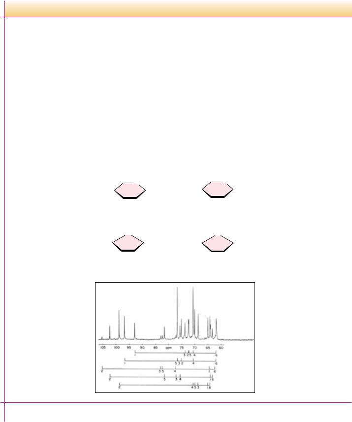
The figure shows a 13C nuclear magnetic resonance spectrum of honey from a mixture of wildflowers in southeastern Pennsylvania. Interestingly, five major hexose species contribute to this spectrum. Although most textbooks show fructose exclusively in its furanose form, the predominant form of fructose (67% of total fructose) is -D-fructopyranose, with the - and - fructofuranose forms accounting for 27% and 6% of the fructose, respectively. In polysaccharides, fructose invariably prefers the furanose form, but free fructose (and crystalline fructose) is predominantly -fructopyranose.
|
|
|
6 |
|
|
|
|
|
1 |
|
|
|
|
|
6 |
|
O |
|
OH |
|
|
|
|
|
|
|
|
|
|
|
|
|
|
|
|
|
|
|
|
|
|
|
|
|
O |
|
CH2OH |
|
|
|
|
|
|
|
5 |
|
|
|
|
OH |
|
|
|
|
5 |
|
|
|
OH |
2 |
|
|
|
|
|
|
|
|
|
|
|
|
|
|
|
|
2 |
|
|
|
|
|
|
|
|
|
|
|
|
|
|
|
|
|
|
|
|
|
|
|
|
|
|
|
|
|
|
|
|
|
|
|
|
|
|
HO |
|
|
|
3 1CH2OH |
HO |
|
|
4 |
|
|
3 |
|
OH |
|
|
4 |
|
|
|
|
OH |
|
|
|
|
|
|
|
|
|
OH |
|
|
|
|
|
|
|
|
|
|
|
|
|
|
|
|
|
|
|
|
|
|
|
|
|
|
|
|
|
|
|
|
|
|
α -D-Fructopyranose |
|
β -D-Fructopyranose |
HOH2C |
|
|
O |
1 |
CH2OH |
|
|
|
O |
|
|
|
|
|
|
|
HOH2C |
|
|
|
|
|
OH |
|
|
|
|
|
|
|
|
|
|
|
|
|
|
|
|
|
|
|
|
|
|
|
|
|
|
|
5 |
|
|
|
|
|
OH |
|
2 |
|
|
5 |
|
|
|
OH |
|
2 |
|
|
|
|
|
|
|
|
|
|
|
|
|
|
|
|
|
|
|
|
|
|
|
|
|
|
|
|
|
|
|
|
|
|
|
|
|
|
|
|
|
|
|
|
|
|
|
OH |
|
|
|
|
|
|
|
|
|
|
CH2OH |
|
|
|
|
4 |
|
3 |
|
|
|
|
|
|
|
4 |
|
3 |
1 |
|
|
|
|
|
|
|
|
|
|
|
|
|
|
|
|
|
|
OH |
|
|
|
|
|
|
|
|
|
OH |
|
|
|
|
|
|
|
|
|
α -D-Fructofuranose |
β -D-Fructofuranose |
Honey
α -D-Glucopyranose
β -D-Glucopyranose
α -D-Fructofuranose
β -D-Fructofuranose
β -D-Fructopyranose
White, J. W., 1978. Honey. Advances in Food Research 24:287–374.
Prince, R. C., Gunson, D. E., Leigh, J. S., and McDonald, G. G., 1982. The predominant form of fructose is a pyranose, not a furanose ring. Trends in Biochemical Sciences 7:239–240.
Melezitose (a constituent of honey) |
Amygdalin (occurs in seeds of Rosaceae; |
|
Laetrile (claimed to be an anticancer |
|
|
|
|
CH2OH |
glycoside of bitter almonds, in kernels of cherries, |
agent, but there is no rigorous scientific |
|
|
|
|
peaches, apricots) |
|
|
|
|
|
|
|
evidence for this) |
|
|
|
|
O |
|
|
|
|
|
|
|
|
|
|
|
|
|
|
|
|
|
|
|
|
|
|
CN |
|
|
|
OH |
|
|
CH2OH |
|
|
|
|
|
|
|
|
|
|
|
COOH |
|
|
|
|
|
|
|
|
|
|
|
|
|
|
|
|
|
|
|
|
|
|
|
|
HO |
|
|
|
O O |
|
|
|
CH2 |
|
|
|
|
|
O O |
|
CH |
|
|
|
|
|
|
|
|
|
|
|
|
|
|
|
|
|
|
|
|
|
|
|
|
|
|
|
|
|
|
|
|
|
|
|
|
|
|
|
|
|
|
|
|
|
|
|
|
|
O O |
C H |
|
|
|
|
|
|
|
|
|
|
|
|
|
|
|
|
CH2OH |
|
OH |
|
OH |
|
|
|
|
|
|
|
|
|
OH |
|
|
|
|
|
|
|
|
|
|
|
|
|
|
O |
|
|
O |
HO |
|
|
|
|
|
OH |
|
CN |
HO |
|
|
|
|
|
|
|
|
|
|
|
|
|
|
|
|
OH |
CH2OH |
|
|
OH |
HO |
|
|
|
|
|
|
|
OH |
|
|
O |
|
|
|
|
|
|
|
|
|
|
|
|
|
HO |
|
|
|
|
|
|
|
|
|
|
|
|
|
|
OH |
|
|
|
|
|
|
|
|
|
|
|
|
|
|
|
O
OH
CH2OH
Stachyose (a constituent of many plants: white jasmine, OH yellow lupine, soybeans, lentils, etc.; causes flatulence
since humans cannot digest it)
Cycloheptaamylose (a breakdown product of starch; |
CH2OH |
|
|
useful in chromatographic separations) |
HO |
O |
|
|
|
|
|
|
|
|
|
CH2OH |
|
|
OH |
|
|
|
|
|
|
|
|
|
|
|
O |
CH2 |
|
|
|
|
|
|
|
|
O |
|
|
|
|
|
|
|
|
|
|
|
|
OH HO |
O |
|
|
|
H |
|
|
|
|
OH |
|
|
|
|
O |
O |
|
|
|
CH |
|
OH |
|
|
|
|
|
|
|
2 |
|
|
CH |
2 |
|
O |
O |
|
|
|
O |
CH2 |
|
|
|
|
O |
|
|
|
|
|
OH |
|
|
|
|
|
|
|
|
H |
|
OH |
|
|
OH |
O |
|
|
|
O |
H |
O |
|
|
|
|
|
|
|
|
|
|
|
|
|
OH |
|
|
|
|
|
|
|
|
|
|
|
|
|
|
|
|
|
|
|
|
|
|
|
|
OH |
O |
|
|
|
|
|
|
|
|
|
|
|
|
|
|
|
|
|
OH |
OH |
|
2 |
CH |
|
|
|
|
|
|
O |
|
|
|
|
|
|
|
|
|
|
|
|
O |
|
|
OH |
|
Dextrantriose (a constituent of saké and honeydew) |
|
|
|
|
|
|
|
|
OH |
|
|
|
|
CH2OH |
CH2 |
CH2 |
|
|
|
|
|
O |
O |
O |
|
|
|
|
|
|
|
|
O |
2CH |
|
|
|
|
OH |
OH |
OH |
HOH |
OH |
|
|
|
HO |
O HO |
O |
HO |
|
|
|
|
|
|
|
OH |
OH |
OH |
|
FIGURE 7.19 ● The structures of some interesting oligosaccharides.
Higher Oligosaccharides
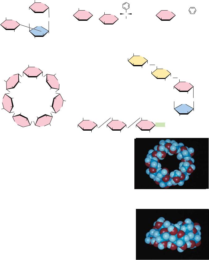
In addition to the simple disaccharides, many other oligosaccharides are found in both prokaryotic and eukaryotic organisms, either as naturally occurring substances or as hydrolysis products of natural materials. Figure 7.19 lists a number of simple oligosaccharides, along with descriptions of their origins and interesting features. Several are constituents of the sweet nectars or saps exuded or extracted from plants and trees. One particularly interesting and useful group of oligosaccharides is the cycloamyloses. These oligosaccharides are cyclic structures, and in solution they form molecular “pockets” of various diameters. These pockets are surrounded by the chiral carbons of the saccharides themselves and are able to form stereospecific inclusion complexes with chiral molecules that can fit into the pockets. Thus, mixtures of stereoisomers of small organic molecules can be separated into pure isomers on columns of cycloheptaamylose, for example.
Stachyose is typical of the oligosaccharide components found in substantial quantities in beans, peas, bran, and whole grains. These oligosaccharides are not digested by stomach enzymes, but are metabolized readily by bacteria in the intestines. This is the source of the flatulence that often accompanies the consumption of such foods. Commercial products are now available that assist in the digestion of the gas-producing components of these foods. These products contain an enzyme that hydrolyzes the culprit oligosaccharides in the stomach before they become available to intestinal microorganisms.
Cycloheptaamylose
Cycloheptaamylose (side view)
226 Chapter 7 ● Carbohydrates
Another notable glycoside is amygdalin, which occurs in bitter almonds and in the kernels or pits of cherries, peaches, and apricots. Hydrolysis of this substance and subsequent oxidation yields laetrile, which has been claimed by some to have anticancer properties. There is no scientific evidence for these claims, and the U.S. Food and Drug Administration has never approved laetrile for use in the United States.
Oligosaccharides also occur widely as components (via glycosidic bonds) of antibiotics derived from various sources. Figure 7.20 shows the structures of a few representative carbohydrate-containing antibiotics. Some of these antibiotics also show antitumor activity. One of the most important of this type is bleomycin A2, which is used clinically against certain tumors.
Bleomycin A2 (an antitumor agent used clinically against specific tumors) |
Aburamycin C (an antibiotic and antitumor agent) |
O |
NH2 O |
|
NH2 O |
H |
|
|
|
|
|
O |
|
|
N |
|
|
|
|
|
H3C |
|
|
|
|
|
|
|
OH |
|
|
|
|
|
|
|
H |
H |
|
|
HO |
H |
|
|
|
|
O |
O |
|
|
|
N |
|
|
|
|
|
|
|
O |
|
|
|
|
NH2 |
O NH |
|
|
|
|
|
HO |
|
|
|
|
|
|
|
|
|
H3C |
|
|
|
|
|
|
|
|
|
|
|
|
CH3 |
N |
|
N |
|
HN |
|
|
|
|
|
|
|
OMe |
|
|
|
H |
H |
|
|
|
|
|
|
|
OH |
HO |
H2N |
CH3 |
N |
|
O |
S |
N |
|
|
OH |
|
|
|
|
|
|
|
OH |
|
|
|
|
|
|
|
|
|
|
|
|
|
|
|
|
O |
|
|
|
|
|
|
|
H3C |
|
OH |
|
|
|
N |
|
|
H |
|
|
|
CH3 |
|
|
O |
OH |
|
|
|
|
|
N |
S |
+ |
S |
O CH |
O |
H3C |
|
|
|
|
|
|
|
|
|
|
|
|
|
|
|
|
|
|
|
|
|
|
|
|
HN |
|
|
O |
|
|
|
CH3 |
OMe |
O |
O |
CH3 |
|
|
|
O |
|
|
|
|
|
|
|
|
|
|
CONH |
|
|
|
|
O |
CH3 |
|
|
CH2OH |
|
|
|
|
H3C |
OOCCH |
|
|
|
|
|
|
|
|
OH |
O |
O |
OH |
HO |
HO |
|
CH2OH |
|
|
|
CH3 |
|
|
|
O |
O |
|
|
|
CH3 |
OOCNH2 |
|
|
|
|
HO |
|
|
|
|
HO
Sulfurmycin B (active against Gram-positive bacteria, mycobacteria, and tumors)
O COOCH3
CH2COCH3
OH
OH O OH O O
CH
O
CH3 HNCH3
Streptomycin (a broad spectrum antibiotic)
NH
NH
H2NCNH
NHCNH2
HO
OH
HO
O
O
CHO
H3C
OH
O
HO O
CH2OH
CH3NH
OH
FIGURE 7.20 ● Some antibiotics are oligosaccharides or contain oligosaccharide groups.
FIGURE 7.21
7.4 ● Polysaccharides |
227 |
7.4 ● Polysaccharides
Structure and Nomenclature
By far the majority of carbohydrate material in nature occurs in the form of polysaccharides. By our definition, polysaccharides include not only those substances composed only of glycosidically linked sugar residues but also molecules that contain polymeric saccharide structures linked via covalent bonds to amino acids, peptides, proteins, lipids, and other structures.
Polysaccharides, also called glycans, consist of monosaccharides and their derivatives. If a polysaccharide contains only one kind of monosaccharide molecule, it is a homopolysaccharide, or homoglycan, whereas those containing more than one kind of monosaccharide are heteropolysaccharides. The most common constituent of polysaccharides is D-glucose, but D-fructose, D-galactose, L-galactose, D-mannose, L-arabinose, and D-xylose are also common. Common monosaccharide derivatives in polysaccharides include the amino sugars (D- glucosamine and D-galactosamine), their derivatives (N-acetylneuraminic acid and N-acetylmuramic acid), and simple sugar acids (glucuronic and iduronic acids). Homopolysaccharides are often named for the sugar unit they contain, so that glucose homopolysaccharides are called glucans, while mannose homopolysaccharides are mannans. Other homopolysaccharide names are just as obvious: galacturonans, arabinans, and so on. Homopolysaccharides of uniform linkage type are often named by including notation to denote ring size and linkage type. Thus, cellulose is a (1n4)- -D-glucopyranan. Polysaccharides differ not only in the nature of their component monosaccharides but also in the length of their chains and in the amount of chain branching that occurs. Although a given sugar residue has only one anomeric carbon and thus can form only one glycosidic linkage with hydroxyl groups on other molecules, each sugar residue carries several hydroxyls, one or more of which may be an acceptor of glycosyl substituents (Figure 7.21). This ability to form branched structures distinguishes polysaccharides from proteins and nucleic acids, which occur only as linear polymers.
CH2OH |
CH2OH |
|
CH2OH |
CH2OH |
CH2OH |
|
O |
O |
|
O |
O |
O |
|
|
O |
O |
O |
O |
|
O . . . |
|
|
|
Amylose |
|
|
|
CH2OH |
CH2OH |
|
CH2OH |
|
|
|
O |
O |
|
O |
|
|
|
|
O |
O |
|
|
|
|
|
|
|
O |
|
|
|
|
CH2OH |
CH2OH |
CH2 |
CH2OH |
|
CH2OH |
|
O |
O |
O |
O |
|
O |
|
O |
|
O |
O |
O |
O . . . |
Amylopectin
● Amylose and amylopectin are the two forms of starch. Note that the linear linkages are (1n4), but the branches in amylopectin are (1n6). Branches in polysaccharides can involve any of the hydroxyl groups on the monosaccharide components.
Amylopectin is a highly branched structure, with branches occurring every 12 to 30 residues.
7.4 ● Polysaccharides |
229 |
CH2OH |
CH2OH |
CH2OH |
CH2OH |
|
O |
O |
O |
O |
|
|
O |
O |
O |
|
|
|
|
OH |
|
|
|
|
n |
|
Nonreducing end |
Amylose |
|
Reducing end |
|
|
|
HPO42– |
|
|
CH2OH |
|
CH2OH |
CH2OH |
CH2OH |
O |
OPO23– + |
O |
O |
O |
|
O |
O |
OH |
|
|
|
|
n–1
α -D-Glucose-1-phosphate
FIGURE 7.23 ● The starch phosphorylase reaction cleaves glucose residues from amylose, producing -D-glucose-1-phosphate.
ucts are one molecule of glucose-1-phosphate and a starch molecule with one less glucose unit. In -amylose, this process continues all along the chain until the end is reached. However, the (1n6) branch points of amylopectin are not susceptible to cleavage by phosphorylase, and thorough digestion of amylopectin by phosphorylase leaves a limit dextrin, which must be attacked by an(1n6)-glucosidase to cleave the 1n6 branch points and allow complete hydrolysis of the remaining 1n4 linkages. Glucose-1-phosphate units are thus delivered to the plant cell, suitable for further processing in glycolytic pathways (see Chapter 19).
In animals, digestion and use of plant starches begins in the mouth with salivary -amylase ( (1n4)-glucan 4-glucanohydrolase), the major enzyme secreted by the salivary glands. Although the capability of making and secreting salivary -amylases is widespread in the animal world, some animals (such as cats, dogs, birds, and horses) do not secrete them. Salivary -amylase is an endoamylase that splits (1n4) glycosidic linkages only within the chain. Raw starch is not very susceptible to salivary endoamylase. However, when suspensions of starch granules are heated, the granules swell, taking up water and causing the polymers to become more accessible to enzymes. Thus, cooked starch is more digestible. In the stomach, salivary -amylase is inactivated by the lower pH, but pancreatic secretions also contain -amylase. -Amylase, an enzyme absent in animals but prevalent in plants and microorganisms, cleaves disaccharide (maltose) units from the termini of starch chains and is an exoamylase. Neither -amylase nor -amylase, however, can cleave the (1n6) branch points of amylopectin, and once again, (1n6)-glucosidase is required to cleave at the branch points and allow complete hydrolysis of starch amylopectin.
G l y c o g e n
The major form of storage polysaccharide in animals is glycogen. Glycogen is found mainly in the liver (where it may amount to as much as 10% of liver mass) and skeletal muscle (where it accounts for 1 to 2% of muscle mass). Liver glycogen consists of granules containing highly branched molecules, with(1n6) branches occurring every 8 to 12 glucose units. Like amylopectin, glycogen yields a red-violet color with iodine. Glycogen can be hydrolyzed by both - and -amylases, yielding glucose and maltose, respectively, as products and can also be hydrolyzed by glycogen phosphorylase, an enzyme present in liver and muscle tissue, to release glucose-1-phosphate.