FIGURE 22.2 ● Schematic diagram of an idealized chloroplast.
is organized into paired folds that extend throughout the organelle, as in Figure 22.1. These paired folds, or lamellae, give rise to flattened sacs or disks, thylakoid vesicles (from the Greek thylakos, meaning “sack”), which occur in stacks called grana. A single stack, or granum, may contain dozens of thylakoid vesicles, and different grana are joined by lamellae that run through the soluble portion, or stroma, of the organelle. Chloroplasts thus possess three membranebound aqueous compartments: the intermembrane space, the stroma, and the interior of the thylakoid vesicles, the so-called thylakoid space (also known as the thylakoid lumen). As we shall see, this third compartment serves an important function in the transduction of light energy into ATP formation. The thylakoid membrane has a highly characteristic lipid composition and, like the inner membrane of the mitochondrion, is impermeable to most ions and molecules. Chloroplasts, like their mitochondrial counterparts, possess DNA, RNA,
(a)
(b)
FIGURE 22.3 ● (a) Spirogyra—a freshwater green alga. (b) A higher plant cell.
(a, Michael Siegel/Phototake NYC; b, Biophoto Associates/Science Source.)
712 Chapter 22 ● Photosynthesis
and ribosomes and consequently display a considerable amount of autonomy. However, many critical chloroplast components are encoded by nuclear genes, so autonomy is far from absolute.
Photosynthesis Consists of Both Light Reactions and Dark Reactions
If a chloroplast suspension is illuminated in the absence of carbon dioxide, oxygen is evolved. Furthermore, if the illuminated chloroplasts are now placed in the dark and supplied with CO2, net hexose synthesis can be observed
temporally ● with regard to time (Figure 22.4). Thus, the evolution of oxygen can be temporally separated from CO2 fixation and also has a light dependency that CO2 fixation lacks. The light reactions of photosynthesis, of which O2 evolution is only one part, are associated with the thylakoid membranes. In contrast, the light-independent reactions, or so-called dark reactions, notably CO2 fixation, are located in the stroma. A concise summary of the photosynthetic process is that radiant electromagnetic energy (light) is transformed by a specific photochemical system located in the thylakoids to yield chemical energy in the form of reducing potential (NADPH) and high-energy phosphate (ATP). NADPH and ATP can then be used to drive the endergonic process of hexose formation from CO2 by a series of enzymatic reactions found in the stroma (see Equation 22.3, which follows).
Water Is the Ultimate e Donor for Photosynthetic NADP Reduction
In green plants, water serves as the ultimate electron donor for the photosynthetic generation of reducing equivalents. The reaction sequence
nh
2 H2O 2 NADP x ADP x Pi 88n
O2 2 NADPH 2 H x ATP x H2O (22.2)
describes the process, where nh symbolizes light energy (n is some number of photons of energy h , where h is Planck’s constant and is the frequency of
FIGURE 22.4 ● The light-dependent and light-independent reactions of photosynthesis. Light reactions are associated with the thylakoid membranes, and light-independent reactions are associated with the stroma.
22.2 ● Photosynthesis Depends on the Photoreactivity of Chlorophyll
713
the light). Light energy is necessary to make the unfavorable reduction of NADP by H2O ( 1.136 V; G 219 kJ/mol NADP ) thermodynamically favorable. Thus, the light energy input, nh , must exceed 219 kJ/mol NADP . The stoichiometry of ATP formation depends on the pattern of photophosphorylation operating in the cell at the time and on the ATP yield in terms of the chemiosmotic ratio, ATP/H , as we will see later. Nevertheless, the stoichiometry of the metabolic pathway of CO2 fixation is certain:
12 NADPH 12 H 18 ATP 6 CO2 12 H2O 88n
C6H12O6 12 NADP 18 ADP 18 Pi (22.3)
A M o re Generalized Equation for Photosynthesis
In 1931, comparative study of photosynthesis in bacteria led van Niel to a more general formulation of the overall reaction:
CO2
Light
(CH2O) 2A H2O
2 H2A 88n
(22.4)
Hydrogen
Hydrogen
Reduced
Oxidized
acceptor
donor
acceptor
donor
In photosynthetic bacteria, H2A is variously H2S (photosynthetic green and purple sulfur bacteria), isopropanol, or some similar oxidizable substrate. [(CH2O) symbolizes a carbohydrate unit.]
CO2 2 H2S 88n (CH2O) H2O 2 S
O
CO2 2 CH3 CHOH CH3 8n (CH2O) H2O 2 CH3 C CH3
In cyanobacteria and the eukaryotic photosynthetic cells of algae and higher plants, H2A is H2O, as implied earlier, and 2 A is O2. The accumulation of O2 to constitute 20% of the earth’s atmosphere is the direct result of eons of global oxygenic photosynthesis.
22.2 ● Photosynthesis Depends on the
Photoreactivity of Chlorophyll
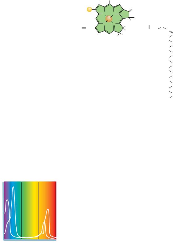
Chlorophylls are magnesium-containing substituted tetrapyrroles whose basic structure is reminiscent of heme, the iron-containing porphyrin (Chapters 5 and 21). Chlorophylls differ from heme in a number of properties: magnesium instead of iron is coordinated in the center of the planar conjugated ring structure; a long-chain alcohol, phytol, is esterified to a pyrrole ring substituent; and the methine bridge linking pyrroles III and IV is substituted and crosslinked to ring III, leading to the formation of a fifth five-membered ring. The structures of chlorophyll a and b are shown in Figure 22.5.
Chlorophylls are excellent light absorbers because of their aromaticity. That is, they possess delocalized electrons above and below the planar ring structure. The energy differences between electronic states in these orbitals correspond to the energies of visible light photons. When light energy is absorbed, an electron is promoted to a higher orbital, enhancing the potential for transfer of this electron to a suitable acceptor. Loss of such a photo-excited electron to an acceptor is an oxidation–reduction reaction. The net result is the transduction of light energy into the chemical energy of a redox reaction.
714 Chapter 22 ● Photosynthesis
CH3
H
CH2
CH3
R=
Chlorophyll a —CH3
R
II
III
O
Chlorophyll b —CHO
O
N
N
V
OCH3
H
Mg
H
C
H2
N
N
O
H2C CH
I
IV
H
C
CH2
CH2
C
O HC
H
C
CH3
FIGURE 22.5
●
Structures of chlorophyll a
CH3
H
CH3
H2C
CH2
and b. Chlorophylls are structurally related to
hemes, except Mg2 replaces Fe2 and ring II
H2C
is more reduced than the corresponding ring
CH
CH3
of the porphyrins. The chlorophyll tetrapyrrole
H2C
ring system is known as a chlorin. R CH3 in
CH2
chlorophyll a; R CHO in chlorophyll b. Note
that the aldehyde CPO bond of chlorophyll b
H2C
introduces an additional double bond into con-
CH
CH3
jugation with the double bonds of the tetrapyr-
H2C
role ring system. Ring V is the additional ring
created by interaction of the substituent of the
CH2
methine bridge between pyrroles III and IV
H2C
with the side chain of ring III. The phytyl side
CH
CH3
chain of ring IV provides a hydrophobic tail to
H3C
anchor the chlorophyll in membrane protein
complexes.
Hydrophobic phytyl side chain
b
Absorbance
a
b
a
400
500
600
700
Wavelength (nm)
FIGURE 22.6 ● Absorption spectra of chlorophylls a and b.
Chlorophylls and Accessory Light-Harvesting Pigments
The absorption spectra of chlorophylls a and b (Figure 22.6) differ somewhat. Plants that possess both chlorophylls can harvest a wider spectrum of incident energy. Other pigments in photosynthetic organisms, so-called accessory lightharvesting pigments (Figure 22.7), increase the possibility for absorption of incident light of wavelengths not absorbed by the chlorophylls. These accessory pigments, such as carotenoids and phycobilins, are also responsible for the magnificent colors of autumn. They persist longer after leaf death than the green chlorophylls, finally imparting their particular hues to the plant. These pigments, like chlorophyll, possess many conjugated double bonds and thus absorb visible light.
The Fate of Light Energy Absorbed by Photosynthetic Pigments
Each photon represents a quantum of light energy. A quantum of light energy absorbed by a photosynthetic pigment has four possible fates (Figure 22.8):
A.Loss as heat. The energy can be dissipated as heat through redistribution into atomic vibrations within the pigment molecule.
B.Loss of light. Energy of excitation reappears as fluorescence (light emission); a photon of fluorescence is emitted as the e returns to a lower orbital.
This fate is common only in saturating light intensities. For thermodynamic reasons, the photon of fluorescence is of longer wavelength and hence lower energy than the quantum of excitation.
C.Resonance energy transfer. The excitation energy can be transferred by resonance energy transfer, a radiationless process, to a neighboring molecule if their energy level difference corresponds to the quantum of excitation energy. In this process, the quantum, or so-called exciton, is transferred,
22.2 ● Photosynthesis Depends on the Photoreactivity of Chlorophyll
715
(a)
H3C
H3C
CH3
CH3
CH3
H3C CH3
CH3
CH3
CH3
β -Carotene
(b)
H
H
N
H
O N
N
N
O
H
CH3
CH
CH3 CH2
CH2
CH3 CH3
CH2
CH3
CH3
CH2
CH2
C
O C
O
OH OH
FIGURE 22.7 ● Structures of representative accessory light-harvesting pigments in photosynthetic cells. (a) -Carotene, an accessory light-harvesting pigment in leaves. Note the many conjugated double bonds. (b) Phycocyanobilin, a blue pigment found in cyanobacteria. It is a linear or open pyrrole.
Light energy (hν )
e–
Pigment molecule (P)
+
Excited state (P*)
+
e–
Qox
e–
Q–red
Thermal
Fluorescence
Exciton
e– Transfer
dissipation
transfer
+
+
+
+
e–
e–
e–
hν
Oxidized P
e
–
(P+)
Heat
Photon of
fluorescence
+
P*
Exciton transfer to neighboring P molecule
FIGURE 22.8 ● Possible fates of the quantum of light energy absorbed by photosynthetic pigments.
FIGURE 22.9
hν
716 Chapter 22 ● Photosynthesis
Light-harvesting pigment (antenna molecules)
Reaction center
● Schematic diagram of a photosynthetic unit. The light-harvesting pigments, or antenna molecules (green), absorb and transfer light energy to the specialized chlorophyll dimer that constitutes the reaction center
(orange).
raising an electron in the receptor molecule to a higher energy state as the photo-excited e in the original absorbing molecule returns to ground state. This so-called Förster resonance energy transfer is the mechanism whereby quanta of light falling anywhere within an array of pigment molecules can be transferred ultimately to specific photochemically reactive sites.
D.Energy transduction. The energy of excitation, in raising an electron to a higher energy orbital, dramatically changes the standard reduction poten-
tial, , of the pigment such that it becomes a much more effective electron donor. That is, the excited-state species, by virtue of having an elec-
tron at a higher energy level through light absorption, has become a potent electron donor. Reaction of this excited-state electron donor with an electron acceptor situated in its vicinity leads to the transformation, or transduction, of light energy (photons) to chemical energy (reducing power, the potential for electron-transfer reactions). Transduction of light energy into chemical energy, the photochemical event, is the essence of photosynthesis.
Photosynthetic Units Consist of Many Chlorophyll
Molecules but Only a Single Reaction Center
In the early 1930s, Emerson and Arnold investigated the relationship between the amount of incident light energy, the amount of chlorophyll present, and the amount of oxygen evolved by illuminated algal cells (this relationship is called the quantum yield of photosynthesis). Their studies gave an unexpected result: When algae were illuminated with very brief light flashes that could excite every chlorophyll molecule at least once, only one molecule of O2 was evolved per 2400 chlorophyll molecules. This result implied that not all chlorophyll molecules are photochemically reactive, and it led to the concept that photosynthesis occurs in functionally discrete units. Chlorophyll serves two roles in photosynthesis. It is involved in light harvesting and the transfer of light energy to photoreactive sites by exciton transfer, and it participates directly in the photochemical events whereby light energy becomes chemical energy. A photosynthetic unit can be envisioned as an antenna of several hundred lightharvesting chlorophyll molecules plus a special pair of photochemically reactive chlorophyll a molecules called the reaction center. The purpose of the vast majority of chlorophyll in a photosynthetic unit is to harvest light incident within the unit and funnel it, via resonance energy transfer, to special reaction center chlorophyll molecules that are photochemically active. Most chlorophyll thus acts as a large light-collecting antenna, and it is at the reaction centers that the photochemical event occurs (Figure 22.9). Oxidation of chlorophyll leaves a cationic free radical, Chl , whose properties as an electron acceptor have important consequences for photosynthesis. Note that the Mg2 ion does not change in valence during these redox reactions.
22.3 ● Eukaryotic Phototrophs Possess
Two Distinct Photosystems
The existence of two separate but interacting photosystems in photosynthetic eukaryotes was demonstrated through analysis of the photochemical action spectrum of photosynthesis, in which the oxygen-evolving capacity as a function of light wavelength was determined (Figure 22.10).
Although chlorophyll a has some capacity to absorb 700-nm light, light of this wavelength is relatively inefficient in driving photosynthesis. However, if light of shorter wavelength (less than 680 nm) is used to supplement 700-nm
evolution)
2
(as O
Photosynthetic efficiency
Red drop
660
680
700
720
Wavelength, nm
22.3 ● Eukaryotic Phototrophs Possess Two Distinct Photosystems
717
Absorption spectrum
Photochemical efficiency with light supplement at 650 nm
Photochemical efficiency
FIGURE 22.10 ● The photochemical action spectrum of photosynthesis. The quantum yield of photosynthesis as a function of wavelength of incident light shows an abrupt decrease above 680 nm, the so-called red drop.
light, an enhancement of photosynthetic quantum yield, the so-called Emerson enhancement effect, is observed. In other words, these two wavelengths are synergistic: When given together, these wavelengths elicit more O2 evolution than expected from the sum of the amounts when each wavelength of light is given alone. One interpretation is that two light reactions participate in oxygenevolving photosynthetic cells, one using light of 700 nm and the other using light of wavelength 680 nm or less. The existence of two light reactions established the presence of two photosystems, I and II. Photosystem I (PSI) is defined as containing reaction center chlorophylls with maximal red light absorption at 700 nm; PSI is not involved in O2 evolution. Photosystem II (PSII) functions in O2 evolution, using reaction centers that exhibit maximal red light absorption at 680 nm.
All photosynthetic cells contain some form of photosystem. Photosynthetic bacteria, unlike cyanobacteria and eukaryotic phototrophs, have only one photosystem. Interestingly, bacterial photosystems resemble eukaryotic PSII more than PSI, even though photosynthetic bacteria lack O2-evolving capacity.
P700 and P680 Are the Reaction Centers of PSI and PSII, Respectively
Precise spectrophotometric measurements showed that a small amount of pigment absorbing 700-nm light (P700) is bleached when light of this wavelength is used to illuminate suspensions of eukaryotic photosynthetic cells. Because bleaching, or disappearance, of the 700-nm absorbance can be mimicked by adding an electron acceptor such as ferricyanide, bleaching is correlated with electron loss from P700. The concentration of P700 is small, only 0.25% of the total amount of chlorophyll in plants. However, this low concentration is consistent with the notion of reaction centers (specific photoreactive sites). P700 is the reaction center of photosystem I. Similar studies using shorter-wavelength light identified an analogous pigment, P680, which constitutes the reaction center of photosystem II. Both P700 and P680 are chlorophyll a dimers situated within specialized protein complexes.
Chlorophyll Exists in Plant Membranes in Association with Proteins
Detergent treatment of a suspension of thylakoids dissolves the membranes, releasing complexes containing both chlorophyll and protein. These chloro- phyll–protein complexes represent integral components of the thylakoid membrane, and their organization reflects their roles as either light-harvesting com-
718 Chapter 22 ● Photosynthesis
FIGURE 22.11 ● Roles of the two photosystems, PSI and PSII.
PS II
PS I
“blue” light < 680 nm
“red” light 700 nm
P680
P700
Strong oxidant
Weak reductant
Weak oxidant
Strong reductant
° > + 0.8 V
“Q” ° 0 V
° 0.45 V
° < –0.6 V
H2O
1
O
ADP + P
ATP
NADP
+
NADPH
2
2
plexes (LHC), PSI complexes, or PSII complexes. All chlorophyll is apparently localized within these three macromolecular assemblies.
The Roles of PSI and PSII
What are the roles of the two photosystems, and what is their relationship to each other? Photosystem I provides reducing power in the form of NADPH. Photosystem II splits water, producing O2, and feeds the electrons released into an electron transport chain that couples PSII to PSI. Electron transfer between PSII and PSI pumps protons for chemiosmotic ATP synthesis. As summarized by Equation (22.2), photosynthesis involves the reduction of NADP , using electrons derived from water and activated by light, h . ATP is generated in the process. The standard reduction potential for the NADP /NADPH couple is 0.32 V. Thus, a strong reductant with more negative than 0.32 V is required to reduce NADP under standard conditions. By similar reasoning, a very strong oxidant will be required to oxidize water to oxygen because(1 O2/H2O) is 0.82 V. Separation of the oxidizing and reducing aspects of°Equation2 (22.2) is accomplished in nature by devoting PSI to NADP reduction and PSII to water oxidation. PSI and PSII are linked via an electron transport chain so that the weak reductant generated by PSII can provide an electron to reduce the weak oxidant side of P700 (Figure 22.11). Thus, electrons flow from H2O to NADP , driven by light energy absorbed at the reaction centers. Oxygen is a by-product of the photolysis, literally “light-splitting,” of water. Accompanying electron flow is production of a proton gradient and ATP synthesis (see Section 22.7). This light-driven phosphorylation is termed photophosphorylation.
22.4 ● The Z Scheme of Photosynthetic Electron Transfer
Photosystems I and II contain unique complements of electron carriers, and these carriers mediate the stepwise transfer of electrons from water to NADP . When the individual redox components of PSI and PSII are arranged as an e transport chain according to their standard reduction potentials, the zigzag result resembles the letter Z laid sideways (Figure 22.12). The various electron carriers are indicated as follows: “Mn complex” symbolizes the manganese-con- taining oxygen-evolving complex; D is its e acceptor and the immediate e donor to P680 ; QA and Q B represent special plastoquinone molecules (see Figure 22.15) and PQ the plastoquinone pool; Fe-S stands for the Rieske iron– sulfur center, and cyt f, cytochrome f. PC is the abbreviation for plastocyanin, the immediate e donor to P700 ; and FA, FB, and FX represent the membrane-
22.4 ● The Z Scheme of Photosynthesis Electron Transfer
719
associated ferredoxins downstream from A0 (a specialized Chl a) and A1 (a specialized PSI quinone). Fd is the soluble ferredoxin pool that serves as the e donor to the flavoprotein (Fp), called ferredoxin–NADP reductase, which catalyzes reduction of NADP to NADPH. Cyt(b6)n,(b6)p symbolizes the cytochrome b6 moieties functioning to transfer e from FA/FB back to P700 during cyclic photophosphorylation (the pathway symbolized by the dashed arrow).
Overall photosynthetic electron transfer is accomplished by three mem- brane-spanning supramolecular complexes, composed of intrinsic and extrinsic polypeptides (shown as shaded boxes bounded by solid black lines in Figure 22.12). These complexes are the PSII complex, the cytochrome b6/cytochrome f complex, and the PSI complex. The PSII complex is aptly described as a lightdriven water:plastoquinone oxidoreductase; it is the enzyme system responsible for photolysis of water, and as such, it is also referred to as the oxygenevolving complex, or OEC. Within this complex, a manganese-containing protein is intimately involved in the evolution of oxygen, perhaps through formation of a tetrametallic center consisting of 4 Mn2 coordinating two equivalents of water. Both protons and electrons are abstracted from these water molecules, and O2 is released as P680 undergoes four cycles of oxidation (Figure 22.13).
Oxygen Evolution Requires the Accumulation of
Four Oxidizing Equivalents in PSII
When isolated chloroplasts that have been held in the dark are illuminated with very brief flashes of light, O2 evolution reaches a peak on the third flash and every fourth flash thereafter (Figure 22.14a). The oscillation in O2 evolved dampens over repeated flashes and converges to an average value. These data are interpreted to mean that the P680 reaction center complex cycles through five different oxidation states, numbered S0 to S4. One electron and one proton are removed photochemically in each step. When S4 is attained, an O2 molecule is released (Figure 22.14b) as PSII returns to oxidation state S0 and two new water molecules bind. The reason the first pulse of O2 release occurred on the third flash (Figure 22.14a) is that the PSII reaction centers in the isolated chloroplasts were already poised at S1 reduction level.
Light-Driven Electron Flow from H2O Through PSII
The events intervening between H2O and P680 involve D, the name assigned to a specific protein tyrosine residue that mediates e transfer from H2O via the Mn complex to P680 (Figure 22.12). The oxidized form of D is a tyrosyl free radical species, D . To begin the cycle, an exciton of energy excites P680 to P680*, whereupon P680* donates an electron to a special molecule of pheophytin, symbolized by “Pheo” in Figure 22.12. Pheophytin is like chlorophyll a, except 2 H replace the centrally coordinated Mg2 ion. This special pheophytin is the direct electron acceptor from P680*. Loss of an electron from P680* creates P680 , the electron acceptor for D. Electrons flow from Pheo via specialized molecules of plastoquinone, represented by “Q” in Figure 22.12, to a pool of plastoquinone within the membrane. Because of its lipid nature, plastoquinone is mobile within the membrane and hence serves to shuttle electrons from the PSII supramolecular complex to the cytochrome b6/cytochrome f complex. Alternate oxidation–reduction of plastoquinone to its hydroquinone form involves the uptake of protons (Figure 22.15). The asymmetry of the thylakoid membrane is designed to exploit this proton uptake and release so that protons (H ) accumulate within the thylakoid vesicle, establishing an electrochemical gradient. Note that plastoquinone is an analog of coenzyme Q, the mitochondrial electron carrier (Chapter 21).


 Q
Q Q
Q
