

Garrett R.H., Grisham C.M. - Biochemistry (1999)(2nd ed.)(en)
.pdfFURTHER READING
Anderson, J. M., 1986. Photoregulation of the composition, function and structure of the thylakoid membrane. Annual Review of Plant Physiology 37:93–136.
Anderson, J. M., and Andersson, B., 1988. The dynamic photosynthetic membrane and regulation of solar energy conversion. Trends in Biochemical Sciences 13:351–355.
Arnon, D. I., 1984. The discovery of photosynthetic phosphorylation. Trends in Biochemical Sciences 9:258–262. A historical account of photophosphorylation by its discoverer.
Bendall, D. S., and Manasse, R. S., 1995. Cyclic photophosphorylation and electron transport. Biochimica et Biophysica Acta 1229:23–38.
Berry, S., and Rumberg, B., 1996. H /ATP coupling ratio at the unmodulated CF1CF0–ATP synthase determined by proton flux measurements.
Biochimica et Biophysica Acta 1276:51–56.
Blankenship, R. E., and Hartman, H., 1998. The origin and evolution of oxygenic photosynthesis. Trends in Biochemical Sciences 23:94–97.
Burnell, J. N., and Hatch, M. D., 1985. Light-dark modulation of leaf pyruvate, Pi dikinase. Trends in Biochemical Sciences 10:288–291. Regulation of a key enzyme in C4 CO2 fixation.
Chapman, M. S., et al., 1988. Tertiary structure of plant rubisco: Domains and their contacts. Science 241:71–74. Structural details of rubisco.
Cramer, W. A., et al., 1985. Topography and function of thylakoid membrane proteins. Trends in Biochemical Sciences 10:125–129.
Cramer, W. A., and Knaff, D. B., 1990. Energy Transduction in Biological Membranes—A Textbook of Bioenergetics. New York: Springer-Verlag, 545 pp. A textbook on bioenergetics by two prominent workers in photosynthesis.
Cramer, W. A., et al., 1996. Some new structural aspects and old controversies concerning the cytochrome b6 f complex of oxygenic photosynthesis. Annual Review of Plant Physiology and Plant Molecular Biology 47:477–508.
Deisenhofer, J., and Michel, H., 1989. The photosynthetic reaction center from the purple bacterium Rhodopseudomonas viridis. Science 245:1463– 1473. Published version of the Nobel laureate address by the researchers who first elucidated the molecular structure of a photosynthetic reaction center.
Deisenhofer, J., Michel, H., and Huber, R., 1985. The structural basis of light reactions in bacteria. Trends in Biochemical Sciences 10:243–248.
Deisenhofer, J., et al., 1985. Structure of the protein subunits in the photosynthetic reaction center of Rhodopseudomonas viridis at 3 Å resolution.
Nature 318:618–624; also The Journal of Molecular Biology (1984) 180:385– 398. These papers are the original reports of the crystal structure of a photosynthetic reaction center.
Ghanotakis, D. F., and Yocum, C. F., 1990. Photosystem II and the oxygenevolving complex. Annual Review of Plant Physiology and Plant Molecular Biology 41:255–276.
Glazer, A. N., 1983. Comparative biochemistry of photosynthetic light-har- vesting pigments. Annual Review of Biochemistry 52:125–157.
Glazer, A. N., and Melis, A., 1987. Photochemical reaction centers: Structure, organization and function. Annual Review of Plant Physiology 38:11– 45.
Golbeck, J. H., 1992. Structure and function of photosystem I. Annual Review of Plant Physiology and Plant Molecular Biology 43:292–324.
Green, B. R., and Durnford, D. G., 1996. The chlorophyll-carotenoid proteins of oxygenic photosynthesis. Annual Review of Plant Physiology and Plant Molecular Biology 47:685–714.
Further Reading |
741 |
Hankamer, B., Barber, J., and Boekema, E. J., 1997. Structure and membrane organization of photosystem II in green plants. Annual Review of Plant Physiology and Plant Molecular Biology 48:641–671.
Harold, F. M., 1986. The Vital Force: A Study of Bioenergetics. Chapter 8: Harvesting the Light. San Francisco: Freeman & Company.
Hatch, M. D., 1987. C4 photosynthesis: A unique blend of modified biochemistry, anatomy and ultrastructure. Biochimica et Biophysica Acta 895:81– 106. A review of C4 biochemistry by its discoverer.
Hoffman, E., et al., 1996. Structural basis of light harvesting by carotenoids: Peridinin-chlorophyll-protein from Amphidinium carterae. Science 272:1788– 1791.
Hoganson, C. W., and Babcock, G. T., 1997. A metalloradical mechanism for the generation of oxygen in photosynthesis. Science 277:1953–1956.
Jagendorf, A. T., and Uribe, E., 1966. ATP formation caused by acid–base transition of spinach chloroplasts. Proceedings of the National Academy of Sciences, USA 55:170–177. The classic paper providing the first experimental verification of Mitchell’s chemiosmotic hypothesis.
Knaff, D. B., 1989. The regulatory role of thioredoxin in chloroplasts.
Trends in Biochemical Sciences 14:433–434.
Knaff, D. B., 1991. Regulatory phosphorylation of chloroplast antenna proteins. Trends in Biochemical Sciences 16:82–83. Additional discussion of the structure of light-harvesting antenna complexes associated with photosynthetic reaction centers can be found in Trends in Biochemical Sciences 11:414 (1986), 14:72 (1989), and 16:181 (1991).
Knight, S., Andersson, I., and Branden, C. I., 1990. Crystallographic analysis of ribulose 1,5-bisphosphate carboxylase from spinach at 2.4 Å resolution. Subunit interactions and active site. Journal of Molecular Biology 215:113–160.
Krauss, N., et al., 1993. Three-dimensional structure of system I of photosynthesis at 6 Å resolution. Nature 361:326–331.
Krauss, N., et al., 1996. Photosystem I at 4 Å resolution represents the first structural model of a joint photosynthetic reaction centre and core antenna system. Nature Structural Biology 3:965–973.
Miziorko, H. M., and Lorimer, G. H., 1983. Ribulose-1,5-bisphosphate carboxylase/oxygenase. Annual Review of Biochemistry 52:507–535. An early review of the enzymological properties of rubisco.
Murata, N., and Miyao, M., 1985. Extrinsic membrane proteins in the photosynthetic oxygen-evolving complex. Trends in Biochemical Sciences 10:122– 124.
Nitschke, W., and Rutherford, A. W., 1991. Photosynthetic reaction centers: Variations on a common structural theme. Trends in Biochemical Sciences 16:241–245.
Ogren, W. L., 1984. Photorespiration: Pathways, regulation and modification. Annual Review of Plant Physiology 35:415–442.
Portis, A. R., Jr., 1992. Regulation of ribulose 1,5-bisphosphate carboxylase/oxygenase activity. Annual Review of Plant Physiology and Plant Molecular Biology 43:415–437.
Rögner, M., Boekema, E. J., and Barber, J., 1996. How does photosystem 2 split water? The structural basis of energy conversion. Trends in Biochemical Sciences 21:44–49.
Ting, I. P., 1985. Crassulacean acid metabolism. Annual Review of Plant Physiology 36:595–622.

8883nc23_742-774 4/12/02 12:40 PM Page 742
Con pan y vino se anda el camino. (With bread and wine you can walk your road.)
Spanish proverb
OUTLINE
23.1 |
● |
Gluconeogenesis |
23.2 |
● |
Regulation of Gluconeogenesis |
23.3 |
● |
Glycogen Catabolism |
23.4 |
● |
Glycogen Synthesis |
23.5 |
● |
Control of Glycogen Metabolism |
23.6 |
● |
The Pentose Phosphate Pathway |
742
Chapter 23
Gluconeogenesis, Glycogen
Metabolism, and the
Pentose Phosphate Pathway
Bread and pasteries on a rack at a French bakery, Paris. Carbohydrates such as these provide a significant portion of human caloric intake.
Tony Stone Images)
As shown in Chapters 18 and 19, the metabolism of sugars is an important source of energy for cells. Animals, including humans, typically obtain significant amounts of glucose and other sugars from the breakdown of starch and glycogen in their diets. Glucose can also be supplied via breakdown of cellular reserves of glycogen (in animals) or starch (in plants). Significantly, glucose also can be synthesized from noncarbohydrate precursors by a process known as gluconeogenesis. Each of these important pathways, as well as the synthesis of glycogen from glucose, will be examined in this chapter.
Another pathway of glucose catabolism, the pentose phosphate pathway, is the primary source of NADPH, the reduced coenzyme essential to most reductive biosynthetic processes. For example, NADPH is crucial to the biosynthesis of

8883nc23_742-774 4/12/02 12:40 PM Page 743
fatty acids (Chapter 25) and amino acids (Chapter 26). The pentose phosphate pathway also results in the production of ribose-5-phosphate, an essential component of ATP, NAD , FAD, coenzyme A, and particularly DNA and RNA. This important pathway will also be considered in this chapter.
23.1 ● Gluconeogenesis
The ability to synthesize glucose from common metabolites is very important to most organisms. Human metabolism, for example, consumes about 160 20 grams of glucose per day, about 75% of this in the brain. Body fluids carry only about 20 grams of free glucose, and glycogen stores normally can provide only about 180 to 200 grams of free glucose. Thus, the body carries only a little more than a one-day supply of glucose. If glucose is not obtained in the diet, the body must produce new glucose from noncarbohydrate precursors. The term for this activity is gluconeogenesis, which means the generation (genesis) of new (neo) glucose.
Further, muscles consume large amounts of glucose via glycolysis, producing large amounts of pyruvate. In vigorous exercise, muscle cells become anaerobic and pyruvate is converted to lactate. Gluconeogenesis salvages this pyruvate and lactate and reconverts it to glucose.
The Substrates of Gluconeogenesis
In addition to pyruvate and lactate, other noncarbohydrate precursors can be used as substrates for gluconeogenesis in animals. These include most of the amino acids, as well as glycerol and all the TCA cycle intermediates. On the other hand, fatty acids are not substrates for gluconeogenesis in animals, because most fatty acids yield only acetyl-CoA upon degradation, and animals cannot carry out net synthesis of sugars from acetyl-CoA. Lysine and leucine are the only amino acids that are not substrates for gluconeogenesis. These amino acids produce only acetyl-CoA upon degradation. Note also that acetylCoA is a substrate for gluconeogenesis when the glyoxylate cycle is operating (Chapter 20).
Nearly All Gluconeogenesis Occurs in the Liver
and Kidneys in Animals
Interestingly, the mammalian organs that consume the most glucose, namely, brain and muscle, carry out very little glucose synthesis. The major sites of gluconeogenesis are the liver and kidneys, which account for about 90% and 10% of the body’s gluconeogenic activity, respectively. Glucose produced by gluconeogenesis in the liver and kidney is released into the blood and is subsequently absorbed by brain, heart, muscle, and red blood cells to meet their metabolic needs. In turn, pyruvate and lactate produced in these tissues are returned to the liver and kidney to be used as gluconeogenic substrates.
Gluconeogenesis Is Not Merely the Reverse of Glycolysis
In some ways, gluconeogenesis is the reverse, or antithesis, of glycolysis. Glucose is synthesized, not catabolized; ATP is consumed, not produced; and NADH is oxidized to NAD , rather than the other way around. However, gluconeogenesis cannot be merely the reversal of glycolysis, for two reasons. First, glycolysis is exergonic, with a G° of approximately 74 kJ/mol. If gluconeogenesis were merely the reverse, it would be a strongly endergonic process and could not occur spontaneously. Somehow the energetics of the process must be aug-
23.1 ● Gluconeogen
Gluconeogenesis, Glycogen
Metabolism, and the Pentose
Phosphate Pathway
8883nc23_742-774 4/12/02 12:40 PM Page 744
744 Chapter 23 ● Gluconeogenesis, Glycogen Metabolism, and the Pentose Phosphate Pathway
FIGURE 23.1 ● The pathways of gluconeogenesis and glycolysis. Species in blue, green, and peach-colored shaded boxes indicate other entry points for gluconeogenesis (in addition to pyruvate).
mented so that gluconeogenesis can proceed spontaneously. Second, the processes of glycolysis and gluconeogenesis must be regulated in a reciprocal fashion so that when glycolysis is active, gluconeogenesis is inhibited, and when gluconeogenesis is proceeding, glycolysis is turned off. Both of these limitations are overcome by having unique reactions within the routes of glycolysis and gluconeogenesis, rather than a completely shared pathway.
Gluconeogenesis—Something Borrowed, Something New
The complete route of gluconeogenesis is shown in Figure 23.1, side by side with the glycolytic pathway. Gluconeogenesis employs three different reactions, catalyzed by three different enzymes, for the three steps of glycolysis that are
|
|
|
This reaction occurs in the ER |
|
|
ATP |
Glucose |
P |
|
|
|
|
|
|
|
|
|
Glucose-6-phosphatase |
|
|
ADP |
Glucose-6-P |
H2O |
|
|
ATP |
|
Fructose-6-P |
P |
|
|
|
||
|
|
|
|
Fructose-1,6-bisphosphatase |
|
ADP |
Fructose-1,6-bisP |
H2O |
|
|
Glyceraldehyde-3-P |
Dihydroxyacetone-P |
||
|
NAD+ |
|
NAD+ |
|
|
NADH |
NADH |
|
|
Glycolysis |
1,3-Bisphosphoglycerate |
Glycerol |
||
ADP |
ADP |
Gluconeogenesis |
||
|
|
|||
|
ATP |
|
ATP |
|
|
3-Phosphoglycerate |
|
||
2-Phosphoglycerate
|
GDP |
PEP carboxykinase |
|
|
|
|
|
ADP |
PEP |
GTP |
|
|
CO2 |
Oxaloacetate |
|
ATP |
Pyruvate |
ADP + P |
Mitochondrial |
matrix |
|||
|
ATP |
Pyruvate |
|
|
carboxylase |
|
|
|
|
|
|
|
|
Amino |
|
|
Lactate |
acids |
|
|
|
|

8883nc23_742-774 4/12/02 12:40 PM Page 745
23.1 ● Gluconeogen
highly exergonic (and highly regulated). In essence, seven of the ten steps of glycolysis are merely reversed in gluconeogenesis. The six reactions between fructose-1,6-bisphosphate and PEP are shared by the two pathways, as is the isomerization of glucose-6-P to fructose-6-P. The three exergonic regulated reac- tions—the hexokinase (glucokinase), phosphofructokinase, and pyruvate kinase reactions—are replaced by alternative reactions in the gluconeogenic pathway.
The conversion of pyruvate to PEP that initiates gluconeogenesis is accomplished by two unique reactions. Pyruvate carboxylase catalyzes the first, converting pyruvate to oxaloacetate. Then, PEP carboxykinase catalyzes the conversion of oxaloacetate to PEP. Conversion of fructose-1,6-bisphosphate to fructose-6-phosphate is catalyzed by a specific phosphatase, fructose-1,6-bis- phosphatase. The final step to produce glucose, hydrolysis of glucose-6-phos- phate, is mediated by glucose-6-phosphatase. Each of these steps is considered in detail in the following paragraphs. The overall conversion of pyruvate to PEP by pyruvate carboxylase and PEP carboxykinase has a G° close to zero but is pulled along by subsequent reactions. The conversion of fructose-1,6-bis- phosphate to glucose in the last three steps of gluconeogenesis is strongly exergonic with a G° of about 30.5 kJ/mol. This sequence of two phosphatase reactions separated by an isomerization accounts for most of the free energy release that makes the gluconeogenesis pathway spontaneous.
The Unique Reactions of Gluconeogenesis
(1) Pyruvate Carboxylase—A Biotin-Dependent Enzyme
Initiation of gluconeogenesis occurs in the pyruvate carboxylase reaction—the conversion of pyruvate to oxaloacetate (Figure 23.2). The reaction takes place in two discrete steps, involves ATP and bicarbonate as substrates, and utilizes biotin as a coenzyme and acetyl-coenzyme A as an allosteric activator. Pyruvate carboxylase is a tetrameric enzyme (with a molecular mass of about 500 kD). Each monomer possesses a biotin covalently linked to the -amino group of a lysine residue at the active site (Figure 23.3). The first step of the reaction involves nucleophilic attack of a bicarbonate oxygen at the -P of ATP to form carbonylphosphate, an activated form of CO2, and ADP (Figure 23.4). Reaction of carbonylphosphate with biotin occurs rapidly to form N-carboxybiotin, liberating inorganic phosphate. The third step involves abstraction of a proton from the C-3 of pyruvate, forming a carbanion which can attack the carbon of N-carboxybiotin to form oxaloacetate.
PYRUVATE CARBOXYLASE IS ALLOSTERICALLY ACTIVATED BY ACYL-COENZYME A
Two particularly interesting aspects of the pyruvate carboxylase reaction are
(a) allosteric activation of the enzyme by acyl-coenzyme A derivatives and (b) compartmentation of the reaction in the mitochondrial matrix. The carboxylation of biotin requires the presence (at an allosteric site) of acetyl-coenzyme A or other acylated coenzyme A derivatives. The second half of the carboxylase reaction—the attack by pyruvate to form oxaloacetate—is not affected by CoA derivatives.
|
|
|
|
|
|
|
|
|
|
|
|
|
|
|
+ ADP |
|
O |
|
|
|
|
|
|
|
O |
||||||||
+ |
|
HCO3– |
+ ATP |
|
|
|
+ |
P |
||||||||
|
|
|
|
|
|
|
|
|
|
|
|
|||||
CH3C |
|
COO– |
|
|
|
–OOC |
CH2C |
|
COO– |
|||||||
|
|
|
|
|
|
|
|
|
|
|||||||
Pyruvate |
|
Bicarbonate |
|
|
Oxaloacetate |
|
|
|||||||||
FIGURE 23.2 ● The pyruvate carboxylase reaction.
E
CH2CH2CH2CH2 NH Lysi
C  O
O
CH2
CH2
CH2 B
CH2 H
N
S
N
H
FIGURE 23.3 ● Covalent linkage an active-site lysine in pyruvate carb

8883nc23_742-774 4/12/02 12:40 PM Page 746
746 Chapter 23 ● Gluconeogenesis, Glycogen Metabolism, and the Pentose Phosphate Pathway
|
|
|
|
|
|
|
|
|
|
|
|
|
|
|
|
|
|
|
|
|
|
|
|
|
|
|
|
|
|
|
|
|
|
|
|
|
|
|
|
|
|
|
|
|
|
|
|
|
|
B |
|
|
E |
|
|
|
|
|
|
|
|
|
|
|
|
|
|
|
|
|
|
|
|
|
|
|
|
|
|
|
|
|
|
|
|
|
|
|
|
|
|
|
|
|
|
|
|
|
|
|
|
|
|
|
|
||
|
|
|
|
|
|
|
|
|
|
|
|
|
|
|
|
|
|
|
|
|
|
|
|
|
|
|
|
|
|
|
|
|
|
|
|
|
|
|
|
|
|
|
|
|
|
|
|
|
|
O |
|
|
|
|
|
|
|
|
|
|
|
|
|
|
|
|
|
|
|
|
|
|
|
|
|
|
|
|
|
|
|
|
|
|
|
|
|
|
|
|
|
|
|
|
H |
|
|
CH |
2 |
|
|
C |
|
|
COO– |
||
|
|
|
|
|
|
|
|
|
|
|
|
|
|
|
|
|
|
|
|
|
|
|
|
|
|
|
|
|
|
|
|
|
|
|
|
|
|
|
|
|
|
|
|
|
|
|
|||||||
|
|
|
|
|
|
|
|
|
|
|
|
|
|
|
|
|
|
|
|
|
|
|
|
|
|
|
|
|
|
|
|
|
|
|
|
|
|
|
|
|
|
|
|
|
|
|
|
|
|
|
|
||
|
|
|
|
|
|
|
|
|
|
|
|
|
|
|
|
|
|
|
|
|
|
|
|
|
|
|
|
|
|
|
|
|
|
|
|
|
|
|
|
|
|
|
|
|
|
|
|
|
|
|
|
|
|
|
|
|
O |
|
O |
|
|
O |
|
|
|
|
|
|
|
|
|
|
|
|
|
|
|
|
|
|
|
|
|
|
|
|
|
|
|
|
|
|
|
O |
|
|
|
||||||||||
|
|
|
|
|
|
|
|
|
|
|
|
|
|
|
|
|
|
|
|
|
|
|
|
|
|
|
|
|
|
|
|
|
|
|
|||||||||||||||||||
|
–O |
|
|
|
|
|
|
|
|
|
|
|
|
|
|
|
O– |
|
|
Adenosine |
|
|
|
|
|
|
|
|
|
|
|
|
|
|
|
|
|
|
|
|
|
|
|
|
|
|
|||||||
|
|
|
|
|
|
|
|
|
|
|
|
|
|
|
|
|
|
|
|
|
|
|
|
|
|
|
|
|
|
|
|
|
|
|
|
|
|
|
|
|
|
|
|
|
|||||||||
|
P |
|
O |
|
P |
|
O |
|
|
P |
|
|
|
|
|
|
|
|
|
|
|
|
|
|
|
|
|
|
|
–CH |
|
|
|
|
|
|
COO– |
||||||||||||||||
|
|
|
|
|
|
|
|
|
|
|
|
|
|
|
|
|
|
|
|
|
|
|
|
|
|
|
|
|
C |
|
|
||||||||||||||||||||||
|
|
|
O– |
|
O– |
|
|
O– |
|
|
|
|
|
|
|
|
|
|
|
|
|
|
|
|
|
|
|
|
|
|
|
|
2 |
|
|
|
|||||||||||||||||
|
|
|
|
|
|
|
|
|
|
|
|
|
|
|
|
|
|
|
|
|
|
|
|
|
|
|
|
|
|
|
|
|
|
|
|
|
|
|
|||||||||||||||
|
|
|
|
|
|
|
|
|
|
|
|
|
|
|
|
|
|
|
|
|
|
P |
|
|
|
|
|
|
|
|
|
|
|
|
|
|
|
|
|
||||||||||||||
|
|
|
|
|
|
|
|
|
|
|
|
|
|
|
|
|
ADP |
|
|
|
|
|
|
|
|
|
|
|
|
|
|
|
|
|
|
|
|
|
|
|
|
|
|
|
|
|
|
||||||
|
|
|
|
|
|
|
|
|
|
|
|
|
|
|
|
|
|
|
O |
|
|
|
O |
|
|
O |
|
|
|
|
|
|
|
|
|
|
|
|
|
||||||||||||||
–O |
|
|
|
|
|
|
|
|
|
|
|
|
|
|
|
O |
|
|
|
|
|
|
|
|
|||||||||||||||||||||||||||||
|
|
|
|
|
|
|
|
|
|
|
|
|
|
|
|
–O |
|
|
|
|
|
|
|
|
|
|
O– |
|
|
|
–O |
|
|
|
|
|
|
|
|
|
|
|
|
|
|||||||||
|
|
C |
|
O |
|
|
|
|
|
|
|
|
|
|
|
|
|
|
|
|
|
|
|
|
|
|
|
|
|
|
|
|
|
|
|
|
|
|
|
|
|
|
|
|
|
|
|||||||
|
|
|
|
|
|
|
|
|
|
|
|
|
|
|
|
|
|
|
|
|
|
|
|
|
|
|
|
|
|
|
|
|
|
|
|
|
|
|
|
|
|
|
|
|
|
|
|
||||||
|
|
|
|
|
|
|
|
|
|
|
|
|
|
|
|
|
|
|
|
C |
|
|
O |
|
P |
|
|
|
|
|
C |
|
|
|
|
|
|
|
|
|
|
|
|
|
|||||||||
|
|
|
|
|
|
|
|
|
|
|
|
|
|
|
|
|
|
|
|
|
|
|
|
|
|
|
|
|
|
|
|
|
|
||||||||||||||||||||
|
|
|
|
|
|
|
|
|
|
|
|
|
|
|
|
|
|
|
|
|
|
|
|
|
|
|
|
|
|
|
|
|
|
|
|
|
|
|
|
|
|||||||||||||
|
|
|
|
|
|
|
|
|
|
|
|
|
|
|
|
|
|
|
|
|
|
|
|
|
|
|
|
|
|
|
|
|
|
|
|
|
|
|
|
||||||||||||||
HO |
|
|
|
|
|
|
|
|
|
|
|
|
|
|
|
|
|
|
|
|
|
|
|
|
|
|
|
|
|
|
|
|
|
|
|
N |
|
|
NH |
|
|
|
|
||||||||||
|
|
|
|
|
|
|
|
|
|
|
|
|
|
|
|
|
|
|
|
|
|
|
|
|
|
|
|
|
|
|
|
|
|
||||||||||||||||||||
|
|
|
|
|
|
|
|
|
|
|
|
|
|
|
|
|
|
|
|
|
|
|
|
O– |
|
|
|
|
|
|
|
|
|
|
|
||||||||||||||||||
|
|
|
|
|
|
|
|
|
|
|
|
|
|
|
|
|
|
|
|
|
|
|
|
|
|
|
|
|
|
|
|
|
|
|
|
|
|
|
|
|
|
|
|
|
|
|
|
|
|
||||
|
|
|
|
|
|
|
|
|
|
|
|
|
|
|
|
|
|
|
|
|
|
|
|
|
|
|
|
|
|
O |
|
|
|
|
|
|
|
|
|
|
|
|
|
|
|
|
|
|
|
|
|||
|
|
|
|
|
|
|
|
|
|
|
|
|
|
|
|
|
|
|
|
|
|
|
|
|
|
|
|
|
|
|
|
|
|
|
|
|
|
|
|
|
|
|
|
|
|
|
|
|
|
||||
|
|
|
|
|
|
|
|
|
|
|
|
|
|
|
|
|
|
|
|
|
|
|
|
|
|
|
|
|
|
|
|
|
|
|
|
|
|
|
|
|
|
|
|
|
|
|
|
|
|
||||
S |
Lys |
HN NH
FIGURE 23.4 ● A mechanism for the pyruvate carboxylase reaction. Bicarbonate must be activated for attack by the pyruvate carbanion. This activation is driven by ATP and involves formation of a carbonylphosphate intermediate —a mixed anhydride of carbonic and phosphoric acids. (Carbonylphosphate and carboxyphosphate are synonyms.)
Biotin
S |
Lys |
O |
|
|
O |
–O C |
CH |
2 |
C COO– |
|
|
|
Activation of pyruvate carboxylase by acetyl-CoA provides an important physiological regulation. Acetyl-CoA is the primary substrate for the TCA cycle, and oxaloacetate (formed by pyruvate carboxylase) is an important intermediate in both the TCA cycle and the gluconeogenesis pathway. If levels of ATP and/or acetyl-CoA (or other acyl-CoAs) are low, pyruvate is directed primarily into the TCA cycle, which eventually promotes the synthesis of ATP. If ATP and acetyl-CoA levels are high, pyruvate is converted to oxaloacetate and consumed in gluconeogenesis. Clearly, high levels of ATP and CoA derivatives are signs that energy is abundant and that metabolites will be converted to glucose (and perhaps even glycogen). If the energy status of the cell is low (in terms of ATP and CoA derivatives), pyruvate is consumed in the TCA cycle. Also, as noted in Chapter 20, pyruvate carboxylase is an important anaplerotic enzyme. Its activation by acetyl-CoA leads to oxaloacetate formation, replenishing the level of TCA cycle intermediates.
COMPARTMENTALIZED PYRUVATE CARBOXYLASE DEPENDS ON METABOLITE CONVERSION AND TRANSPORT The second interesting feature of pyruvate carboxylase is that it is found only in the matrix of the mitochondria. By contrast, the next enzyme in the gluconeogenic pathway, PEP carboxykinase, may be localized in the cytosol or in the mitochondria or both. For example, rabbit liver PEP carboxykinase is predominantly mitochondrial, whereas the rat liver enzyme is strictly cytosolic. In human liver, PEP carboxykinase is found both in the cytosol and in the mitochondria. Pyruvate is transported into the mitochondrial matrix, where it can be converted to acetyl-CoA (for use in the TCA cycle) and then to citrate (for fatty acid synthesis; see Figure 25.1). Alternatively, it may be converted directly to OAA by pyruvate carboxylase and used in glu-

8883nc23_742-774 4/12/02 12:40 PM Page 747
coneogenesis. In tissues where PEP carboxykinase is found only in the mitochondria, oxaloacetate is converted to PEP, which is then transported to the cytosol for gluconeogenesis (Figure 23.6). However, in tissues that must convert some oxaloacetate to PEP in the cytosol, a problem arises. Oxaloacetate cannot be transported directly across the mitochondrial membrane. Instead, it must first be transformed into malate or aspartate for transport across the mitochondrial inner membrane (Figure 23.5). Cytosolic malate and aspartate must be reconverted to oxaloacetate before continuing along the gluconeogenic route.
(2) PEP Carboxykinase
The second reaction in the gluconeogenic pyruvate-PEP bypass is the conversion of oxaloacetate to PEP. Production of a high-energy metabolite such as PEP requires energy. The energetic requirements are handled in two ways here. First, the CO2 added to pyruvate in the pyruvate carboxylase step is removed in the PEP carboxykinase reaction. Decarboxylation is a favorable process and helps to drive the formation of the very high-energy enol phosphate in PEP. This decarboxylation drives a reaction that would otherwise be highly endergonic. Note the inherent metabolic logic in this pair of reactions: pyruvate carboxylase consumed an ATP to drive a carboxylation, so that the PEP carboxykinase could use the decarboxylation to facilitate formation of PEP. Second, as shown in Figure 23.6, another high-energy phosphate is consumed by the carboxykinase. Mammals and several other species use GTP in this reaction, rather than ATP. The use of GTP here is equivalent to the consumption of an ATP, due to the activity of the nucleoside diphosphate kinase (see Figure 20.4). The substantial free energy of hydrolysis of GTP is crucial to the synthesis of PEP in this step. The overall G for the pyruvate carboxylase and PEP carboxykinase reactions under physiological conditions in the liver is 22.6 kJ/mol. Once PEP is formed in this way, the phosphoglycerate mutase, phosphoglycerate kinase, glyceraldehyde-3-P dehydrogenase, aldolase, and triose phosphate isomerase reactions act to eventually form fructose-1,6-bisphos- phate, as in Figure 23.1.
(3) Fructose-1,6-Bisphosphatase
The hydrolysis of fructose-1,6-bisphosphate to fructose-6-phosphate (Figure 23.7), like all phosphate ester hydrolyses, is a thermodynamically favorable (exergonic) reaction under standard-state conditions ( G° 16.7 kJ/mol). Under physiological conditions in the liver, the reaction is also exergonic ( G 8.6 kJ/mol). Fructose-1,6-bisphosphatase is an allosterically regulated enzyme. Citrate stimulates bisphosphatase activity, but fructose-2,6-bisphosphate is a potent allosteric inhibitor. AMP also inhibits the bisphosphatase; the inhibition by AMP is enhanced by fructose-2,6-bisphosphate.
23.1 ● Gluconeogen
Pyruvate
Pyruvate
Oxaloacetate
NADH
 NAD+
NAD+
Malate
Malate
NAD+
 NADH
NADH
Oxaloacetate
Gluconeogenesis
● Pyruvate carboxyl compartmentalized reaction. Pyruva verted to oxaloacetate in the mitoch Because oxaloacetate cannot be tra across the mitochondrial membrane reduced to malate, transported to t and then oxidized back to oxaloace gluconeogenesis can continue.
O |
+ GTP |
|
|
2–O3PO |
||||||
|
|
|
COO– |
|
||||||
C |
|
PEP carboxykinase |
C |
|
COO– |
|||||
|
|
|||||||||
|
|
|
COO– |
|
|
|
|
|||
H2C |
|
|
|
|
|
|
|
|
||
|
|
|
|
|
CH2 |
|||||
Oxaloacetate |
|
|
|
PEP |
||||||
+
+
GDP
CO2
● The PEP carboxykinase reaction. GTP formed in this reaction can be converted to ATP by nucleoside diphosphate kinase, although liver cells in some species may not contain this enzyme.

8883nc23_742-774 4/12/02 12:40 PM Page 748
748 Chapter 23 ● Gluconeogenesis, Glycogen Metabolism, and the Pentose Phosphate Pathway
2– |
O |
|
|
|
|
2– |
|
O |
|
CH2O |
2– |
|
|
|
|||||
O3POH2C |
|
PO3 |
+ H2O |
|
O3POH2C |
CH2OH |
+ P |
||
H |
HO |
|
|
|
|
H |
HO |
||
|
|
Fructose-1,6- |
|||||||
H |
|
OH |
|
H |
OH |
|
|||
|
|
bisphosphatase |
|
||||||
HO |
H |
|
|
|
HO |
H |
|
||
|
|
|
|
|
|
||||
Fructose-1,6-bisphosphate |
|
|
|
Fructose-6-phosphate |
|
||||
|
|
|
|
∆ G '=–16.7 kJ/mol |
|
|
|
||
FIGURE 23.7 ● The fructose-1,6-bisphosphatase reaction.
(4) Glucose-6-Phosphatase
The final step in the gluconeogenesis pathway is the conversion of glucose-6- phosphate to glucose by the action of glucose-6-phosphatase. This enzyme is present in the membranes of the endoplasmic reticulum of liver and kidney cells, but is absent in muscle and brain. For this reason, gluconeogenesis is not carried out in muscle and brain. Its membrane association is important to its function because (Figure 23.8) the substrate is hydrolyzed as it passes into the endoplasmic reticulum itself. Vesicles form from the endoplasmic reticulum membrane and diffuse to the plasma membrane and fuse with it, releasing their glucose contents into the bloodstream. The glucose-6-phosphatase reaction involves a phosphorylated enzyme intermediate, which may be a phosphohistidine (Figure 23.9). The G for the glucose-6-phosphatase reaction in liver is 5.1 kJ/mol.
COUPLING WITH HYDROLYSIS OF ATP AND GTP DRIVES GLUCONEOGENESIS The net reaction for the conversion of pyruvate to glucose in gluconeogenesis is
2 Pyruvate 4 ATP 2 GTP 2 NADH 2 H 6 H2O g
glucose 4 ADP 2 GDP 6 Pi 2 NAD
The net free energy change, G° , for this conversion is 37.7 kJ/mol. The consumption of a total of six nucleoside triphosphates drives this process forward. If glycolysis were merely reversed to achieve the net synthesis of glucose from pyruvate, the net reaction would be
2 Pyruvate 2 ATP 2 NADH 2 H 2 H2O g
glucose 2 ADP 2 Pi 2 NAD
FIGURE 23.8 ● Glucose-6-phosphatase is localized in the endoplasmic reticulum membrane. Conversion of glucose-6-phosphate to glucose occurs during transport into the ER.
and the overall G° would be about 74 kJ/mol. Such a process would be highly endergonic, and therefore thermodynamically unfeasible. Hydrolysis of four additional high-energy phosphate bonds makes gluconeogenesis thermodynamically favorable. Under physiological conditions, however, gluconeogenesis is somewhat less favorable than at standard state, with an overall G of15.6 kJ/mol for the conversion of pyruvate to glucose.
Cytosol |
Endoplasmic reticulum |
|
Glucose-6- |
Glucose |
|
phosphate |
||
P |
||
|
Glucose-6- |
|
|
phosphatase |

8883nc23_742-774 4/12/02 12:40 PM Page 749
|
|
|
|
|
|
|
23.1 ● |
Gluconeogen |
|
|
O |
|
H2O |
O |
|
|
|
|
|
|
P O– |
|
|
|
|
|
|
|
|
O |
NH E |
–O |
|
|
NH E |
HOPO23– |
+ N |
N |
|
|
N |
P N |
|||||||
H2C |
O– |
|
CH2OH |
|
|
+ |
|
|
|
|
O |
– |
|
|
|
|
|||
O |
|
|
O |
|
|
|
|
|
|
|
|
|
|
|
|
|
|
||
OH CHOH
HO
OH CHOH
HO
OH |
OH |
FIGURE 23.9 ● The glucose-6-ph reaction involves formation of a ph dine intermediate.
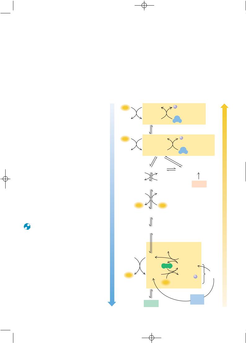
LACTATE FORMED IN MUSCLES IS RECYCLED TO GLUCOSE IN THE LIVER A final point on the redistribution of lactate and glucose in the body serves to emphasize the metabolic interactions between organs. Vigorous exercise can lead to oxygen shortage (anaerobic conditions), and energy requirements must be met by increased levels of glycolysis. Under such conditions, glycolysis converts NAD to NADH, yet O2 is unavailable for regeneration of NAD via cellular respiration. Instead, large amounts of NADH are reoxidized by the reduction of pyruvate to lactate. The lactate thus produced can be transported from muscle to the liver, where it is reoxidized by liver lactate dehydrogenase to yield pyruvate, which is converted eventually to glucose. In this way, the liver shares in the metabolic stress created by vigorous exercise. It exports glucose to muscle, which produces lactate, which can be processed by the liver into new glucose. This is referred to as the Cori cycle (Figure 23.10). Liver, with a typically high NAD /NADH ratio (about 700), readily produces more glucose than it can use. Muscle that is vigorously exercising will enter anaerobiosis and show a decreasing NAD /NADH ratio, which favors reduction of pyruvate to lactate.
Gluconeogenesis |
FIGURE 23.10 ● The Cori cycle. |
Glucose |
6 NTP
Pyruvate
 NADH
NADH
LDH
NAD+
Lactate
Liver
(high [NAD+] ) [NADH]
Blood
Glycolysis
Glucose
 2 NTP
2 NTP
Pyruvate
NADH
LDH
NAD+ 
Lactate
Muscle
(low [NAD+] ) [NADH]

8883nc23_742-774 4/12/02 12:40 PM Page 750
750 Chapter 23 ● Gluconeogenesis, Glycogen Metabolism, and the Pentose Phosphate Pathway
C R I T I C A L D E V E L O P M E N T S I N B I O C H E M I S T R Y
The Pioneering Studies of Carl and Gerty Cori
The Cori cycle is named for Carl and Gerty Cori, who received the Nobel Prize in physiology or medicine in 1947 for their studies of glycogen metabolism and blood glucose regulation. Carl Ferdinand Cori and Gerty Theresa Radnitz were both born in Prague (then in Austria). They earned medical degrees from the German University of Prague in 1920 and were married later that year. They joined the faculty of the Washington University School of Medicine in St. Louis in 1931. Their remarkable collaboration resulted in many fundamental advances in carbohydrate and glycogen metabolism. They were credited with the discovery of glucose-1-phosphate, also known at the time as the “Cori ester.”
They also showed that glucose-6-phosphate was produced from glucose-1-P by the action of phosphoglucomutase. They isolated and crystallized glycogen phosphorylase and elucidated the pathway of glycogen breakdown. In 1952, they showed that absence of glucose-6-phosphatase in the liver was the enzymatic defect in von Gierke’s disease, an inherited glycogen-storage disease. Six eventual Nobel laureates received training in their laboratory. Gerty Cori was the first American woman to receive a Nobel prize. Carl Cori said of their remarkable collaboration: “Our efforts have been largely complementary and one without the other would not have gone so far. . . .”
23.2 ● Regulation of Gluconeogenesis
Nearly all of the reactions of glycolysis and gluconeogenesis take place in the cytosol. If metabolic control were not exerted over these reactions, glycolytic degradation of glucose and gluconeogenic synthesis of glucose could operate simultaneously, with no net benefit to the cell and with considerable consumption of ATP. This is prevented by a sophisticated system of reciprocal control, so that glycolysis is inhibited when gluconeogenesis is active, and vice versa. Reciprocal regulation of these two pathways depends largely on the energy status of the cell. When the energy status of the cell is low, glucose is rapidly degraded to produce needed energy. When the energy status is high, pyruvate and other metabolites are utilized for synthesis (and storage) of glucose.
In glycolysis, the three regulated enzymes are those catalyzing the strongly exergonic reactions: hexokinase (glucokinase), phosphofructokinase, and pyruvate kinase. As noted, the gluconeogenic pathway replaces these three reactions with corresponding reactions that are exergonic in the direction of glucose synthesis: glucose-6-phosphatase, fructose-1,6-bisphosphatase, and the pyruvate carboxylase–PEP carboxykinase pair, respectively. These are the three most appropriate sites of regulation in gluconeogenesis.
Gluconeogenesis Is Regulated by Allosteric and
Substrate-Level Control Mechanisms
The mechanisms of regulation of gluconeogenesis are shown in Figure 23.11. Control is exerted at all of the predicted sites, but in different ways. Glucose- 6-phosphatase is not under allosteric control. However, the Km for the substrate, glucose-6-phosphate, is considerably higher than the normal range of substrate concentrations. As a result, glucose-6-phosphatase displays a near-linear dependence of activity on substrate concentrations and is thus said to be under sub- strate-level control by glucose-6-phosphate.
Acetyl-CoA is a potent allosteric effector of glycolysis and gluconeogenesis. It allosterically inhibits pyruvate kinase (as noted in Chapter 19) and activates pyruvate carboxylase. Because it also allosterically inhibits pyruvate dehydrogenase (the enzymatic link between glycolysis and the TCA cycle), the cellular fate of pyruvate is strongly dependent on acetyl-CoA levels. A rise in