

- •VOLUME 3
- •CONTRIBUTOR LIST
- •PREFACE
- •LIST OF ARTICLES
- •ABBREVIATIONS AND ACRONYMS
- •CONVERSION FACTORS AND UNIT SYMBOLS
- •EDUCATION, COMPUTERS IN.
- •ELECTROANALGESIA, SYSTEMIC
- •ELECTROCARDIOGRAPHY, COMPUTERS IN
- •ELECTROCONVULSIVE THERAPHY
- •ELECTRODES.
- •ELECTROENCEPHALOGRAPHY
- •ELECTROGASTROGRAM
- •ELECTROMAGNETIC FLOWMETER.
- •ELECTROMYOGRAPHY
- •ELECTRON MICROSCOPY.
- •ELECTRONEUROGRAPHY
- •ELECTROPHORESIS
- •ELECTROPHYSIOLOGY
- •ELECTRORETINOGRAPHY
- •ELECTROSHOCK THERAPY.
- •ELECTROSTIMULATION OF SPINAL CORD.
- •ELECTROSURGICAL UNIT (ESU)
- •EMERGENCY MEDICAL CARE.
- •ENDOSCOPES
- •ENGINEERED TISSUE
- •ENVIRONMENTAL CONTROL
- •EQUIPMENT ACQUISITION
- •EQUIPMENT MAINTENANCE, BIOMEDICAL
- •ERGONOMICS.
- •ESOPHAGEAL MANOMETRY
- •EVENT-RELATED POTENTIALS.
- •EVOKED POTENTIALS
- •EXERCISE FITNESS, BIOMECHANICS OF.
- •EXERCISE, THERAPEUTIC.
- •EXERCISE STRESS TESTING
- •EYE MOVEMENT, MEASUREMENT TECHNIQUES FOR
- •FETAL MONITORING
- •FETAL SURGERY.
- •FEVER THERAPY.
- •FIBER OPTICS IN MEDICINE
- •FICK TECHNIQUE.
- •FITNESS TECHNOLOGY.
- •FIXATION OF ORTHOPEDIC PROSTHESES.
- •FLAME ATOMIC EMISSON SPECTROMETRY AND ATOMIC ABSORPTION SPECTROMETRY
- •FLAME PHOTOMETRY.
- •FLOWMETERS
- •FLOWMETERS, RESPIRATORY.
- •FLUORESCENCE MEASUREMENTS
- •FLUORESCENCE MICROSCOPY.
- •FLUORESCENCE SPECTROSCOPY.
- •FLUORIMETRY.
- •FRACTURE, ELECTRICAL TREATMENT OF.
- •FUNCTIONAL ELECTRICAL STIMULATION
- •GAMMA CAMERA.
- •GAMMA KNIFE
- •GAS AND VACUUM SYSTEMS, CENTRALLY PIPED MEDICAL
- •GAS EXCHANGE.
- •GASTROINTESTINAL HEMORRHAGE
- •GEL FILTRATION CHROMATOGRAPHY.
- •GLUCOSE SENSORS
- •HBO THERAPY.
- •HEARING IMPAIRMENT.
- •HEART RATE, FETAL, MONITORING OF.
- •HEART VALVE PROSTHESES
- •HEART VALVE PROSTHESES, IN VITRO FLOW DYNAMICS OF
- •HEART VALVES, PROSTHETIC
- •HEART VIBRATION.
- •HEART, ARTIFICIAL
- •HEART–LUNG MACHINES
- •HEAT AND COLD, THERAPEUTIC
- •HEAVY ION RADIOTHERAPY.
- •HEMODYNAMICS
- •HEMODYNAMIC MONITORING.
- •HIGH FREQUENCY VENTILATION
- •HIP JOINTS, ARTIFICIAL
- •HIP REPLACEMENT, TOTAL.
- •HOLTER MONITORING.
- •HOME HEALTH CARE DEVICES
- •HOSPITAL SAFETY PROGRAM.
- •HUMAN FACTORS IN MEDICAL DEVICES
- •HUMAN SPINE, BIOMECHANICS OF
514HIP JOINTS, ARTIFICIAL
65.Mammel MC, et al. Acute airway injury during high-fre- quency jet ventilation and high-frequency oscillatory ventilation. Crit Care Med 1991;19:394–398.
66.Kirpilani H, et al. Diagnosis and therapy of necrotizing tracheobronchitis in ventilated neonates. Crit Care Med 1985; 13:792–797.
67.Keszler M, Durand DJ. Neonatal high-frequency ventilation. Past, present, and future. Clin Perinatol 2001;28:579–607.
68.Spitzer AR, Butler S, Fox WW. Ventilatory response to combined HFJV and conventional mechanical ventilation for the rescue treatment of severe neonatal lung disease. Ped Pulmonol 1989;7:244–250.
69.Carlon GC, et al. High-frequency jet ventilation. A prospective randomized evaluation. Chest 1983;84:551–559.
See also CONTINUOUS POSITIVE AIRWAY PRESSURE; RESPIRATORY MECHANICS AND GAS EXCHANGE; VENTILATORY MONITORING.
HIP JOINTS, ARTIFICIAL
Z. M. JIN
J. L. TIPPER
M. H. STONE
E. INGHAM
J. FISHER
University of Leeds
Leeds, United Kingdom
INTRODUCTION
Natural synovial joints, such as hips, are remarkable bearings in engineering terms. They can transmit a large dynamic load of several times bodyweight during steadystate walking, yet with minimal friction and wear achieved through effective lubrication and with little maintenance. However, diseases such as osteoarthritis and rheumatoid arthritis or trauma sometimes necessitate the replacement of these natural bearings. Artificial hip joints replace the damaged natural bearing material, articular cartilage. As a result, pain in the joint is relieved and joint mobility and functions are restored. Total hip joint replacement has been considered as one of the greatest successes in orthopaedic surgery in the last century in improving the quality of life of the patients. Currently, > 1 million hip joints are replaced worldwide each year, with ever increasing use of these devices in a wider range of patients.
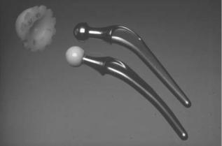
The majority of current artificial hip joints consist of an ultrahigh molecular weight polyethylene (UHMWPE) acetabular cup against a metallic or ceramic femoral head as illustrated in Fig. 1. These devices can generally last 10–15 years in the body without too many problems. However, after this period of implantation, loosening of prosthetic components becomes the major clinical problem. It is now generally accepted that the loosening is caused by the osteolysis as a result of biological reactions to particulate wear debris mainly released from the articulating surfaces. Therefore, one of the main strategies to avoid the loosening problem and to extend the clinical life of the hip prosthesis is to minimize wear and wear particles. Application of tribology, defined as ‘‘the branch of science and technology concerned with interacting surfaces in relative motion and
Figure 1. A typical Charnley hip prosthesis consisting of an UHMWPE acetabular cup against either a metallic (stainless steel) or a ceramic (alumina) femoral head.
with associated matters (as friction, wear, lubrication, and the design of bearings’’ (Oxford English Dictionary), to biological systems (biotribology) such as artificial hip joints, can play an important role in this process. Coupled tribological studies of friction, wear and lubrication of the bearing surfaces, and biological studies of wear debrisinduced adverse reactions become necessary.
HISTORICAL DEVELOPMENT
Early History: Hemiarthroplasty, Interposition Arthroplasty,
and Total Hip Replacement
The first recognizable ball and socket joint was reported in Germany by Professor Gluck in 1890 in a dog with an ivory ball and socket hip joint. This did not gain popular support for use in humans until Hey Groves in Bristol reported his ivory hemiarthroplasty for fractured neck of femur in 1926. Attempts to use metal at this stage were unsuccessful. A significant breakthrough came in 1923. It began with a chance observation that a piece of glass left in an individual’s back for 1 year stimulated a fibrous tissue and fluid producing reaction. It formed a fluid-filled synovial sac (Smith Peterson 1948). Smith Peterson went on to insert a glass cup-shaped mould between the surfaces of an ankylosed hip. Although the glass broke, at the time of its removal the acetabulum and the head of the femur were found to be covered with a smooth lining of fibrous tissue. Over the next few years a number of different materials were used including Viscaloid, Pyrex, Bakelite, and finally Vitallium (chromium–cobalt–molybdenum alloy) in 1938. This material worked well and was used for 1000 interposition arthroplasties at Massachusetts General Hospital alone over the next 10 years. It remained the standard treatment for hip arthritis until the advent of total hip replacement.
Charnley Era
Jean and Robert Judet reported their use of a replacement femoral head made of poly (methyl methacrylate) (PMMA). Although the prosthesis failed, it survived
long enough to squeak within the human body. It was this squeaking prosthesis that set Charnley on his quest for a low friction-bearing surface. He began with Teflon in 1958 and throughout the 1950s Charnley experimented with two thin cups of Teflon, one in the acetabulum and one over a reshaped femoral head. They failed within a year due to loosening of the cup and avascular necrosis of the femoral head. He abandoned this surface replacement for an endoprosthesis and as the acetabular cups wore through Charnley sought better wearing materials. He moved to high density PE and later to UHMWPE.
Low Friction Arthroplasty. Charnley began his cemented total hip replacement era with a large femoral head (Moore’s hemiarthroplasties). He argued that distributing the load over a large area of contact would decrease wear. However, after loosening of these large head components, he began working on the low frictional torque prosthesis, which reduced the torque at the cement-bone and prosthesis interfaces. He achieved this by reducing the diameter of the femoral head from 41 to 22 mm. In this way, Charnley developed his prosthesis of a 22 mm head on a metal stem, UHMWPE cup and PMMA cement. Charnley hips still have a survival today of > 90% at 10 years (1).
There continues to be debate as to the cause of up to 10% failures of Charnley hips. Early belief that it was due to the cement and cement particles led to the development of uncemented prostheses. Concern that the production of PE particles was producing bone lysis, led to the development of alternative bearing surfaces, for example, ceramics that wear less than metal against PE, and metal-on-metal prostheses, which produce lower volumes of wear debris. Impingement of the femoral prosthesis on the cup leading to loosening led to the narrowing of the neck of the femoral component and the use of cut away angle bore sockets. Concern about access of fluid to the femoral cement– prosthesis interface and subsequently to the cement–bone interface through cement deficiencies, with the production of osteolysis, have led some manufacturers to produce polished femoral stems. These self-locking tapers prevent fluid flowing between the cement and the femoral stem. The debate continues. It can be difficult to separate the improvements in surgical techniques that have improved the clinical results from the effect of modification and changes in materials used to produce joint replacements.
Current Developments
There is currently much interest in reducing the trauma of the surgery itself. These include surgical techniques of minimal incision surgery with the skin wound < 8 cm and a more conservative approach to femoral bone use at the time of surgery. Surface replacement has returned with ‘‘better’’ metal-on-metal surfaces, and a shortstemmed Judet type of metaphyseal fix femoral prosthesis is also available. Hydroxyapatite as a method of component fixation is also gaining popularity. The current short-term results of these techniques are interesting, and may lead to significant benefits especially for the younger patient in the future. However, the proof will only come from long-term clinical results that are not yet available.
HIP JOINTS, ARTIFICIAL |
515 |
JOINT ANATOMY AND ENVIRONMENT
The bearing material in the natural hip joint is articular cartilage, firmly attached to the underlying bone. Articular cartilage is an extremely complex material, consisting of both fluid (interstitial water) and solid (primarily collagen and proteoglycan) phases. Such a biphasic or poroelastic feature determines the time-dependent deformation of articular cartilage, and largely governs the lubrication mechanism of the natural hip joint. The lubricant present in the natural hip joint is synovial fluid, which is similar to blood plasma with hyaluronic acid added and becomes periprosthetic synovial fluid after total hip replacement. Rheological studies of these biological lubricants have shown shear-thinning characteristics, particularly at low shear rates and for the joint fluid taken from diseased or replaced joints (2).
The load experienced in the hip joint during steady-state walking varies both in direction and magnitude. The maximum load can reach five times bodyweight during the stance phase after heel-strike and is largely reduced in the swing phase after the toe-off. On the other hand, the speed is relatively low, particularly in the stance phase and during the motion reversal. However, the hip contact force can be substantially increased under other conditions. For example, the hip contact force has been reported to be 5.8 times bodyweight up a ramp, 6.6 times up and down stairs, and 7.6 times on fast level walking at a speed of 2.01 m s 1 (3).
CURRENT BEARING SURFACES
Biomaterials used for current artificial hip joints include UHMWPE, stainless steel, cobalt chromium alloy (CoCr), and ceramics (alumina and zirconia). A number of combinations for the bearing surfaces using these materials have been introduced since 1950s in order to minimize wear and wear particle generation. These can generally be classified as soft-on-hard and hard-on-hard as summarized in Table 1.
Two main parameters that govern the tribology of the articulating surfaces are geometrical and mechanical properties. The geometrical parameters of the bearing surfaces
are the diameters of the acetabular cup (Dcup) and the femoral head (Dhead). The size of the hip prosthesis is usually characterized by its diameter, which is important
for both clinical and tribological considerations, such as stability, dislocation, and sliding distance. In addition to size, the diametral mismatch or clearance between the
cup and the head (d ¼ Dcup Dhead) is also important, particularly for hard-on-hard bearing surfaces. From a
tribological point of view, these geometric parameters can often be approximated as a single equivalent diameter (D) defined as
D |
¼ |
ðDheadDcupÞ |
1 |
Þ |
|
d |
ð |
Typical values of equivalent diameter used in current hip prostheses are summarized in Table 2. In addition, geometric deviations from perfectly spherical surfaces, such as nonsphericity and surface toughness, are also very

516 HIP JOINTS, ARTIFICIAL
Table 1. Typical Biomaterials and Combinations for the Bearing Surfaces of Current Artificial Hip Joint Replacements
|
|
|
Acetabular Cup |
|
|
||
|
|
|
|
|
|
|
|
|
|
|
Soft |
|
|
|
Hard |
|
|
|
|
|
|
|
|
Femoral Head (Hard) |
UHMWPE |
Cross-linked UHMWPE |
Polyurethane |
CoCr |
Alumina |
||
|
|
|
|
|
|
|
|
Stainless Steel |
H |
|
H |
|
|
|
|
CoCr |
H |
|
H |
H |
H |
|
|
Alumina |
H |
|
H |
H |
H |
H |
|
Zirconia |
H |
|
H |
H |
|
H |
|
|
|
|
|
||||
Table 2. Typical Geometric Parameters of Various Bearing Couples for Artificial Hip Jointsa |
|
|
|||||
Bearing Couples |
Femoral Head Diameter, mm |
Diametral Clearance, mm |
Equivalent Diameter, m |
||||
|
|
|
|
|
|
|
|
UHMWPE-on-metal |
28 (22–40) |
|
|
300 (160–1000) |
|
2.6 (1.0–5.0) |
|
Metal-on-metal |
28 (28–60) |
|
|
60 (60–300) |
|
10 |
|
Ceramic-on-ceramic |
28 (28–36) |
|
|
80 (20–80) |
|
10 |
|
aSee Ref. 4.
important factors in determining the tribological performance of the prosthesis.
The mechanical properties of the bearing surfaces are also important tribological determinants. Typical values of elastic modulus and Poisson’s ratio are given in Table 3 for the biomaterials used in artificial hip joints. Other parameters, such as hardness, particularly in the soft-on-hard combinations, are also important, in that the hard surface should be resistant to third-body abrasion to minimize the consequences of polymeric wear.
COUPLED TRIBOLOGICAL AND BIOLOGICAL METHODOLOGY
The vast majority of studies to evaluate the wear performance of hip prostheses have simply measured the volumetric wear rate (6–8). There are very few groups who have also investigated the characteristics of the wear particles generated in in vitro simulations (9–11). The cellular response to prosthetic wear particles, and thus the functional biological activity of implant materials, is complex and is dependent not only on the wear volume, but also the mass distribution of particles as a function of size, their concentration, morphology, and chemistry (see the section, Biological Response of Wear Debris).
During the latter 1990s, methods were developed for the isolation and characterization of UHMWPE particles from
Table 3. Typical Mechanical Properties in Terms of Elastic Modulus and Poisson’s Ratio of the Bearing Materials for Artificial Hip Jointsa
Bearing Materials |
Elastic Modulus, GPa |
Poisson’s Ratio |
|
|
|
UHMWPE |
0.5–1. |
0.4 |
Cross-Linked UHMWPE |
0.2–1.2a |
0.4 |
Stainless steel |
210 |
0.3 |
CoCr |
230 |
0.3 |
Zirconia |
210 |
0.26 |
Alumina |
380 |
0.26 |
aSee Ref. 5.
retrieved tissues and serum lubricants from simulators that allow discrimination between the particles generated in different patient samples and from different types of polyethylene tested in vitro (12–18). The basis of this method is to determine the mass distribution of the particles as a function of size. Determination of the number distribution as a function of size fails to discriminate between samples since the vast majority of the number of particles are invariably in the smallest size range detectable by the resolution of the imaging equipment.
In our laboratories we have pioneered cell culture studies with clinically relevant UHMWPE wear particles generated in experimental wear simulation systems operated under aseptic conditions (17–21). These studies have been extended to cell culture studies of clinically relevant metal (22), ceramic (23), and bone cement wear particles (24,25).
By combining volumetric wear determinations in hip joint simulations with experiments to determine the direct biological activity of the particles generated, we have developed novel methodologies to evaluate the functional biocompatibility of different materials used in prosthetic joint bearings. The functional biocompatibility can be used as a preclinical estimate of the in vivo performance of the material under test compared to historical materials. We have adopted two different approaches to determining functional biocompatibility. Our choice of method is dependent on the bearing material and the type of prosthesis.
The first approach is indirect, but can be applied to all materials and devices. It utilizes data obtained from the direct culture of UHMWPE wear particles in three different size ranges: 0.1–1, 1–10, and > 10 mm at different volumetric concentrations with human peripheral blood macrophages. Measurements of the biological activity for unit volumes of particles in the different size ranges are generated (20). The use of TNF-a as a determinant is justified since, in our experience the major cytokines concerned in osteolysis (TNF-a, IL-1, IL-6, GM-csf) all show the same pattern of response to clinically relevant wear particles (19–21). By using our methods to determine the

volumetric concentration of particles generated in simulations as a function of size (see above), it is then possible to integrate the volume concentration and biological activity function to produce a relative index of specific biological activity (SBA) per unit volume of wear. The functional biological activity (FBA) has been defined as the product of volumetric wear and SBA (26). This has allowed us to compare the functional biological activity of different types of PE in hip joint simulators (27) and different types of bearing materials (23).
The second approach is to directly culture wear debris from wear simulators with primary macrophages. For metal and ceramic particles, we can directly culture wear particles from standard simulation systems after isolation, sterilisation, and removal of endotoxin by heat treatments (23). However, for PE this is not feasible since the heat treatment at elevated temperature required to remove endotoxin cannot be applied. For these materials, we have developed a sterile endotoxin free multidirectional wear simulator in which wear particles are generated in macrophage tissue culture medium. While this does not test whole joints, it allows the application of different kinematics to represent the hip and the knee. The advantage of this approach is that all the wear products are directly cultured with the cells, and there is no risk of modification during the isolation procedure. This method has recently been used to compare the biological reactivity of particles from PEs of different molecular weights and different levels of crosslinking. Higher molecular weight of GUR 1050 and higher levels of cross-linking of both GUR 1020 and 1050 produced particles that were more biologically reactive (18).
Tribology of Bearing Surfaces
Tribological studies of the bearing surfaces of artificial hip joints include friction, wear, and lubrication, which have been shown to mainly depend on the lubrication regimes involved. There are three lubrication regimes: boundary, fluid-film, and mixed. In the boundary lubrication regime, a significant asperity contact is experienced, and consequently both friction and wear are high. In the fluid film lubrication regime, where the two bearing surfaces are completely separated by a continuous lubricant, minimal friction and wear is expected. The mixed-lubrication regime consists of both fluid film lubricated and boundary contact regions. Friction and lubrication studies are usually performed to understand the wear mechanism involved in artificial hip joints. However, friction forces may be important in determining the stresses experienced at the interface between the implant and the cement bone (28) as well as temperature rise (29).
Friction in artificial hip joints is usually measured in a pendulum-like simulator with a dynamic load in the vertical direction and a reciprocating rotation in the horizontal direction. The coefficient of friction is usually expressed as a friction factor defined as
T |
ð2Þ |
m ¼ wðdhead=2Þ |
where T is the measured friction torque and w is the load.
HIP JOINTS, ARTIFICIAL |
517 |
The measured coefficient of friction in a particular hip prosthesis itself can generally reveal the nature of the lubrication regime, since each mechanism is associated with broad ranges of the coefficient of friction. The variation in the coefficient of friction against a Sommerfeld
number defined as, S ¼ ðhudhead=wÞ, where is viscosity and u velocity, can further indicate the lubrication regime.
If the measured friction factors remain constant, fall or increase as the Sommerfeld number is increased, the associated modes of lubrication are boundary, mixed, or fluidfilm, respectively (30).
Lubrication studies of artificial hip joints are generally carried out using both experimental and theoretical approaches. The experimental measurement is usually involved with the detection of the separation between the two bearing surfaces using a simple resistivity technique. A large resistance would imply a thick lubricant film, while a small resistance is attributed to the direct surface contact. Such a technique is directly applicable to metal- on-metal bearings as well as UHMWPE-on-metal and ceramic-on-ceramic bearings if appropriate coatings are used (31,32). The theoretical analysis is generally involved with the solution to the Reynolds equation, together with the elasticity equation subjected to the dynamic load and speed experienced during walking. The predicted film thickness (hmin) is then compared with the average surface roughness (Ra) using the following simple criterion.
l ¼ |
hmin |
ð3Þ |
½Rahead2 þ Racup2 &1=2 |
The lubrication regime is then classified as fluid film, mixed, or boundary if the predicted ratio is > 3, between 1 and 3, or < 1, respectively.
Wear of artificial hip joints has been investigated extensively, due to its direct relevance to biological reactions and clinical problems of osteolysis and loosening. Volumetric wear and wear particles can be measured using the following machines, among others:
Pin-on-disk machines.
Pin-on-plate machines.
Joint simulators.
A unidirectional sliding motion is usually used in the pin-on-disc machine, and the reciprocating motion is added to the pin-on-plate machine. Both of these machines are used to screen potential bearing materials under well controlled, and often simplified conditions. Generally, it is necessary to introduce additional motion in order to produce a multidirectional motion. The next stage of wear testing is usually carried out in joint simulators with a varied degree of complexity of the 3D loading and motion patterns experienced by hip joints, while immersing the test joints in a lubricant deemed to be physically and chemically similar to synovial fluid. Wear can be evaluated by either dimensional or gravimetric means.
Contact mechanics analysis is often performed to predict the contact stresses within the prosthetic components and to compare with the strength of the material. However, other predicted contact parameters such as the contact
518 HIP JOINTS, ARTIFICIAL
area and the contact pressure at the bearing surfaces have been found to be particularly useful in providing insights into friction, wear, and lubrication mechanisms. Contact mechanics can be investigated either experimentally using pressure-sensitive film and sensors, or theoretically using the finite element method.
Biological Response of Wear Debris
Our current understanding of the mechanisms of wear particle-induced osteolysis has developed from > 30 years experience with UHMWPE-on-metal. The major factor limiting the longevity of initially well-fixed UHMWPE total joint replacements is osteolysis resulting in late aseptic loosening (33). There is extremely strong evidence from in vivo and in vitro studies that osteolysis is a UHMWPE particle related phenomenon.
Following total hip arthroplasty, a pseudocapsule forms around the joint and this may have a pseudosynovial lining. A fibrous interfacial tissue may also form at the bone–cement or bone–prosthesis interface that is normally thin with few vessels or cells (34–36). At revision surgery for aseptic loosening, the fibrous membrane is thickened, highly vascularized, and contains a heavy infiltrate of UHMWPE-laden macrophages and multinucleated giant cells (37,38). There is a correlation between the number of macrophagesandthe volumeof UHMWPEweardebrisinthe tissues adjacent to areas of aggressive osteolysis (39–45). Analyses of interfacial membranes have demonstrated the presence of a multitude of mediators of inflammation including cytokines that may directly influence osteoclastic bone resorption:-TNF-a(46),IL-1b(47), IL-6 (48), andM-CSF(49). There is a direct relationship between the particle concentration and the duration the implant, and there are billions of particles generated per gram of tissue (9,15,50,51). Osteolysis is likely to occur when the threshold of particles exceeds 1 1010/g of tissue (45). Each milligram of PE wear has been estimated to generate 1.3 1010 particles (15).
The UHMWPE particles isolated from retrieved tissues vary in size and morphology, from large platelet-like particles, up to 250 mm in length, fibrils, shreds, and submicrometer globule-shaped spheroids 0.1–0.5 mm in diameter (15,52–54). The vast majority of the numbers of particles are the globular spheroids and the mode of the frequency distribution is invariably 0.1–0.5 mm, although the larger particles may account for a high proportion of the total volume of wear debris. Analysis of the mass distribution as a function of size is therefore necessary to discriminate between patient samples (15,55).
UHMWPE wear particles generated in vitro in hip joint simulators have a larger proportion of the mass of particles in the 0.01–1 mm sized range than those isolated from periprosthetic tissues (27,55). This may indicate that in vivo, the smaller particles are disseminated more widely away from the implant site. Recently, improvements to particle imaging techniques have revealed nanometer sized UHMWPE particles generated in hip joint simulators. These particles have yet to be identified in vivo. These nanometer size particles account for the greatest number of particles generated, but a negligible proportion of the total volume (18).
Studies of the response of macrophages to clinically relevant, endotoxin-free polyethylene particles in vitro have clearly demonstrated that particle stimulated macrophages elaborate a range of potentially osteolytic mediators (IL-1, IL-6, TNF-a, GM-CSF, PGE2) and bone resorbing activity (19–21,56–58). Induction of bone resorbing activity in particle stimulated macrophage supernatants has been shown to be critically dependent on particle size and concentration with particles in the 0.1–1.0 mm size range at a volumetric concentration of 10–100 mm3/cell being the most biologically reactive (19,56). The importance of UHMWPE particle size has also been demonstrated in animal studies (59). These findings have enabled the preclinical prediction of the functional biological activity of different polyethylenes by analysis of the wear rate and mass distribution of the particles as a function of particle size (26,27). For a review of the biology of osteolysis, the reader is referred to Ingham and Fisher (60).
In metal-on-metal bearings in the hip, an abundance of small nanometer size particles are generated (61,62). It is believed that the majority of metal debris is transported away from the periprosthetic tissue. While only isolated instances of local osteolysis have been found around metal- on-metal hips, this is most commonly associated with high concentrations of metal debris and tissue necrosis. In vitro cell culture studies have shown that these nanometer size metal particles are highly toxic to cells at relatively low concentrations (22). These particles have a very limited capacity to activate macrophages to produce osteolytic cytokines at the volumes likely to be generated in vivo (63), however, metal particles are not bioinert and concerns exist regarding their potential genotoxicity (22).
Ceramic-on-ceramic prostheses have been shown to have extremely low wear rates. Ceramic wear particles generated in hip joint simulations under clinically relevant conditions in the hip joint simulator (64) and in vivo (65) have a bimodal size distribution with nanometer sized (5– 20 nm) and larger particles (0.2–> 10 mm). Alumina ceramic particles have been shown to be capable of inducing osteolytic cytokine production by human mononuclear phagocytes in vitro (23). However, the volumetric concentration of the particles needed to generate this response was 100–500 mm3/cell. Given the extremely low wear rates of modern ceramic-on-ceramic bearings, even under severe conditions, it is unlikely that this concentration will arise in the periprosthetic tissues in vivo (60).
APPLICATIONS
UHMWPE-on-Metal and UHMWPE-on-Ceramic
The friction in UHMWPE hip joints has been measured using a pendulum-type simulator with a flexionsol–exten- sion motion and a dynamic vertical load. The friction factor has been found to be generally in the range 0.02–0.06 for 28 mm diameter metal heads and UHMWPE cups (66), broadly representative of mixed lubrication, and this has been confirmed from the variation in the friction factor with the Sommerfeld number. These experimental observations are broadly consistent with the theoretical prediction of typical lubricant film thicknesses between 0.1 and

HIP JOINTS, ARTIFICIAL |
519 |
Table 4. Volumetric Wear Rate, % wear volume < 1 mm, SBA, and FBA for Nonirradiated and Irradiated UHMWPEs and Alumina Ceramic-on-Ceramic Hip Joint Prosthesesa
Material |
Volumetric Wear rate, mm3/106 cycles 95% CL |
% Volume < 1 mm |
SBA |
FBA |
|
Nonirradiated UHMWPE |
50 8 |
23 |
0.32 |
16 |
|
Gamma in air UHMWPE, 2.5 Mrad GUR1120 |
49 9 |
46 |
0.55 |
55 |
|
Stabilized UHMWPE (2.5–4 Mrad) GUR1020 |
35 9 |
43 |
0.5 |
17.5 |
|
Highly cross-linked UHMWPE, 10 Mrad GUR1050 |
8.6 |
3.1 |
95 |
0.96 |
8 |
Alumina ceramic-on-ceramic (microseparation) |
1.84 |
0.38 |
100 |
0.19 |
0.35 |
aSee Refs. 60,69.
0.2 mm and the average surface roughness of UHMWPE bearing surface between 0.1 and 1 mm. Therefore, wear of UHMWPE acetabular cups is largely governed by the boundary lubrication mechanism. An increase in the femoral head diameter can lead to an increase in sliding distance and consequently wear (41). As a result, 28 mm diameter femoral heads appear to be a better choice. Furthermore, reducing the surface roughness of the metallic femoral head or using harder alumina to resist thirdbody abrasion and to maintain the smoothness is also very important. For example, the wear factor in UHMWPE-on- ceramic implants is generally 50% of that in UHMPWE-on- metal (67). The introduction of cross-linked UHMWPE has been shown to reduce wear significantly in simulator studies. However, the degree of wear reduction appears to depend on cross-linking, kinematics, counterface roughness, and bovine serum concentration (68). It should be pointed out that volumetric changes are often accompanied by morphology changes, which may have different biological reactions as discussed below.
First, let us consider the effect of irradiation and crosslinking on the osteolytic potential of UHMWPE bearings. Historically, UHMWPE acetabular cups were gamma irradiated in air until it became clear that oxidative degeneration of the PE was occurring. This oxidative damage was caused by the release of free radicals, which produced strand scission of the long PE chains. Research has indicated that deterioration to important mechanical properties such as tensile strength, impact strength, toughness, fatigue strength, and Young’s modulus occurs (12). These time-dependent changes have been shown to affect the volumetric wear of the UHMWPE and typical values are in the region of 100 mm3/million cycles. In addition, UHMWPE that had been gamma irradiated in air produced a greater volumetric concentration of wear particles that were in the most biologically active size range, 0.1– 1 mm (46% of the wear volume compared to 24% for nonirradiated UHMWPE). When the specific biological activity (biological activity per unit volume of wear; SBA) of the wear particles was calculated this gave an SBA that was 1.7-fold higher than the SBA of the nonirradiated material, which translated into a functional biological activity (FBA), which was 3.5-fold higher than the FBA of the nonirradiated material (Table 4).
Currently, UHMWPE is sterilized by gamma irradiation (2.5– 4 Mrad) in an inert atmosphere. This material undergoes partial cross-linking as a result of this processing, and is often referred to as moderately cross-linked or stabilized PE. This material produces lower wear rates than the nonirradiated UHMWPE, but has a higher
volumetric concentration of wear particles < 1 mm compared to the nonirradiated material as shown in Table 4 (69). Consequently, the specific biological activity of the wear particles is higher at 0.5 compared to 0.32 for the nonirradiated material. However, as the wear volume is substantially lower, the FBA value for the stabilized UHMWPE is very similar to the nonirradiated material.
As the level of cross-linking increases, the wear volume decreases (69). The highly cross-linked UHMWPE is GUR 1050, irradiated at 10 Mrad and remelted, and has very low wear volumes at 8.6 3.1 mm3/million cycles. However, as can be seen from Table 4, 95% of the wear volume is comprised of particles in the most biologically active size range, leading to an extremely high SBA. However, as the wear volume is significantly lower than the other UHMWPEs, the FBA is one-half of those of the nonirradiated and stabilized materials (Table 4).
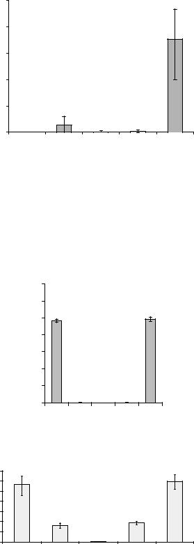
In addition, the wear particles from the cross-linked materials have increased biological activity per unit volume of wear (Fig. 2). A recent study by Ingram et al. (18) has shown that when worn against a scratched counterface, PE irradiated with 5 and 10 Mrad of gamma irradiation produced higher volumetric concentrations of wear particles in the 0.01–1.0 mm size range compared to noncross-linked material. This increased volumetric concentration of wear particles in the 0.01–1.0 mm size range meant that both cross-linked materials were able to stimulate the release of elevated levels of TNF-a, an osteolytic cytokine, at a 10-fold lower volumetric dose than the
(ng/OD) |
0.45 |
|
|
|
0.4 |
|
|
|
|
0.35 |
|
|
|
|
Activity |
|
|
|
|
0.3 |
|
|
|
|
0.25 |
|
|
|
|
Specific |
0.2 |
|
|
|
0.15 |
|
|
|
|
l |
|
|
|
|
-α |
0.1 |
|
|
|
TNF |
0.05 |
|
|
|
0 |
|
|
|
|
|
|
|
|
|
|
Cells only |
NXL |
5 MRad |
10 MRad |
Cell Challenge
Figure 2. TNF-a release (specific activity 95% confidence limits) as a result of challenge with UHMWPE particles, which were noncross-linked (NXL), cross-linked with 5 Mrad of irradiation, cross-linked with 10 Mrad of irradiation compared to the cell only control.
520 HIP JOINTS, ARTIFICIAL
noncross-linked polyethylene (0.1 mm3 debris/cell compared to 1–10 mm3 debris/cell). So, while the cross-linked materials produced lower wear volumes, the particles produced from these materials were more reactive compared to the noncross-linked PE.
However, when the same materials were worn against a smooth counterface, analysis of the wear particles showed that both cross-linked and noncross-linked PE produced very high numbers of nanometer-sized wear particles. In addition, the cross-linked and noncross-linked materials produced similar low volumes of particles in the 0.1– 1.0 mm size range, which resulted in wear debris that was only stimulatory a the highest volumetric dose of 50 mm3 debris/cell. This offers further explanation as to why the FBA or osteolytic potential of the highly cross-linked polyethylene’s are lower than the moderately cross-linked and noncross-linked materials (Table 4).
Metal-on-Metal
The friction factor measured in metal-on-metal hip joints with different sizes and clearances in simple pendulum type machines is generally much higher than for UHMWPE-on-metal articulations, in the range between 0.1 and 0.2, indicating a mixed-boundary lubrication regime (66). However, the lubrication regime in metal- on-metal bearings has been shown to be sensitive to the surface roughness, loading and velocity, and design parameters (70–74). Consequently, different friction factors or wear factors are possible. Therefore, it is important to optimize the bearing system, in terms of the femoral head diameter, the clearance and the structural support (75,76). From a lubrication point of view, the femoral head diameter is the most important geometric parameter, since it is directly related to both the equivalent diameter defined in Eq. 1 and the sliding velocity (70). If the lubrication improvement is such that a fluid-film dominant lubrication regime is present, the increase in the sliding distance becomes unimportant. Such an advantage has been utilized in large-diameter metal-on-metal hip resurfacing prostheses (77). However, it should be pointed out that the lubrication improvement in large-diameter metal-on- metal hip resurfacing prostheses can only be realized with adequate clearances (78). A too large clearance can reduce the equivalent diameter, shifting the lubrication regime toward mixed–boundary regions. In addition, the increase in the sliding distance associated with the large diameter means that the bedding-in wear becomes important. The wear in optimised systems can be quite low, of the order of a few millimeters cubed.
The biological activity in terms of osteolytic potential of metal-on-metal hip prostheses is difficult to define. If macrophages and fibroblasts are challenged with clinically relevant cobalt chrome wear particles, there is some release of the osteolytic cytokine TNF-a (Fig. 3), however, this only takes place at very high levels of particulate load (50 mm3 debris/cell), and the level of cytokine produced is at lower levels compared to UHMWPE particles [see Fig. 2(79)]. The predominant biological reaction is cytotoxicity or a reduction in cell viability (Fig. 4). Macrophage and fibroblast cell viability is significantly reduced when
|
0.25 |
|
activity |
0.2 |
|
0.15 |
||
specific |
||
0.1 |
||
TNF-α |
||
0.05 |
||
|
||
|
* |
|
|
0 |
Cells only CoCr 50:1 CoCr 5:1 CoCr 0.5:1 LPS Particle volume ( m3) to cell number ratio
Figure 3. TNF-a production by human peripheral blood mononuclear cells stimulated with clinically relevant cobalt–chromium particles.
challenged with 50 or 5 mm3 debris/cell (22). The specific biological activity of metal wear particles is difficult to assess as the cells may release cytokines, such as TNF-a as a consequence of cell death. In addition, the high levels of particulate load required to stimulate cytokine release
|
140 |
Macrophages |
|
||
|
|
|
|||
|
120 |
|
|
|
|
CL) |
100 |
|
|
|
|
95% |
80 |
|
|
|
|
(± |
|
|
|
|
|
%Viability |
60 |
|
|
|
|
40 |
|
|
|
|
|
20 |
|
|
|
|
|
|
** |
* |
|
|
|
|
|
|
|
||
|
0 |
|
|
|
0.005:1 |
|
Latex |
Campt |
50:1 |
5:1 |
|
|
|
Wear debris ( m3/cell) |
|
||
CL) |
140 |
|
Fibroblasts |
|
|
|
|
|
|
||
120 |
|
|
|
|
|
95% |
100 |
|
|
|
|
80 |
|
|
|
|
|
(± |
|
|
|
|
|
60 |
|
|
|
|
|
Viability |
* |
|
|
|
|
40 |
|
|
|
||
|
|
|
|
||
20 |
|
* |
|
|
|
% |
0 |
|
50:1 |
5:1 |
0.005:1 |
Latex |
Campt |
||||
|
|
Wear debris ( m3/cell) |
|
|
|
Figure 4. Effect of metal-on-metal cobalt–chromium wear particles on macrophage and fibroblast cell viability ( Significant (p < 0.05, ANOVA) reduction in cell viability).

HIP JOINTS, ARTIFICIAL |
521 |
|
7 |
|
|
|
|
|
|
|
|
|
|
|
|
|
|
|
6 |
|
|
|
|
|
|
|
|
|
|
|
|
|
|
|
5 |
|
|
|
|
|
|
|
|
|
|
|
|
|
|
FBA |
4 |
|
|
|
|
|
|
|
|
|
|
|
|
||
3 |
|
|
20-fold |
|
|
|
|
|
|
|
|
|
|
||
|
2 |
|
|
lower |
|
|
|
|
|
|
|
|
|
|
|
|
1 |
|
|
|
|
|
|
|
|
|
|
|
|
|
|
|
0 |
|
|
|
|
|
|
|
|
|
|
|
|
|
|
|
|
|
|
|
|
|
Alumina |
Highly cross- |
ceramic |
linked |
microsep |
polyethylene |
Figure 5. Predicted functional biological activity or osteolytic potential of alumina ceramic-on-ceramic and highly cross-linked UHMWPE-on-metal hip prostheses.
may only be achieved in vivo if a pairing is particularly high wearing.
Ceramic-on-Ceramic
The friction factor in ceramic-on-ceramic hip implants is quite low, particularly when the nonbiological type lubricant is used (66). However, when biological lubricants such as bovine serum are tested, the friction factor can be quite high due to the complex interactions with proteins. The wear under normal ideal conditions is low, but can be increased substantially under adverse conditions such as microseparation (80). Despite this, the wear in ceramic-on- ceramic hip implants is generally the lowest among current hip prostheses available clinically.
The introduction of microseparation conditions into the in vitro simulation model replicates clinically relevant wear rates, wear patterns, and wear particles. Alumina wear particles have a lower biological activity than UHMWPE particles. A 10-fold higher concentration of alumina wear particles is required to stimulate the release of osteolytic cytokine TNF-a from macrophages compared to UHMWPE wear particles (23). It is questionable whether the volume of alumina wear particles will reach this threshold in vivo given the extremely low wear rates of ceramic-on-ceramic prostheses even under severe microseparation conditions. Consequently, alumina wear particles have a lower specific biological activity than UHMWPE particles (Table 4). When this lower SBA is integrated with the comparatively small volumetric wear rates that are produced by ceramic-on-ceramic couples compared to metal-on-polyethylene, a substantially lower functional biological activity or osteolytic potential is pro-
duced (Table 4; Fig. 5). In fact, alumina ceramic-on-ceramic couples produce a 20-fold lower FBA than the currently used highly cross-linked UHMWPEs.
Summary
Typical values of friction factor, wear factor, and biological reactions are summarized in Tables 5, 6, and 7 for various hip implants with different bearing couples.
FUTURE DEVELOPMENTS
Cross-Linked Polyethylene
The introduction of highly cross-linked PE into clinical use in the last 5 years has been extensive. Standard UHMWPE is irradiated at high dose levels ( 5–10 Mrad), which produces chain scission and cross-linking between the molecular chains. Subsequent heating and remelting recombines the free radicals producing a more stable material (81). The additional cross-links provide improved wear resistance, particularly during kinematic conditions with high levels of cross-shear as found in the hip. A number of early simulator studies showed no wear for these materials (81), while other studies demonstrated measurable wear (82). Initial clinical studies, however, do show penetration and wear (83). Wear and surface cracking has been identified in a few isolated retrievals. The wear rates reported more recently in simulator studies have been found to be in the range 5–10 mm3/million cycles, which is four to five times less than with conventional material (69). Recent work has also shown that cross-linked PE produces a greater proportion of smaller particles, per unit volume of wear debris and has been found to be up to three times more biologically active than conventional material (18). This leads to a functional reduction in osteolytic potential of about twofold compared to conventional PE. This improvement in functional osteolytic potential may not be sufficient for high demand patients, and in patients who require large head sizes. In these patients, larger diameter hard-on-hard, such as ceramic-on-ceramic or metal-on-metal, may be a more appropriate bearing choice.
Ceramic-on-Metal Bearing
Currently used hard-on-hard bearings are comprised of similar materials, such as alumina ceramic-on-ceramic or metal-on-metal. When lubrication conditions are depleted, like bearing materials can produce elevated adhesive friction and wear. The ceramic-on-metal hip was developed to produce a deferential hardness hard bearing (84). Laboratory simulation studies have shown a reduction in wear of
Table 5. Typical Friction Factors and Lubrication Regimes in Various Bearings for Hip Implantsa
|
|
Variation of Friction Factor Against |
Indicated Lubrication |
Bearing Couples |
Friction Factor |
Increasing Sommerfeld Number |
Regimes |
|
|
|
|
UHMWPE-on-Metal |
0.06–0.08 |
Constant/decreasing |
Boundary/mixed |
Metal-on-metal |
0.22–0.27 |
Decreasing |
Mixed |
Ceramic-on-ceramic |
0.002–0.2 |
Increasing |
Fluid-film/mixed |
aSee Ref. 4.

522 |
HIP JOINTS, ARTIFICIAL |
|
|
Table 6. Typical Volumetric and Linear Wear Rates for Different Bearings for Hip Implantsa |
|||
Bearing Couples |
Volumetric Wear Rate, mm3/million cycles |
Linear Wear Rate, mm/million cycles |
|
|
|
|
|
UHMWPE-on-metal |
30–100 |
100–300 |
|
UHMWPE-on-ceramic |
15–50 |
50–150 |
|
Metal-on-metal |
0.1–1 |
2–20 |
|
Ceramic-on-ceramic |
0.05–1 |
1–20 |
|
|
|
|
|
aSee Ref. 4. |
|
|
|
Table 7. Typical Particle Sizes and Biological Responses in Different Bearings for Hip Implantsa |
|||
Bearing Couples |
Dominant Particle Diameters, mm |
Biological Responses |
|
|
|
|
|
UHMWPE-on-metal/ceramic |
UHMWPE, 0.01–1 |
Macrophages/osteoclasts/osteolysis |
|
Metal-on-metal |
Metallic, 0.02–0.1 |
Low osteolysis, cytotoxicity |
|
Ceramic-on-ceramic |
Ceramic, 0.01–0.02 |
Bioinert, low cytotoxicity |
|
|
|
Ceramic, 0.1–10 |
Macrophages/osteoclasts/osteolysis |
aSee Ref. 69.
up to 100-fold compared to metal on metal. The ceramic head does not wear and remains smooth, improving lubrication and reducing wear of the metallic cup. This new concept is currently entering clinical trials.
Surface Replacement Bearings
There is considerable interest in surface replacement solutions in the hip (85). In this approach, a large diameter metallic shell is placed over the reamed femoral head, preserving femoral bone stock, and this articulates against a large diameter acetabular cup. Both cobalt chrome cast and wrought alloys have been used in different bearing designs. The larger diameter head improves lubrication and reduces wear compared to smaller head sizes (70). However, it is important to maintain a low radical clearance between the components to ensure low bedding-in wear (78,86,87). Surface replacement metal on metal bearings are not suitable for all patients, due to the nature of the femoral bone, but are currently used in 10% of patients receiving hip prostheses.
Surface Engineered Metal-on-Metal Bearings SUREHIP
Concerns still remain about wear particles in metal on metal bearings and elevated metal ion levels. Surface engineering solutions are an attractive option for reducing wear and metal ion levels, and can be readily applied to surface replacement hips. Recent research with thick AEPVD chromium nitride and chromium carbon nitride surface engineered coatings of thicknesses between 8 and 12 mm have shown a 30-fold reduction in wear and metal ion levels (88,89). These coatings are now undergoing product development in preparation for clinical studies.
Compliant Materials, Cushion Form Bearings
In recent years, the trend has been to move toward harder bearing materials that wear less, and away from the lower elastic modulus properties of articular cartilage. Compliant materials such as polyurethane have been investigated as bearing materials in acetabular cups. The cups have been formed as a composite structure with a higher modulus substrate to give structural support (90). The bearing has
shown improved lubrication and reduced wear compared to conventional polyethylene bearings. However, concerns remain about the long-term stability of these low modulus materials. More recently an experimental polyurethane surface replacement cup has been investigated (91).
Hemiarthroplasty and Hemisurface Replacements
Interest in more conservative bone preserving, and minimally invasive surgery has generated renewed interest in surgical treatments that replace only one side of the diseased joint or potentially just the diseased surface itself. In these scenarios, consideration has not only to be given to the biomaterial replacing the degenerated tissue, but also the function of the apposing articulating surface.
In the hip hemiarthroplasty using compliant materials has just entered clinical trials, where the femoral head covered with a layer of polyurethane articulates against the natural cartilage in the acetabulum (http://www.impliant.com/home/index.html). Future designs will focus on full or partial replacement of the diseased cartilage on one side of the joint.
SUMMARY
This article summarizes the biotribology of artificial hip joints and development over the last four decades. Although adequate solutions exist for the elderly less active patients > 65 years old with average life expectances < 20 years, considerable technological advances are required to meet the demands and improved performance of younger patients. Recent moves toward large diameter heads to give greater function, stability and range of motion are placing greater demands on tribological performances and increasing use of the hard-on-hard bearings.
Nomenclature
d Diametral clearance
DDearing diameter or equivalent diameter defined in Eq. 1
FBA Functional biological activity
hmin Minimum lubricant film thickness PMMA Poly(methyl methacrylate)
Ra Average surface roughness
S Summerfeld number
SBA Specific biological activity
T Frictional torque u Siding velocity
UHMWPE Ultrahigh molecular weight polyethylene w Load
hViscosity
lRatio defined in Eq.3
mFrictional factor defined in Eq. 2
Subscripts:
Head Femoral head
Cup Acetabular cup
BIBLIOGRAPHY
Cited References
1.Malchau H, Herberts P, Ahnfelt L. Prognosis of total hip replacement in Sweden. Acta Orthop Scand 1993;64–65.
2.Yao JQ, Laurent MP, Johnson TS, Blanchard CR, Crowinshield RD. The influence of lubricant and material on polymer/CoCr sliding friction. Wear 2003;255:780–784.
3.Paul JP. Strength requirements for internal and external prostheses. J Biomech 1999;32(4):381–393.
4.Jin ZM, Medley JB, Dowson D. Fluid Film Lubrication In Artificial Hip Joints. Proc 29th Leeds-Lyon Symp Tribology; 2003. p 237–256.
5.Lewis G. Properties of crosslinked ultra-high-molecular- weight polyethylene. Biomaterials 2001;22(4):371–401.
6.Chiesa R, Tanzi MC, Alfonsi S, Paracchini L, Moscatelli M, Cigada A. Enhanced wear performance of highly cross-linked UHMWPE for artificial joints. J Biomed Mat Res 2000;50:381–387.
7.Bowsher JG, Shelton JC. A hip simulator study of the influence of patient activity level on the wear of cross-linked polyethylene under smooth and roughened counterface conditions. Wear 2001;250:167–179.
8.Hermida JC, Bergula A, Chen P, Colwell CW, D’Lima DD. Comparison of wear rates of twenty-eight and thirty-two millimeter femoral heads on cross-linked polyethylene acetabular cups in a wear simulator. J Bone Joint Surg 2003;85A:2325–2331.
9.McKellop HA, Campbell P, Park SH, Schmalzried TP, Grigoris P, Amstutz HC, Sarmiento A. The origin of submicron polyethylene wear debris in total hip arthroplasty. Clin Orthopaed Rel Res 1995;311:3–20.
10.Tipper JL, Ingham E, Fisher J. Characterisation of wear debris from UHMWPE, metal on metal and ceramic on ceramic hip prostheses. Wear 2001;250:120–128.
11.Saikko V, Calonius O, Keranen J. Wear of conventional and cross-linked ultra-high molecular -weight polyethylene acet-
HIP JOINTS, ARTIFICIAL |
523 |
abular cups against polished and roughened CoCr femoral heads in a biaxial hip simulator. J Biomed Res Appl Biomat 2002;63:848–853.
12.Besong AA, Tipper JL, Ingham E, Stone MH, Wroblewski BM, Fisher J. Quantitative comparison of wear debris from UHMWPE that has and has not been sterilized by gamma irradiation. J Bone Joint Sur 1998;80B:340–344.
13.Endo MM, Barbour PSM, Barton DC, Wroblewski BM, Fisher J, Tipper JL, Ingham E, Stone MH. A comparison of the wear and debris generation of GUR 1120 (compression moulded) and GUR 4150HP (ram extruded) ultra high molecular weight polyethylene. Bio Med Mat Eng 1999;9:113–124.
14.Endo MM, Barbour PS, Barton DC, Fisher J, Tipper JL, Ingham E, Stone MH. Comparative wear and wear debris under three different counterface conditions of cross-linked and non-cross-linked ultra high molecular weight polyethylene. Bio Med Mat Eng 2001;11:23–35.
15.Tipper JL, Ingham E, Hailey JL, Besong AA, Fisher J, Wroblewski BM, Stone MH. Quantitative analysis of polyethylene wear debris, wear rate and head damage in retrieved Charnley hip prostheses. J Mat Sci, Mat Med 2000;11:117–124.
16.Bell J, Besong AA, Tipper JL, Ingham E, Wroblewski BM, Stone MH, Fisher J. Influence of gelatin and bovine serum lubrication on ultra-high molecular weight polyethylene wear debris generated in in vitro simulations. Proc Inst Mech Eng J Eng Med 2000;214H:513–518.
17.Ingram J, Matthews JB, Tipper JL, Stone MH, Fisher J, Ingham E. Comparison of the biological activity of grade GUR 1120 and GUR 415 HP UHMWPE wear debris. Bio Med Mat Eng 2002;12:177–188.
18.Ingram JH, Stone MH, Fisher J, Ingham E. The influence of molecular weight, crosslinking and counterface roughness on TNF-alpha production by macrophages in response to ultra high molecular weight polyethylene particles. Biomaterials 2004;25:3511–3522.
19.Green TR, Fisher J, Matthews JB, Stone MH, Ingham E. Effect of size and dose on bone resorption activity of macrophages in vitro by clinically relevant ultra high molecular weight polyethylene particles. J Biomed Mat Res Appl Biomat 2000;53:490–497.
20.Matthews JB, Green TR, Stone MH, Wroblewski BM, Fisher J, Ingham E. Comparison of the response of primary human peripheral blood mononuclear phagocytes from different donors to challenge with polyethylene particles of known size and dose. Biomaterials 2000;21:2033–2044.
21.Matthews JB, Stone MH, Wroblewski BM, Fisher J, Ingham E. Evaluation of the response of primary human peripheral blood mononuclear phagocytes challenged with in vitro generated clinically relevant UHMWPE particles of known size and dose. J Biomed Mat Res Appl Biomat 2000;44:296–307.
22.Germain MA, Hatton A, Williams S, Matthews JB, Stone MH, Fisher J, Ingham E. Comparison of the cytotoxicity of clinically relevant cobalt-chromium and alumina ceramic wear particles in vitro. Biomaterials 2003;24:469–479.
23.Hatton A, Nevelos JE, Matthews JB, Fisher J, Ingham E. Effects of clinically relevant alumina ceramic wear particles on TNF-a production by human peripheral blood mononuclear phagocytes. Biomaterials 2003;24:1193–1204.
24.Ingham E, Green TR, Stone MH, Kowalski R, Watkins N, Fisher J. Production of TNF-a and bone resorbing activity by macrophages in response to different types of bone cement particles. Biomaterials 2000;21:1005–1013.
25.Mitchell W, Matthews JB, Stone MH, Fisher J, Ingham E. Comparison of the response of human peripheral blood mononuclear cells to challenge with particles of three bone cements in vitro. Biomaterials 2003;24:737–748.
524HIP JOINTS, ARTIFICIAL
26.Fisher J, Bell J, Barbour PSM, Tipper JL, Matthews JB, Besong AA, Stone MH, Ingham E. A novel method for the prediction of functional biological activity of polyethylene wear debris. J Eng Med Proc Inst Mech Eng 2001; 215H: 127–132.
27.Endo MM, Tipper JL, Barton DC, Stone MH, Ingham E, Fisher J. Comparison of wear, wear debris and functional biological activity of moderately crosslinked and non-cross- linked polyethylenes in hip prostheses. Proc Instn Mech Eng J Eng Med 2002;216:111–122.
28.Nassutt R, Wimmer MA, Schneider E, Morlock MM. The influence of resting periods on friction in the artificial hip. Clin Orthop 2003;407:127–38.
29.Bergmann G, Graichen F, Rohlmann A, Verdonschot N, van Lenthe GH. Frictional heating of total hip implants. Part 2: finite element study. J Biomech 2001;34(4):429–435.
30.Dowson D. New joints for the Millennium: wear control in total replacement hip joints. Proc Instn Mech Eng J Eng Med 2001;215(H4):335–358.
31.Smith SL, Dowson D, Goldsmith AJ, Valizadeh R, Colligon JS. Direct evidence of lubrication in ceramic-on-ceramic total hip replacements. Proc Inst Mech Eng J Mech Eng Sci 2001;215(3):265–268.
32.Murakami T, Sawae Y, Nakashima K, Sakai N, Doi S, Sawano T, Ono M, Yamamoto K, Takahara A. Roles of Materials and Lubricants in Joint Prostheses. Proc 4th Int Biotribol Forum 24th Biotribol Symp 2003;1–4.
33.Archibeck MJ, Jacobs JJ, Roebuck KA, Glant TT. The basic science of periprosthetic osteolysis. J Bone Joint Surg 2000;82A:1478–1497.
34.Goldring SR, Schiller AL, Roelke M, Rourke CM, O’Neil DA, Harris WH. The synovial-like membrane at the bone-cement interface in loose total hip replacements and its proposed role in bone lysis. J Bone Joint Surg 1983;65A:575–584.
35.Goodman SB, Chin RC, Chou SS, Schurman DJ, Woolson ST, Masada MP. A clinical-pathologic-biochemical study of the membrane surrounding loosened and non-loosened total hip arthroplasties. Clin Orthopaed 1989;244:182–187.
36.Bullough PG, DiCarlo EF, Hansraj KK, Neves MC. Pathologic studies of total joint replacement. Orthopaed Clin North Am 1988;19:611–625.
37.Mirra JM, Marder RA, Amstutz HC. The pathology of failed total joint arthroplasty. Clin Orthopaed Rel Res 1982; 170:175–183.
38.Willert HG, Buchhorn GH. Particle disease due to wear of ultrahigh molecular weight polyethylene. Findings from retrieval studies. In: Morrey BF, editor. Biological, Material, and Mechanical Considerations of Joint Replacement. New York: Raven Press; 1993.
39.Maloney WJ, Jasty M, Harris WH, Galante MD, Callaghan JJ. Endosteal erosion in association with stable uncemented femoral components. J Bone Joint Surg 1990;72A:1025–1034.
40.Santavirta S, Holkka V, Eskola A, Kontinen YT, Paavilainen T, Tallroth K. Aggressive granulomatous lesions in cementless total hip arthroplasty. J Bone Joint Surg 1990;72B:980–985.
41.Livermore J, Ilstrup D, Morrey B. Effect of femoral head size on the wear of the polyethylene acetabular component. J Bone Joint Surg 1990;72A:518–528.
42.Schmalzried TP, Jasty M, Harris WH. Periprosthetic bone loss in total hip arthroplasty: polyethylene wear debris and the concept of the effective joint space. J Bone Joint Surg 1992;74A:849–863.
43.Howie AW, Haynes DR, Rogers SD, McGee MA, Pearcey MJ. The response to particulate debris. Orthopaed Clinics North Am 1993;24:571–581.
44.Bobyn JD, Jacobs JJ, Tanzer M, Urban RM, Arbindi R, Summer DR, Turner TM, Brooks CE. The susceptibility of smooth implant surfaces to peri-implant fibrosis and
migration of polyethylene wear debris. Clin Orthopaed Rel Res 1995;311:21–39.
45.Revell PA. Biological reaction to debris in relation to joint prostheses. Proc Inst Mech Eng J Eng Med 1997;211:187–197.
46.Xu JW, Kotinnen T, Lassus J, Natah S, Ceponis A, Solovieva S, Aspenberg P, Santavirta S. Tumor necrosis factor-alpha (TNF-a) in loosening of total hip replacement (THR). Clin Exp Rheumatol 1996;14:643–648.
47.Kim KJ, Rubash H, Wilson SC, D’Antonio JA, McClain EJ. A histologic and biochemical comparison of the interface tissues in cementless and cemented hip prostheses. Clin Orthopaed Rel Res 1993;287:142–152.
48.Sabokbar A, Rushton Role of inflammatory mediators and adhesion molecules in the pathogenesis of aseptic loosening in total hip arthroplasties. J Arthropl 1995;10:810–815.
49.Takei I, Takagi M, Ida H, Ogino S, Santavirta, Konttinen YT. High macrophage-colony stimulating factor levels in synovial fluid of loose artificial hip joints. J Rheumatol 2000;27:894–899.
50.Campbell P, Ma S, Yeom B, McKellop H, Schmalzried TP, Amstutz HC. Isolation of predominantly sub-micron sized UHMWPE wear particles from periprosthetic tissues. J Biomed Mat Res 1995;29:127–131.
51.Hirakawa K, Bauer TW, Stulberg BN, Wilde AH. Comparison and quantitation of wear debris of failed total hip and knee arthroplasty. J Biomed Mat Res 1998;31:257–263.
52.Maloney WJ, Smith RL, Hvene D, Schmalzried TP, Rubash
H.Isolation and characterization of wear debris generated in patients who have had failure of a hip arthroplasty without cement. J Bone Joint Surg 1994;77A:1301–1310.
53.Margevicius KT, Bauer TW, McMahon JT, Brown SA, Merritt
K.Isolation and characterization of debris from around total joint prostheses. J Bone Joint Surg 1994;76A: 1664–1675.
54.Shanbhag AS, Jacobs JJ, Glant T, Gilbert JL, Black J, Galante JO. Composition and morphology of wear debris in failed uncemented total hip replacement. J Bone Joint Surg 1994;76B:60–67.
55.Howling GI, Barnett PI, Tipper JL, Stone MH, Fisher J, Ingham E. Quantitative characterization of polyethylene debris isolated from periprosthetic tissue in early failure knee implants and early and late failure Charnley hip implants. J Biomed Mat Res Appl Biomater 2001;58:415–420.
56.Green TR, Fisher J, Stone MH, Wroblewski BM, Ingham E. Polyethylene particles of a critical size are necessary for the induction of cytokines by macrophages in vitro. Biomaterials 1998;19:2297–2302.
57.Matthews JB, Green TR, Stone MH, Wroblewski BM, Fisher J, Ingham E. Comparison of the response of primary murine peritoneal macrophages and the U937 human histiocytic cell line to challenge with in vitro generated clinically relevant UHMWPE particles. Biomed Mat Eng 2000;10:229–240.
58.Matthews JB, Green TR, Stone MH, Wroblewski BM, Fisher J, Ingham E. Comparison of the response of three human monocytic cell lines to challenge with polyethylene particles of known size and dose. J Mat Sci: Mat Med 2001;12:249–258.
59.Goodman SB, Fornasier VL, Lee J, Kei J. The histological effects of the implantation of different sizes of polyethylene particles in the rabbit tibia. J Biomed Mat Res 1990;24:517–524.
60.Ingham E, Fisher J. The role of macrophages in the osteolysis of total joint replacement. Biomaterials (In Press, 2005).
61.Doorn PF, Campbell PA, Worrall J, Benya PD, McKellop HA, Amstutz HC. Metal wear particle characterization from metal on metal total hip replacements: transmission electron microscopy study of periprosthetic tissues and isolated particles. J Biomed Mat Res 1998;42:103–111.
62.Firkins PJ, Tipper JL, Saadatzadeh MR, Ingham E, Stone MH, Farrar R, Fisher J. Quantitative analysis of wear and wear debris from metal-on-metal hip prostheses tested in a