

Molecular and Cellular Signaling - Martin Beckerman
.pdf10.4 Most Integrins Bind to Proteins Belonging to the ECM |
225 |
FIGURE 10.3. Integrin crystal structure: (a) V-shaped, or bent, closed conformation of the aVb3 integrin. The alpha chain is shown in light gray and the beta chain in black. (b) Complex formed between two aL inserted (I) domains (black) and a pair of ICAM-1 ligands (light gray). The figure was prepared using Protein Explorer with atomic coordinates deposited in the PDB under accession numbers 1jv2 (a) and 1mq8 (b).
function-associated antigen-1 (LFA-1) integrin, to contact ICAM-1 proteins on opposing endothelial cells (Figure 10.3). LFA-1 is also a main component of the pSMAC rings of immunological synapses.
Contacts between LFA-1 integrins and ICAM-1s trigger the formation of signaling complexes and signaling through Rho GTPases to downstream kinases, most notably myosin light chain kinase (MLCK) and Rho kinase (ROCK). These kinases are restricted to specific locations in the cell. MLCK is concentrated near the leading edge of the cell, and ROCK is localized to the tailing edge. These serine/threonine kinases act on myosin light chains (MLCs) which when phosphorylated promote actin-myosin-fiber contractions. When coordinated over time, signaling through the adhesion receptors produces leading edge attachments, cell body contractions, tailing edge detachments, and T cell movement.
10.4Most Integrins Bind to Proteins Belonging to the ECM
The extracellular matrix (ECM) is intercellular material, mostly glycoproteins and proteoglycans, that surrounds cells and forms the connective tissue in the body. Examples of ECM connective tissue include teeth and bone, cartilage and tendons, and skin. The ECM encompasses the various basement membranes that provide structural support for tissues and organs. A typical basement membrane has a basal lamina formed by glycoproteins secreted by the attached cells and a reticular lamina formed by protein
fibers from the underlying connective tissue. Collagens, glycine-rich glyco-

226 10. Cell Adhesion and Motility
proteins, are the primary ECM elements. Collagens are long ropelike linear molecules composed of three chains arranged in a triple helix. Typical lengths of collagens are 300 to 400 nm. Two families of adapter molecules are present in the basal lamina—laminins and fibronectins. These molecules connect ECM collagens to adhesive cellular proteins.
The extracellular matrix has considerable influence over the metazoan cell. Both mechanical and chemical signals are sent from the ECM to the cell surface, and from there they are transduced into the cell interior. Integrins have as their ligands the aforementioned ECM adapter molecules laminin and fibronectin. Binding to these proteins triggers the formation of adhesive contacts at specific locations along the plasma membrane. Binding to the matrix proteins stimulates integrin clustering, signaling to proteins embedded in the plasma membrane,and to proteins located in the cytoplasm.
The cytoplasmic tails of integrin beta chains contain motifs of the form NPxY (arginine-proline-X-tyrosine) that are recognized by phosphotyro- sine-binding (PTB) domains of cytoplasmic adapter molecules, nonreceptor tyrosine kinases such as Src, focal adhesion kinase (FAK), and the actin cytoskeleton-binding protein talin. The binding of the beta chains to talin and other proteins establishes mechanical linkages called focal adhesions between the ECM, the cell surface, and the cytoskeleton that facilitates clustering of integrins and the onset of signaling. Once the focal adhesions are formed positive feedback signals to the integrin receptors stimulate their further clustering and additional signaling from them to the focal adhesions. The result of this two-way signaling is the tying together, or integration, of the ECM with the cytoskeleton of the cell. The integrative properties of these receptors led to their being given the name “integrins.”
10.5 Cadherins Are Present on Most Cells of the Body
Cadherins (calcium dependent adherins) are a large family of transmembrane proteins. In mammalian species there are at least 80 family members. A large number of these proteins is expressed preferentially in the nervous system. Cadherins contain an extracellular domain, a transmembrane segment, and a cytoplasmic tail. The defining characteristic of this family is the presence of a cadherin motif, or EC domain, in the extracellular domain. These motifs are tandemly repeated anywhere from 2 times to 30 or more times. The most N-terminal of these domains is the adhesive site, while other portions of the extracellular domain serve as spacers and supply multiple binding sites for calcium ions. Like integrins, cadherins form clusters when they bind their ligands, in this case, counterreceptors on opposing surfaces.
The juxtamembrane portion of the cytoplasmic domain—the part of the cytoplasmic domain nearest the plasma membrane—interacts and contributes to the clustering and adhesive strengthening taking place upon ligand binding.
10.5 Cadherins Are Present on Most Cells of the Body |
227 |
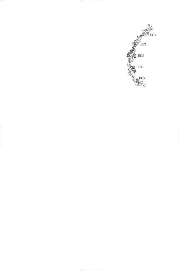
FIGURE 10.4. Ectodomain of a classical cadherin: Shown are the five domains, labeled EC1 through EC5, from the N-terminus towards the C-terminus, which is nearest plasma membrane. Small single and paired spheres represent calcium ions, which are necessary for proper functioning of the receptor. Clusters of spheres denote the locations of N-linked and O-linked sugars. The figure was prepared using Protein Explorer with atomic coordinates deposited in the PDB under accession number 1l3w.
Classical cadherins such as E-cadherin (epithelial cadherin) and N- cadherin (neural cadherin), contain about 750 amino acid residues. Their extracellular domain consists of five repeats of a 100-amino acid EC domain, which extends out 250 Åfrom the cell surface (Figure 10.4). These cadherins attach to the actin cytoskeleton by means of a linker protein called catenin. The cytoplasmic domain of these cadherins is approximately 150 amino acid residues in size, and contains a catenin-binding site. There are two catenin subunits. The b subunit binds to the catenin domain of the cadherin and to the a subunit of the catenin. The a subunit, in turn, connects to the actin cytoskeleton. This cadherin-catenin complex is referred to as a zonula adherens junction in epithelial cells and as an adherens junction in neurons. In epithelia, these junctions link cells together laterally to permit them to form stable sheets, and they separate the apical (top) region from the basolateral (bottom) region of each of the cells. In neurons, the adherens junctions connect the preand postsynaptic cells, thereby providing mechanical stability and a means of conveying signals in both directs across the synapse.
Cadherins operate in a homophilic manner, and have a role in maintaining tissue boundaries and stabilizing synapses. Cells that segregate into distinct tissues express different cadherins, so that a cadherin receptor on the surface of one cell binds to a cadherin receptor of the same type on the surface of an opposing cell. Calcium binding is necessary for clustering, or oligomerization, of cadherin receptors as well as for ligand binding. An example of how this requirement might serve a useful function is provided by N-cadherin binding in synaptic junctions. Intense synaptic activity generates long-lasting changes in synaptic transmission. Remodeling of the synapse underlies these changes. These changes might occur through the following sequence of steps: Intense activity depletes the supply of calcium in the vicinity of the synapse. This lowering in the local calcium concentration weakens the links between cadherin molecules allowing remodeling

228 10. Cell Adhesion and Motility
activities to proceed. This step is followed by reestablishement of firm contact when the calcium concentration returns to its basal levels.
10.6IgCAMs Mediate Cell–Cell and Cell–ECM Adhesion
Cell adhesion molecules (CAMs) of the immunogloulin superfamily (Ig-SF) mediate cell–cell and cell–ECM adhesion. Cell surface receptors belonging to this superfamily, the IgCAMs, are characterized by the presence of one or more immunoglobulin-like domains in their extracellular region. These adhesion molecules mediate cell-to-cell contact by binding to cell surface counterreceptors, and help establish and maintain contact with the extracellular matrix by binding to ECM constituents. Unlike cadherins, calcium binding is not required either for ligand binding or for clustering to occur. Some IgCAMs bind in a homophilic manner while others bind in a heterophilic way.
One of the main kinds of IgCAM is the neural IgCAM, or NCAM, expressed on neurons (Table 10.1). These receptors contain one or more repeats of the Ig fold plus a number of fibronectin type III folds, in their extracellular domain as depicted in Figure 10.1. The NCAMs are grouped into families according to the organization of the extracellular domains. NCAM contains five immunoglobulin-fold repeats plus a pair of fibronectin type III (FnIII) repeats proximal to the plasma membrane. It is thus classified as a 5/2 IgCAM. Another important and well-studied neural IgCAM is L1. This cell adhesion molecule is a 6/5 IgCAM family member. A third
TABLE 10.1. Members of the IgCAM group of cell adhesion receptors: Abbreviations—ICAM (intercellular cell adhesion molecule); LFA (lymphocyte function-associated antigen), NCAM (neural cell adhesion molecule), PECAM (platelet endothelial call adhesion molecule), VCAM (vascular cell adhesion molecule), CD (cluster of differentiation).
Ig-SF CAM |
CD designation |
Ligand |
Distribution/role |
ICAM-1 |
CD54 |
LFA-1, Mac-1, fibrinogen |
Lymphocytes, endothelial |
|
|
|
cells when inflammation is |
|
|
|
present |
ICAM-2 |
CD102 |
LFA-1, Mac-1 |
Recirculating leukocytes |
ICAM-3 |
CD50 |
LFA-1 |
Leukocytes |
LFA-2 |
CD2 |
LFA-3 |
Lymphocytes |
LFA-3 |
CD58 |
LFA-2 |
Broad distribution |
NCAM |
CD56 |
NCAM, collagen, heparin |
Neural tissue |
PECAM-1 |
CD31 |
PECAM-1 |
Platelets, leukocytes |
VCAM-1 |
CD106 |
a4b1 and a4b7 integrins |
Lymphocytes, endothelial |
|
|
|
cells when inflammation is |
|
|
|
present |
|
|
|
|

10.7 Selectins Are CAMs Involved in Leukocyte Motility |
229 |
group of IgCAMs are the DCC family members. Named for the protein DCC (deleted in colorectal cancer) these are 4/6 IgCAMs. Molecules closely related to DCC have a prominent role in the development of the nervous system. They are found on growth cones where they bind a family of diffusible chemoattractants called netrins that guide growing axons during embryonic development, as will be discussed later in this chapter.
The two most prominent families of IgCAMs are the NCAMs and ICAMs (intercellular cell adhesion molecules). The ICAMs bind to integrins and thus mediate heterophilic binding. NCAMs bind to identical receptors on opposing cells and thus mediate homophilic binding. Besides their role in the immune/inflammatory system, these proteins play an important role during embryonic development and in the nervous system both during development and in adult life. NCAMs are also expressed in the heart, gonads, and skeletal muscle. Because of their role in signaling and maintaining tissue integrity they have a prominent role in several forms of cancer.
10.7 Selectins Are CAMs Involved in Leukocyte Motility
Leukocytes circulate in blood vessels and the lymphatic system, and when an infection is detected they converge to infection sites. Selectins mediate the movement of leukoctyes within blood vessels to sites of infection, and out of the blood vessel into the surrounding inflamed tissue. They are expressed on the surface of leukoctes, the endothelium lining the blood vessels, and platelets that form adhesive plugs, or clots, at sites of wounds. There are three kinds of selectins: L-selectins are expressed on leukocytes, E-selectins are expressed on the endothelial cells, and P-selectins are expressed on platelets on the endothelium. These adhesion molecules mediate the capture of the circulating leukocytes by the walls of the blood vessels and make possible their subsequent rolling.
Selectins are mosaic proteins with a common structural organization. They each have an NH2 terminal lectin domain followed by an epidermal growth factor (EGF) domain followed by a number of consensus repeats (CRs) of a complement-like binding sequence, a single transmembrane segment, and a short cytoplasmic tail. The lectin domain is a carbohydratebinding domain that enables the selectin to bind to carbohydrate structures on its ligand. The CRs are thought to function as spacers that extend the molecule a distance that supports optimal rolling. Deletion of portions of this segment impairs rolling. The three selectins vary in the number of CRs. Human P-selectin has nine CRs, while E-selectin has six and L-selectin just two.
The lectin domain situated near the NH2 terminus is a carbohydratebinding domain. Selectin ligands such as GlyCAM-1 and CD34 are members of the mucin family. These are long rodlike glycoproteins with multiple serine/threonine residues and are heavily glycosylated (O-linked).

230 10. Cell Adhesion and Motility
TABLE 10.2. Selectins, cells, and structures that express them, their functions, and ligands: Ligand abbreviations are as follows: PSGL-1 (P-selectin glycoprotein ligand-1); MAdCAM-1 (mucosal addressin cellular adhesion molecule-1); GlyCAM-1 (glycosylation-dependent cell adhesion molecule-1).
Selectin |
Expression |
Functions |
Ligands |
L-selectin |
Leukocytes |
Leukocyte trafficking, |
PSGL-1, GlyCAM-1, |
|
|
rolling adhesion |
MAdCAM-1, CD34 |
P-selectin |
Platelets, endothelium |
Rolling adhesion |
PSGL-1 |
E-selectin |
Endothelium |
Rolling adhesion induced |
PSGL-1 |
|
|
by inflammation |
|
|
|
|
|
Another ligand, MAdCAM-1, is an IgCAM and directs selectins to mucosal tissues. The primary P-selectin ligand is PSGL-1. Both ligands and selectins support rapid bond formation and dissociation.
Selectins are the first cell adhesion receptors to be activated in an inflammatory response. In the absence of an injury E-selectins and P-selectins are not expressed on the cell surface while L-selectins are constitutively active in order to promote continual leukocyte homing. This activity is guided by the appearance of its ligand in the vicinity of an inflammation. The expression of E-selectin on the surface of endothelial cells is triggered by cytokines such as TNF-a, IL-1, and lipopolysaccharide (LPS). P-selectin is stored in granules inside platelets (a-granules) and endothelial cells (Weibel–Palade bodies). P-selectin can be shipped to the outer surface in minutes in response to histamines and other triggering signals. Cytokines induce synthesis of P-selectin mRNAs.
Platelets are cytoplasmic fragments of bone marrow cells called megakaryocytes. Platelets are released from the bone marrow and circulate in the bloodstream. Their role in the inflammatory response is to form clots or plugs that block blood flow at sites of injury. Adhesion and aggregation are central to platelet function, and they express integrins, selectins, and IgCAM receptors. Microvilli are adhesion molecule-rich extensions of the cell surface. They are formed at the outside facing, or apical, surface in a variety of different cell types in order to increase the effective surface area. The primary platelet selectin ligand is the P-selectin glycoprotein-1 (PSG1). It is constitutively expressed on the tips of microvilli of leukocytes such as neutrophils and monocytes.
10.8Leukocytes Roll, Adhere, and Crawl to Reach the Site of an Infection
Leukocytes are highly mobile cells that migrate from blood in and out of lymphatic organs and into tissues, and then back out again into circulation. They respond to infections by attacking and destroying the causative agents.

10.9 Bonds Form and Break During Leukocyte Rolling |
231 |
The leukocytes do not passively float along the blood vessels, but instead remain in contact with the cells lining the walls of the blood vessels. They move along the walls by rolling in a controlled way. This mode of locomotion enables them to stop at the right location and then exit by crawling out between the cells. Adhesion is critically important, enabling the leukocytes to first roll, and then crawl, and finally to kill the pathogens.
In an inflammatory response, the physiology of the blood vessels is altered to better enable the leukocytes to reach the site of infection. Postcapillary venules are the main locus of vascular inflammatory activity during an inflammatory response. These structures, some 30 to 40 microns in diameter, consist of a layer of endothelial cells on the inside and a supporting basement membrane on the outside. Blood flow in small vessels such as the postcapillary venules takes place under low flow rates, reasonably high viscosities, and small vessel diameter. Under these conditions the velocity of the blood flow is greatest in the center of the vessel and decreases to zero as the vessel walls are approached. During inflammation the blood vessels dilate and the overall flow rate is slowed. Red blood cells aggregate into large assemblies called rouleaux that collect into the center of the vessels displacing white blood cells. In response the leukocytes move from the center of the blood vessel to the vicinity of the vessel walls. When this happens cell surface receptors expressed on the plasma membranes of the endothelial cells and leukocytes can engage one another to promote rolling and then hard adhesion.
10.9 Bonds Form and Break During Leukocyte Rolling
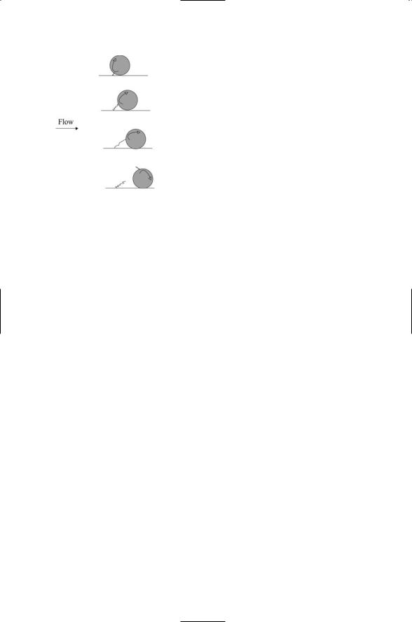
Shear stresses are forces generated when adjacent layers of a fluid slip over each other. Because of the shear stresses that are present, leukocytes will either tumble or roll in the direction of the blood flow. Tumbling motion is a rotary movement done without any contact with the cell wall. Rolling, in contrast, is a rotary movement carried out while in contact with the endothelial cells of the vessel wall. The observed motions of leukocytes are jerky. Periods of rapid motion are interrupted by periods in which the motions are far slower. The periods of slow motion correspond to tethering of the leukocytes to the membranes of the endothelium and the periods of rapid motion to contact-free flow.
Bonds form and break during leukocyte rolling (Figure 10.5). Bond lifetimes must be long enough to permit formation of multiple bonds. If the off (bond dissociation) rates are too high multiple bonds cannot form. One bond will dissociate before another can be formed. At the other extreme, too tight a bonding will either immobilize the cell or will lead to situations where large forces can pull a receptor molecule out of the membrane. The association and dissociation rates should be rapid, but not too rapid. In leukocyte rolling, the bonds that tether leukocytes to the endothelial wall
232 10. Cell Adhesion and Motility
FIGURE 10.5. Bond under shear forces during leukocyte rolling: Depicted are the compression, stretching, and finally dissociation of bonds between a cell adhesion molecule and its ligand during rolling of a leukocyte along the inner wall of a blood vessel.
are subjected to stretching, or tensile, forces as a result of the shear stresses. Forces have an important effect on the bond lifetimes. They shorten the lifetimes of the bonds. The enhancements can be appreciable—the off-rate rises exponentially with increasing force. The enhanced dissociation rates arising from the tensile forces shift the bond lifetimes into the range needed to ensure proper rolling.
The off-rate amplification is a consequence of the lowering of the energy barriers by the applied forces. The dependence of bond lifetimes on the presence and strength of applied forces is not specific to rolling or leukocytes. Instead, these considerations apply equally well to other bonds between receptors and ligands. The acceleration in dissociation rates due to the presence of applied forces provides a general mechanism for transducing mechanical stresses into signaling responses. Membrane, cytoskeletal, and signaling elements coming together at control points will sense and respond to mechanical stresses by dissociating far more rapidly than would be the case in the absence of stresses.
10.10Bond Dissociation of Rolling Leukocyte as Seen in Microscopy
Rugged energy landscapes describe bond dissociation during leukocyte rolling. The stretching properties of bonds between cell adhesion molecules and their ligands have been explored using atomic force microscopy (AFM) and optical tweezers. In a typical AFM experiment, the adhesion molecules of interest are attached to a sharp tip, one having a diameter of no more than a few microns. The tip is mounted on a flexible cantilever. When the adhesion molecules are brought into close proximity to their ligands, mounted on a separate surface, the cantilever will bend and undergo a displacement. These displacements are measured by sweeping a laser beam
10.11 Slip & Catch Bonds Between Selectins & Carbohydrate Ligands |
233 |
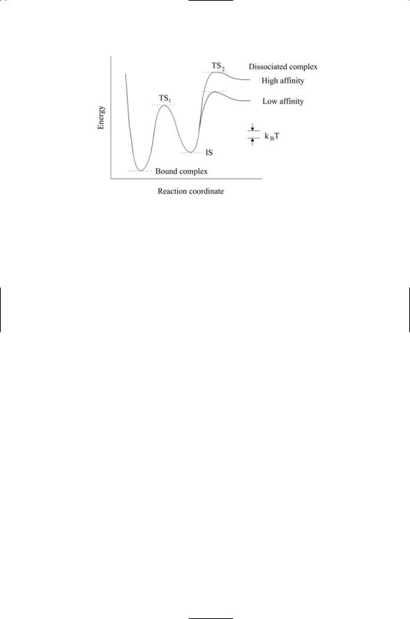
FIGURE 10.6. Energy landscape for dissociation of bonds between adhesive molecules and their ligands: Abbreviations—transition state 1 (TS1); transition state 2 (TS2); intermediate state (IS).
across the cantilever and measuring the angle and orientation of the reflected light. In an optical tweezers experiment, one uses a laser beam to trap dielectric particles. External forces applied to the trapped particles can be determined by monitoring the angular deflection of the laser light. The adhesive molecules are mounted on a pedestal attached to a surface and the ligands are attached to optically trapped beads.
Experiments of several different adhesion molecule/ligand combinations have been studied, and the results placed within an energy-landscape perspective. A representative landscape deduced from these experiments is presented in Figure 10.6. This landscape has two prominent features. First, it is a rugged landscape. The pockets are deep, several times the thermal energy, resulting in the presence of at least one metastable intermediate state. The situation shown in the figure is that of two transition states. Second, two different outer barriers heights are present—one of these corresponds to low affinity binding and the other to high affinity binding. The high inner barrier is responsible for high strengths seen at short time scales and the major portion of the activation energy. The lower barriers located further out extend bond lifetimes when forces are absent.
10.11Slip and Catch Bonds Between Selectins and Their Carbohydrate Ligands
The thrust of the previous section was that externally applied forces reduce the lifetimes of receptor-ligand bonds by lowering the energies of the transition barriers. The force-driven reductions in lifetime allow rolling

234 10. Cell Adhesion and Motility
leukocytes to detach from surface tethers at the right time while maintaining good adhesion to that place on the surface at earlier times. The corresponding bonds are called slip bonds because they allow the rolling cells to slip away. These are not the only kinds of bonds that form. An observation often made not only of rolling leukocytes, but also migrating bacteria, is that shear stresses promote good surface contact. The physical mechanism underlying this phenomenon is captured by the notion of a catch bond—a bond that is strengthened by the external forces rather than weakened. The selectins that mediate leukocyte rolling exhibit both kinds of bonds. The catch bonds are formed first. They enable the leukocytes to be caught by the surface. Once the leukocytes are caught and begin rolling, a transition takes place from a catch to a slip bond regime so that the latter can mediate proper detachment from the surface.
Selectins are not only expressed by migrating leukocytes. They are expressed early during embryonic development and enable embryos to attach to the lining of the uterus. Failure to properly lodge in the uterus is a prominent cause of fertilization and implantation failures. In the attachment process, carbohydrate ligands upregulated and expressed on the surface of the uterus bind the L-selectin molecules expressed on the surface of the embryo. The adhesion takes place under shear stresses created by mucin secretions and, once established, sets off a chain of signaling events.
10.12 Development in Central Nervous System
Development of the central nervous system involves many of the same operations as used in body development. The development of the central nervous system, with its laminar structure and organization into dozens of anatomically and functionally distinct areas, is one of the most remarkable and dramatic processes in nature. It takes place in several stages. It begins with the production of immature neurons, or neurites, and glia, cells that eventually supply neurons with growth factors, nutrients, protection (astrocytes); and electrical insulation (Schwann cells). The neurites multiply, grow, differentiate, and migrate to various sites where they aggregate into distinct cortical layers and regions. During this period support structures such as the ECM are erected from molecules secreted by the developing cells. Cell growth, maturation, and circuit formation follows arrival of the neurites at their cortical destinations. The growing cells within each of the layers and areas develop their morphologically and electrophysiologically distinct axonal and dendritic structures, and they establish initial circuit-forming synaptic contacts. The initial contacts are then refined, and unwanted connections and cells are removed. At the end of this process more than 1010 neurons will have formed some 1015 precise synaptic connections.
Once the neurites reach their cortical destination and their migration ceases, they send out processes that become axons and dendrites that form the synapses and neural circuits. The growth cone (Figure 10.7) is a motile