

161.US Dept of Health and Human Services. Reducing the Health Consequences of Smoking: 25 Years of Progress: A Report of the Surgeon General. US Dept of Health and Human Services, Public Health Service, Centers for Disease Control, Center for Chronic Disease Prevention and Health Promotion, Office on Smoking and Health. 1989. DHHS Publication (CDC); 89–8411.
162.McIntyre KM. Cardiopulmonary resuscitation and the ultimate coronary care unit. JAMA 1980;244:510–511.
See also CARDIAC OUTPUT, THERMODILUTION MEASUREMENT OF; RESPIRATORY MECHANICS AND GAS EXCHANGE; SHOCK, TREATMENT OF; VENTILATORS, ACUTE MEDICAL CARE; VENTILATORY MONITORING.
CARTILAGE AND MENISCUS, PROPERTIES OF
JEREMY J. MAO
ALEXANDER J. TROKEN
NICHOLAS W. MARION
University of Illinois
Chicago, Illinois
LEO Q. WAN
VAN C. MOW
Columbia University
New York, New York
INTRODUCTION
Synovial or diarthrodial joints are created to enable the movement between bones. Articular cartilage on the end of articulating bone, therefore, must accomplish two functions: (1) absorb, distribute, and transmit mechanical loading, and (2) create a low friction and wear surface for movement over decades of mammalian life. Three cartilage phenotypes exist: hyaline cartilage, fibrocartilage, and elastic cartilage. In the older literature, articular cartilage is often referred to as hyaline cartilage due to its glassy appearance; this appearance is derived from its high proteoglycan content. Indeed, articular cartilage has the highest proteoglycan content of all biological tissues, while, at the same time, it has the lowest cellular content. The chondrocytes not only secret and control collagen and proteoglycan contents in the extracellular matrix, but also are responsible for regulating the elaborate molecular architecture of these macromolecules and their ultrastructural organization (1–3). Throughout life, the healthy chondrocytes under normal conditions secrete and elaborate sufficient amounts of the extracellular matrix macromolecules and completely encase themselves in an environment that possesses truly remarkable biomechanical mechanisms that protect them against the mechanical insults associated with joint loading, and thus survive for long periods of time under normal health conditions (4–6).
Cartilage in a small number of joints in humans, such as the knee meniscus, temporo-mandibular joint, and intervertebral discs, is fibrocartilage. The intervertebral disc, besides its complex macromolecular architectural and ultrastructural organization, also has a complex macrostructural organization; the latter is manifested in the
CARTILAGE AND MENISCUS, PROPERTIES OF |
63 |
macro-layering of the outer rings of collagen-rich annulus fibrosis and an inner core of a proteoglycan-rich ‘‘kidneyshaped’’ nucleus pulposus. The cells of these fibrocartilaginous tissues are fibroblasts and chondrocytes, some of which are called fibrochondrocytes. Whereas the genotype and phenotype of cartilage cells determine the biochemical and molecular properties of cartilage, the mechanical properties of articular cartilage are largely dependent on the constituents of extracellular matrix (3). This divergence in the determination of biological and mechanical properties is attributed to the scarce cellularity in adult cartilage, with chondrocytes that account for only less than 10% of the adult cartilage volume (4,7). Comprehensive reviews of hyaline and fibrocartilage can be found elsewhere (1,3,8).
An average human takes approximately 2 million steps per year. The jointsin the lower limbs, therefore, can undergo 1–4 million cyclic loads from physical activities (9,10). These loads can peak 4–5 times body weight (11,12), and can cause both macroand micro-structural changes in articular cartilage that may ultimately lead to degenerative diseases such as osteoarthritis (4,7,13–15). Arthritis, which encompasses more than 100 diseases and conditions, is recognized as among the leading causes of physical disability worldwide (16). Thus, investigation of the properties of normal and arthritic cartilage is essential not only for the understanding of the etiology of arthritis (6), but also devising possible approaches toward the tissue engineering of cartilage and meniscus for clinical treatment modalities (17).
ARTICULAR CARTILAGE AND MENISCUS: COMPOSITION AND STRUCTURE
Chondrocytes and fibrochondrocytes are responsible for the morphogenesis, matrix synthesis, and maintenance of articular cartilage and meniscus as functional tissues. However, these cells only account for approximately 10% of the total cartilage volume in adults (3,7,18,19). Chondrocytes receive nutrients and shed metabolic waste products largely from convective transport and diffusion, either from/to the synovial fluid or from subchondral bone. In the adult, articular cartilage is generally aneural and avascular. Vasculature is present only in the periphery of mature meniscus (20).
Hyaline Cartilage and Articular Cartilage
Hyaline cartilage is present on the articulating surfaces of the bones in most, but not all, synovial joints. Articular cartilage serves to bear and distribute load and contribute to joint lubrication. It serves these different purposes through varying the amount of water relative to the amounts of Type I collagen and proteoglycans and the molecular and ultrastructural organizations of these structural molecules. Healthy articular cartilage appears smooth, bluish white, glistening, and intact. Osteoarthritic articular cartilage appears dull and coarse and may have tears and frays. Hyaline cartilage is also present in the growth plate at the metaphyseal region of long bones and in the cranial base and serves to enable longitudinal bone growth by endochondral ossification (21–23). A review of

64 CARTILAGE AND MENISCUS, PROPERTIES OF
growth plate cartilage is beyond the scope of this chapter, but can be found elsewhere (24–26).
Articular cartilage has two immiscible phases—a solid phase and a fluid phase. Small electrolytes such as Naþ and Cl are dissolved in the fluid phase and are freely mobile by diffusion and convection through the porous-permeable solid phase. The fluid and solid phases have been modeled in the now classic biphasic theory developed by Mow et al. (27). Normal fluid component ranges from 75 to 80% by wet weight, and the remaining 20 to 25% of the organic matrix forms solid material with complex material properties (3,18,19). Up to 65% of the solid ECM by dry weight is made of collagen, whereas proteoglycans constitute up to 25%; other glycoproteins, chondrocytes, and lipids can generally make up 10% (3,28,29). Collagen fibers are classified on the basis of their amino acid composition and molecular structure. Although an assortment of collagens exist in both hyaline cartilage and fibrocartilage, Type II collagen is most prevalent in articular cartilage, whereas Type I collagen is most common in the meniscus (3,30). The collagen fibers are assembled as tight triple-helical structures made from three polypeptide alpha-chains. The triple helices are then arranged as tropocollagen molecules, which are wound in a helical manner to form larger collagen fibers that are, in turn, are organized into a strong cohesive collagen network (3,31,32). This arrangement allows for considerable tensile stiffness and strength (33–38). The collagen also serves to restrain the swelling pressure created from the surrounding embedded proteoglycans (2,18,19,39). Proteoglycans (PGs) are hydrophilic macromolecules with numerous glycosaminoglycans (chondroitin and keratin sulfates) attached to a protein core; the protein core of this bottle-brush-shaped molecular is, in turn, attached to a hyaluronan (mw: molecular weight 0.5 106) resulting in a supra-macromolecule with an approximate molecular weight ranging from (200 to 300) 106 (3,29,40–42). These enormous, negatively charged molecules are trapped in the fine porous meshwork of collagen by frictional and electrostatic forces and by steric exclusion; thus, in the ECM, PGs function largely to generate osmotic pressure (2,39) and to resist the compressive stresses of articulation acting on the cartilaginous surface. Although various PGs exist in cartilage, the one that constitutes up to 80–90% of the total PGs in cartilage is aggrecan (3). As the name implies, aggrecan facilitates the formation of large aggregates. Like collagen,
aggrecan, as with all PGs, maintains a structure that is directly correlated with its function. The general structure of PGs occurs through noncovalent bonding of aggrecans to the hyaluronan via link proteins, thus securing firm linkages. Attached to the protein core are the glycosaminoglycan side chains (GAGs) that are vital for biological and biomechanical functions of the tissues; indeed, they are hallmarks of chondrogenic activity in tissue engineering. These GAGs bear the necessary physical properties that ultimately confer onto these tissues their hydrophyllic tendencies and compressive load-carriage abilities (3,18,19). The presence of large numbers of sulfate and carboxyl groups on the GAGs gives rise to a high negative-charge density in the ECM (2). This anionic nature attracts positively charged ions, creating an osmotic pressure, known as Donnan osmotic pressure, that favors tissue hydration (3,39,43). The fixed-negative charges also create intense repulsive forces of the GAGs against each other. This expansion force of the PG molecules causes tensile stresses to be developed within the surrounding collagen network surrounding the PGs. This swelling pressure thus resists the compressive forces against the cartilage without volume loss.
Articular cartilage is organized into three layers or zones (3,44) as shown in Fig. 1. The superficial zone forms the articular surface, whereas the deep zone is anchored to calcified cartilage and subchondral bone; both zones have well-defined collagen architectures. An intermediate zone exists in between with a random collagen fiber ultrastructural organization. These layered structural arrangements have long been hypothesized to be important in cartilage function (45–47). The overall thickness of articular cartilage, including the three zones, varies between joints, age, individuals, and species from less than a millimeter to a few millimeters, with the thickest being measured at the retro-surface of the human patella and femoral trochlea (3,13,48).
The superficial zone has the highest collagen, water content, and chondrocyte density, but the lowest proteoglycan content among all three zones (2,49–51). The abundant collagen fibrils are aligned parallel to the articular surface and provide the superficial zone with substantial tensile strength in an orthotropic manner (37,38,52,53). The chondrocytes in this zone are flattened and are apparently polarized to be parallel to the surface (Fig. 2a) (7,54). Recently, intricate 3D images have been taken to
Figure 1. Layered structure of cartilage collagen network showing three distinct regions (3).
CARTILAGE AND MENISCUS, PROPERTIES OF |
65 |
Figure 2. (a) Articular cartilage from a mature rabbit femur showing typical zonal arrangement of chondrocytes (polished saw-cut of resin-embedded tissues, surface-stained with basic fuchsine and toluidine blue) (54). (b) Schema of chondrocyte organization in the superficial zone (SZ), middle zone (MZ), and deep zone (DZ) (7).
view the discoid-shaped cells as they are maintained in this layer. Methods have ranged from digital volumetric imaging (55) to atomic force microscopy (56).
The intermediate, or middle, zone is generally the thickest amongst the three uncalcified zones of articular cartilage. Collagen fibrils, although less dense, have a greater diameter than the superficial zone, but appear to be more randomly oriented (47,57). The intermediate zone also has the highest proteoglycan content (3). Chondrocytes are more rounded, although cell density is not as high as in the superficial zone (3,58) (Fig. 2b).
The deep zone is relatively thin and the collagen are intertwined to form larger fiber bundles, and, from this zone, they insert perpendicularly into the calcified zone, and thus anchor the uncalcified tissue to the bony ends as required by joint articulation. This organization allows the bundles to firmly anchor the articular cartilage to the underlying subchondral bone. In general, chondrocyte density decreases from the middle zone to the deep zone, where they are similarly aligned as the collagen bundles, arranging into columns perpendicular to the uncalcified-calcified cartilage intersurface (3,55).
Several recent studies have investigated the pericellular matrix (PCM) and the interterritorial matrix (ITM) of chondrocytes. Using algorithms to account for fluid flow and differences in the relative stiffness between the PCM, the ITM, and the chondrocyte, different elastic moduli between PCM and ITM have been found to have a significant effect on chondrocyte’s mechanical environment (59). Gradient distributions of charges and material densities relative to chondrocyte surface are important in cartilage fluid flow dynamics and deformation behavior (60). Using micropipette isolation of chondrocytes and nuclei, chondrocyte nuclei have been found to be stiffer than intact chondrocytes (59,61,62). Cultured chondrocytes are able to elaborate a PCM rich in Type VI collagen; however, intact chondron pellets accumulate significantly more
Figure 3. A representative height map of the PCM and ITM chondrocytes obtained through force mode of atomic force microscopy. Qualitatively, the ITM showed greater peak and valley contours than the topographic contour of the PCM (a).
(b) presents the average Young’s moduli of the PCM and ITM attained via nanoindentation. The average Young’s modulus of the ITM (636.1 124.91 kPa) was significantly greater than the PCM (265.1 52.76 kPa) ( p <0.01) (N ¼ 19) (64).
proteoglycans and Type II collagen than chondrocytes without a native PCM (63). Following a few weeks of accumulation of the ITM and PCM by isolated chondrocytes, a rapid increase in compressive stiffness occurs in both the chondron and the chondrocyte pellets (63). Using atomic force microscopy (AFM), the ITM is found to be stiffer to nanoindentation than the PCM (Figs. 3a and 3b) (64).
Meniscus and Fibrocartilage
The meniscus in the knee joint is a fibrocartilage. The two menisci (lateral and medial) in each knee joint are crescent or semi-lunar shaped and are attached to the joint capsule. The triangular cross section of the meniscus tapers radially inward from the periphery, and the center of the meniscus is thin and unattached. Thus, the cross section of the meniscus is wedge-shaped. The central region is a vascular and has more proteoglycans, hence more hyaline in appearance. The anterior and posterior horns of the meniscus form the tips of the crescents. The anterior horn of the lateral meniscus is attached to the tibia in front of the intercondylar eminence, partially blending with the
66 CARTILAGE AND MENISCUS, PROPERTIES OF
anterior cruciate ligament. The posterior horn is attached to the tibia near the intercondylar eminence as well as to the femur via the meniscofemoral ligament. The anterior and posterior horns of the medial meniscus are attached to the tibia near their respective intercondylar fossae. The anterior horns of the lateral and medial menisci are connected by the transverse ligament. The thick peripheral borders and associated horns of the meniscus are vascularized by blood supply predominantly from the genicular arteries surrounding the joint. The thinner central portions of the meniscus are aneural and avascular, a region very much like hyaline cartilage (20). The meniscus is lubricated with synovial fluid (65), probably by the same lubrication mechanisms known to exist in articular cartilage (66). Fibrocartilage is found in a small number of other joints. The disk of the temporomandibular joint (TMJ) and the intervertebral disks are both composed of fibrocartilage, although they are drastically different structures with different distributions of cartilage and PGs and ultrastructual organization.
The fibrocartilagenous structure of the meniscus differs from that of hyaline cartilage in many ways. The cells of the meniscus are sometimes called fibrochondrocytes, although it is probable that some cells are more like fibroblasts, whereas others are more like chondrocytes (65,67). The peripheral two-thirds of the meniscus are primarily composed of a randomly oriented mesh-like, coarse, collagen fibrillar matrix (68–71). In deeper portions, large rope-like collagen fiber bundles are arranged circumferentially, retaining the overall semi-lunar shape of the meniscus and providing tensile strength. Smaller fibers are also found radially and connect to the larger circumferential collagen fiber bundles (72). As mentioned above, the inner portion of the meniscus resembles that of hyaline cartilage, containing a higher percentage of proteoglycans enmeshed within a randomly arranged collagen fibrillar matrix (71,73,74).
The function of the meniscus is to enhance higher congruity of the articulating surfaces of the distal femur and proximal tibia, to accommodate the range of motion, in addition to the same functions of load bearing and load distribution to that of articular cartilage (20). The previous assumption that the menisci are functionless, evolutionary remains of leg muscles is erroneous and that menisectomy (a common clinical procedure) is indeed a common procedure in animal models to study the etiology of osteoarthritis (75–78).
CARTILAGE AND MENISCUS: MECHANICAL PROPERTIES
Articular cartilage and meniscus are both important loadbearing tissues and vital to the maintenance of normal joint functions (3,18,19). Articular cartilage can absorb mechanical shock of joint motion and spread the applied load onto the subchondral bone. It also contributes to the lubrication mechanism and provides a surface with low friction, enabling repetitive gliding motion between articulating surfaces (7,66). The meniscus of the knee has important biomechanical functions such as load transmission at the otherwise highly incongruent tibiofemoral
articulation, shock absorption, joint congruity, and stability (18,19). The salient biomechanical functions of articular cartilage and meniscus are dependent on their biological structure, composition, and the intrinsic material properties of the ECM. The knowledge of their material properties such as tensile, compressive, and shear moduli is essential to understand not only their biomechanical functions, but also in the tissue engineering of articular cartilage and meniscus to produce in a biomimetic manner artificial-biological replacements (17,79).
Tensile Properties
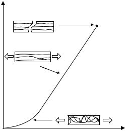
When cartilage is tensed, the tensile stress-strain behavior is nonlinear. A typical nonlinear stress-strain (s-e) curve for cartilage, meniscus, and other soft tissues is depicted in Fig. 4 (3). For small deformations, a ‘toe-region’ is seen in the stress-strain curve, in which the collagen fibrils will primarily realign in the direction of the externally applied force instead of being stretched (elongation per unit length). For larger deformations, the collagen fibrils are stretched, and a larger tensile stress is generated within the collagen fibers (e.g., 35–38,52,53,80–83). In this linear region, the stress is proportional to the applied strain, and their ratio is known as Young’s modulus in tension, or tensile modulus. This tensile modulus is a measure of the stiffness of the collagen-PG solid matrix and is primarily dependent on the density of collagen fibrils, fibril diameter, and type or amount of collagen cross-linking (3,18,19,52). Beyond the linear region, the cartilage strip will rupture abruptly, and the tensile failure stress is a measure of the strength of the collagen fibrillar network.
In general, the tensile modulus of articular cartilage will be in a range of 1–30 MPa, which is much larger than the compressive modulus of cartilage ( 0.5 MPa), which is known as tension-compression nonlinear property of the
Failure |
Linear Region |
TensileStress |
Toe Region |
Strain |
Figure 4. Typical stress-strain curve for articular cartilage and meniscus in a uniaxial and uniform strain rate experiment. The toe region is marked by an increasing slope, whereas the linear region appears to be a straight line (3).
cartilage (84–86). The tensile properties are also known to vary with location, depth, and orientation of test specimens of cartilage and meniscus. Hultkrantz (45) demonstrated the anisotropic organization of the collagen network by puncturing holes in the surface of articular cartilage with a round pin. He found that round puncture holes will form elongated splits, analogous to splits formed in lumber when a large round awl pierces it. The split-line patterns were, to him, evidence of collagen fiber orientation, which is still an enigma today because electron microscopy has not found such surface collagen anisotropy. (Nevertheless, the pattern of split lines is similar to Langer lines formed in the skin in a similar manner.) Much later, Woo et al. (38), Kempson et al. (52), and Roth and Mow (37) showed that the tensile strength and stiffness of the samples cut parallel to the split-line direction were higher than those cut perpendicular to it. The cartilage strips from high weightbearing areas of human knee joints exhibit larger tensile modulus than those from low weight-bearing areas (53,82) because high weight-bearing areas generally have a relatively higher proteoglycan content. The adult human femoral articular cartilage exhibits a gradual decrease in tensile strength and stiffness as the distance from the articular surface increases (36,81), while this functional dependence was not observed for young bovine humeral joints (38). A dependency of cartilage tensile properties with skeletal maturation was found by Roth and Mow (37). These investigators found that, with the closing of the growth plate (indicative of skeletal maturity), the strength and stiffness of cartilage are much less that those properties of immature cartilage (open-physis). The effects of age on the tensile properties of adult cartilage were extensively studied by Kempson (81), and the results showed that the tensile modulus decreases with age, and that the modulus of the hip cartilage decreases more markedly than that of ankle cartilage. This finding may explain the relatively high occurrence of osteoarthritis in the hip compared with the ankle. Like articular cartilage, the tensile properties of meniscus vary with respect to the location (anterior, central, and posterior) and specimen orientation relative to the predominant collagen fiber direction (circumferential and radial) (18,19,74). Specimens from the posterior half of the medial meniscus have been shown to be significantly less stiff and less strong in tension than specimens from all other regions (87). This experimental result agrees with the ultrastructural findings using polarized light; In the posterior half of the medial meniscus, collagen fiber bundles have significantly reduced circumferential organization (87).
Numerous experiments have shown that the tensile modulus is correlated with the collagen content or the ratio of collagen content to proteoglycan content in articular cartilage (e.g., 82). The tensile modulus of articular cartilage decreases to only 1% after disruption of collagen cross-linking by elastase (88). In contrast, no significant correlations have been found between the tensile property of the cartilage and proteoglycan content (36,89). These findings indicate that collagen content, organization, and cross-linking play significant roles in generating high tensile modulus of articular cartilage. For meniscus, although the tensile properties show significant regional
CARTILAGE AND MENISCUS, PROPERTIES OF |
67 |
and directional variations, little difference appeared in the biochemical composition with site, and no significant correlation exists between tensile property and chemical contents (74). The variation of tensile properties seems to reflect local differences in collagen ultrastructure and fiber bundle direction as described above.
Compressive Properties
The compressive behavior of cartilage and meniscus has been extensively studied under various configurations, such as confined compression, unconfined compression, and indentation (see Fig. 5). Most of the earliest studies (e.g., 90–94) used the indentation technique to determine the mechanical property of articular cartilage and modeled the cartilage to be a single-phase, elastic body with the assumption of the Poisson’s ratio ranging between 0.4 and 0.5 (e.g., 91–96). However, this single-phase elastic model cannot describe the time-dependent viscoelastic behavior of the tissue nor the role played by cartilage’s major component (i.e., water). Cartilage and meniscus exhibit a viscoelastic creep in response to a constant load (i.e., its deformation will increase with time). Conversely, if a constant displacement is applied, the force response will decrease gradually with time to a constant value (i.e., a stress-relaxation will be observed).
These viscoelastic behaviors derive from the friction of water flowing through solid matrix (27,97), as well as the flow-independent intrinsic energy dissipation inside the macromolecular solid matrix during mechanical loading (98–101). As mentioned, articular cartilage and meniscus can be regarded as biphasic materials: a fluid phase composed of water and electrolytes, and a solid phase mainly composed of collagen and proteoglycans (27). The solid matrix is considered as being porous and permeable. Water resides in the microscopic pores and flows through the matrix during joint loading. Under a slow ramp loading, the observed viscoelastic behaviors are usually dominated by the large drag forces generated by the flow of interstitial fluid through the porous-permeable solid matrix, and therefore, the flow-independent intrinsic energy dissipation is negligible. However, osteoarthritic cartilage has higher permeability and lower ECM stiffness; in such tissues, the intrinsic viscoelastic behavior becomes the dominating component governing their mechanical behaviors.
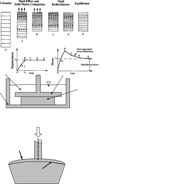
The transient behavior of the tissue under compression is primarily determined by the mechanism of fluid pressurization because of high friction between solid and fluid phases, which is also known as flow-dependent viscoelastic behavior (27). Figure 6 (3) shows the stress-relaxation behavior of a tissue specimen under confined compression. In this experiment, before time t0, the tissue is compressed with a constant rate, and the interstitial fluid inside the tissue will be pushed out through upper porous platen. As a result of the distributive fluid drag force, a larger deformation can be seen at the downstream side (Fig. 6a and 6b). During the relaxation phase (after t0), no fluid exudation occurs, but the fluid needs to redistribute inside the tissue before the equilibrium is reached (Figs. 6c, 6d, and 6e). Although the velocity of fluid flow is very low, the friction force, or the drag force, could be very large because the pore
68 CARTILAGE AND MENISCUS, PROPERTIES OF
Applied load
Saline |
Porous platen |
|
solution |
||
|
||
|
Tissue |
|
|
sample |
|
Container |
|
|
|
(a) |
Applied load
Saline |
Impermeable |
solution |
platen |
|
Tissue |
|
sample |
Container |
|
|
(b) |
Applied load
Articular
cartilage  Porous indenter
Subchondral bone
(c)
Figure 5. Schema of three configurations frequently used to study the compressive properties of articular cartilage. (a) In the confined compression configuration, a load is applied to the cartilage sample via a rigid porous permeable platen. The side walls are assumed to smooth, impermeable, and rigid, thereby preventing lateral expansion and fluid flow. (b) In the unconfined compression configuration, the cartilage sample is compressed between two rigid, smooth, and impermeable platens. The lateral side allows fluid flow. (c) In the indentation configuration, the cartilage is compressed via a rigid porous permeable indenter. The porous indenter allows the fluid exudation to occur freely into the indenter tip and, therefore, creep of the cartilage layer.
size inside the tissue is very small ( 50–65 nm for articular cartilage), and the permeability of the tissue is as low as 10 15 N s/m4. Therefore, the generated fluid pressure can be remarkably high inside the tissue during the transient state, which also means that chondrocytes encased within the ECM will normally be bathed in a highly
Figure 6. A schematic representation of fluid exudation and redistribution within cartilage during a rate-controlled confined compression stress-relaxation test (lower left). The horizontal bars in the upper figures. indicate the distribution of strain in the tissue. The lower graph (right) shows the stress response during the compression phase (O, A, B) and relaxation phase (B, C, D, E) (3).
pressurized fluid. It has been estimated that this fluid pressure could be 30 times more than the elastic stress generated in the solid matrix of articular cartilage (3). Considering that the equilibration process usually takes several hours, no real equilibrium state occurs in joints under physiological conditions because the joints are moving virtually at all times, even during sleep. Thus, the mechanism for fluid pressurization is likely to be the major physiological load-supporting mechanism in diarthroidal joints, and it plays an important role in shielding the solid matrix from large compressive stresses during the joint function (7).
At equilibrium, the fluid flow stops, no fluid pressure gradient exists inside the tissue, and the applied load is entirely supported by the solid matrix of the tissue. Thus, the compressive property can be obtained from the relations between stress and strain. It has been found that the equilibrium strain is proportional to the applied load. Typically, the equilibrium aggregate modulus (27) for normal articular cartilage ranges from 0.4 to 1.5 MPa, whereas the average equilibrium aggregate modulus for the meniscus is about 0.4 MPa. Table 1 shows the equilibrium aggregate moduli of lateral condyle and patellar groove cartilage and meniscus, showing considerable variation among the species and tissue location (3).
Tissue mechanical properties are highly dependent on their composition and structure. It has been shown that the equilibrium aggregate modulus for human articular cartilage correlates in an inverse manner with water content and in a direct manner with PG content (27,102,103). The highly loaded regions of articular cartilage generally have larger compressive modulus and greater PG content (53,104,105). In contrast, no correlation is found between the compressive stiffness and collagen content. Removal of PGs from articular cartilage samples dramatically decreases the compressive modulus, whereas trypsin

|
|
|
CARTILAGE AND MENISCUS, PROPERTIES OF |
69 |
|
Table 1. Equilibrium Aggregate Modulus of Lateral Condyle, Patellar Groove Cartilage and Meniscus (MPa) (3) |
|
||||
|
|
|
|
|
|
|
Humana |
Bovineb |
Caninec |
Monkeyd |
Rabbite |
|
|
|
|
|
|
Lateral condyle |
0.70 |
0.89 |
0.60 |
0.78 |
0.54 |
Patellar groove |
0.53 |
0.47 |
0.55 |
0.52 |
0.51 |
Meniscus |
NA |
0.41 |
NA |
NA |
NA |
aYoung normal.
b18 months to 2 years old. cMature beagles and greyhounds. dMature cynomologus monkeys.
eMature New Zealand white rabbits. fNot available.
digestion of collagen fibrils has little effect on compressive modulus (80,106).
The biphasic theory has been the most successful model for the compressive viscoelastic behaviors of cartilage and meniscus under various conditions (27). This theory assumes that (1) the solid matrix and interstitial fluid are immiscible and incompressible; (2) viscous dissipation is due to the fluid flow between water and the porouspermeable solid matrix; and (3) the frictional drag is proportional to the relative velocity and can be affected by ECM compression. This biphasic theory further assumes that the solid matrix experiences infinitesimal strain and that the stress-strain relations can be described by the generalized Hooke’s law. Despite its simplification, as biological models typically are, the isotropic form of the linear biphasic theory has been shown to provide an accurate description of the compressive creep and stress relaxation behavior of these tissues. In particular, a numerical algorithm based on this biphasic theory was developed and accurately predicted the aggregate modulus, Poisson’s ratio, and permeability of articular cartilage from the indentation creep experiment (13,100,107). The biphasic theory has also been extended by employing higher levels of tissue complexities, including material inhomogeneities (108–110), material symmetries (33,34,85,86,111), and matrix viscoelasticities (33,34,84,98–100).
Shear Properties
The intrinsic viscoelastic properties of the solid matrix of cartilage and meniscus can only be determined in a pure shear experiment and under small strain conditions. In pure shear, the kinematics of deformation does not permit volumetric change, and hence, no interstitial fluid flow is possible when no pressure gradients are applied. Under these three conditions, the tissue deforms without change in volume, and therefore, the interstitial fluid pressure and fluid flow are minimal. As a result, the flow-dependent viscoelastic properties are excluded, and the measured physical parameters will be independent on the friction or drag force between fluid phase and solid phase, which often occurs in compressive configurations, thus directly reflecting the intrinsic viscoelastic property of solid matrix. This flow-independent viscoelastic behavior of the col- lagen-PG matrix derives from the internal friction between collagen and PG molecules (3,101).
The first shear properties measurement was reported by Hayes and Mockros (112), and later, nonlinear viscoelastic and fatigue properties of bovine articular cartilage were
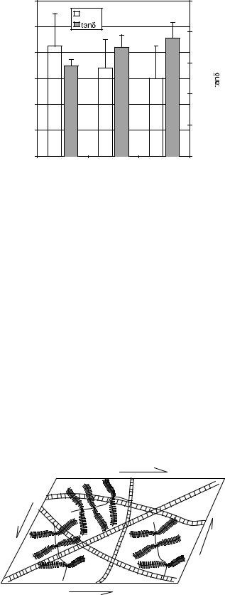
investigated (113,114). However, all these tests were performed in a simple shear configuration, and dynamic shear properties of these studies were reported at frequencies (e.g., 20–1000 Hz) much higher than the physiological range (e.g., 1 Hz). Pure shear tests of articular cartilage and meniscus have been performed under transient, equilibrium, and dynamic conditions to characterize the intrinsic or flow-independent viscoelastic behavior (e.g., (101,115–117)). When a circular cartilage specimen is subject to a sudden change of angular displacement, the shear stress will increase instantaneously, followed by a rapid decay before equilibrium is reached. The quasilinear viscoelastic theory (118) has been shown to provide an excellent description of this intrinsic stress-relaxation behavior of normal human patellar cartilage (116). The equilibrium shear modulus for normal human, bovine, and canine articular cartilage has been found to vary in a range of 0.05–0.25 MPa. Values for the magnitude of the dynamic shear modulus jG j of normal cartilage are in the range of 0.2–20 MPa and vary with both the frequency and magnitude of the normal stress. The phase shift angle (d) for cartilage lies between 98 and 208 over a frequency range of 0.01 Hz to 20 Hz (101). Please note that d is a measure of matrix dissipation, with a loss angle of 08 corresponding to a perfectly elastic material, and 908 to a perfectly dissipative material. The viscoelasticity of meniscus in response to shear is qualitatively similar to that exhibited by articular cartilage, although the magnitudes of the material coefficients of these tissues are significantly different. Meniscus shear properties exhibit an orthotropic symmetry (i.e., the three planes of symmetry defined by its fibrous architecture dominate the shear properties of the meniscus). The equilibrium shear moduli are 36.8 kPa, 29.8 kPa, and 21.4 kPa in the circumferential, axial, and radial directions, respectively (18,19,119). These shear modulus values are ten times less than those observed for articular cartilage. For dynamic tests, the magnitude of the complex shear modulus jG j and phase shift angle d for circumferential, axial, and radial specimens reflect orthotropic collagen fiber organizational symmetry as well (Fig. 7) (3).
The collagen network plays an active mechanical role in contributing to the shear stiffness and energy storage in cartilage (101). Conceptually, the role played by collagen when the specimen is in shear may be visualized as shown in Fig. 8 (101). The tension in the diagonally oriented collagen acts to increase the shear stiffness of the solid matrix. This effect is confirmed by the experimental result that jG j is directly and significantly related to the collagen content of articular cartilage and also by the fact that
70 CARTILAGE AND MENISCUS, PROPERTIES OF
|
0.12 |
|
0.5 |
|
|G*| |
|
|
|
0.1 |
|
0.4 |
|
|
|
|
|
0.08 |
|
|
|G*| (MPa) |
|
|
0.3 |
0.06 |
|
|
|
0.04 |
|
0.2 |
|
|
|
|
|
|
0.02 |
|
0.1 |
|
|
|
|
|
0 |
|
0 |
|
Circumferential |
Axial |
Radial |
Sample Orientation
Figure 7. The magnitude of dynamic shear modulus jG j and tan d for meniscal specimens with normal vectors oriented circumferentially, axially, and radially at 1 rad/s (3).
cartilage has a relatively small loss angle and large shear modulus (101) compared with that of PG solutions at physiological concentrations (107,120,121). The depletion of the PG content has been shown to decrease the dynamic shear modulus up to 55% (101,122), which is considered as a result of the decrease of tensile stress inside the collagen fibrils due to the decrease of PG swelling pressure.
Swelling Properties
Swelling in articular cartilage derives from the presence of negatively charged groups (SO3 and COO ) along the GAG chains of PG molecules. For normal and degenerative femoral head cartilage, the fixed-charge density (FCD) ranges from 0.04 to 0.18 mEq/g wet tissue at physiological pH (2,123). These fixed charges will require a high concentration of counter-ions (Naþ) to maintain electroneutrality, and the concentration, along with that of co-ions (Cl ), is governed by the Donnan equilibrium ion distribu-
Pure shear
τ
τ
τ
τ
Figure 8. A scheme representation of cartilage in pure shear. The tensile stress inside collagen fibrils provides shear stiffness (101).
tion law (124). This excess of freely mobile ions will introduce an imbalance of ion concentration between the fluid compartment inside the tissue and the bathing fluid outside the tissue, giving rise to a higher pressure in the interstitial fluid than the ambient pressure in the external bath, known as the Donnan osmotic pressure. This osmotic pressure decreases with FCD and has a range of 0.1 to 0.25 MPa for normal articular cartilage. With the increase of the external saline concentration, the osmotic pressure will decrease. For the very large external saline concentration (e.g., 2 M), the osmotic pressure is considered to be extremely small, or zero.
This osmotic pressure causes the tissue to swell, as measured by both weight change (2,123) and dimensional change (125,126). These latter experimental results show that the swelling of articular cartilage is inhomogeneous and anisotropic. The swelling ability increases with depth, with the largest dimensional change for the deep zone and almost no change for the superficial layer. The magnitude of swelling is the largest in the thickness direction and the smallest in the split-line direction. In articular cartilage, the osmotic pressure is restrained by the surrounding collagen network. Therefore, residual stress or pre-stress exists inside the solid matrix even before external load is applied. Articular cartilage will warp or curl toward its articular surface upon its removal from the subchondral bone, and the curvature or the extent of curling will decrease with the increase of external saline concentration (126). It has been hypothesized that this curling is caused by the combination effects of swelling pressure and inhomogeneity inside the tissue (126–129). Recently, a three-layer orthotropic model based on triphasic theory (39) has been developed to describe the curling behavior of cartilage strip by considering its layered structure that includes depthdependent collagen fibril orientation and chemical content distributions (128). The predicted curvature change with external saline concentration agrees well with previously published experimental results. This model has also suggested that the large stiffness of the superficial layer and high swelling pressures play key functional roles in the development of pre-stress in cartilage and in its curling behavior.
Quantification of morphological changes has been extensively used to study changes in cartilage swelling with osteoarthritis (OA) (44). With OA, compositional and microstructural changes will occur, which includes the fibrillation of the superficial zone of articular cartilage, the decrease of the PG concentration, and the imbibitions of water; these items are the earliest indicators of the OA degeneration of cartilage (18,19,29,102). The elevated water content or swelling has been shown to be very sensitive to collagen fibrillation. Experimental results also suggest that the water content of the tissue increases after digestion with collagenase (82,83,101). Physically, collagen fibrillation decreases the stiffness of the solid matrix, specifically the elastic bulk modulus, which allows the tissue to imbibe more water (52,104).
Triphasic Mixture Theory
To account for the swelling behavior, Donnan osmotic effects, ion transport, and electrical potentials inside the
tissue, Lai et al. (39) developed the triphasic theory to incorporate the effects of negatively charged groups on the PGs of solid matrix. In this theory, the electrolytes (mainly Naþ and Cl ) within the interstitial fluid are considered as a separate phase, and the solid phase is charged. This triphasic theory was further extended to account for multiple species of ions in the tissue (130). Note that Huyghe and Janssen (131) developed an equivalent theory, named the quadriphasic theory, in which ion species Naþ and Cl were treated as two separate phases.
Using the triphasic theory, the electrokinetic coefficients such as electrical conductivity has also been derived in terms of the physical parameters of charged tissues (39,130,132–134). Furthermore, a theoretical analysis (134,135) showed that the electrical potential inside the tissue comes from two competing sources: a diffusion potential deriving from the FCD inhomogeneity and a streaming potential resulting from the fluid flow within a charged material. These two sources of electrical potential have different polarity and compete against each other. Within the physiological range of material properties of articular cartilage, the polarity of electrical potential inside the tissue depends on the stiffness of the tissue. For softer tissues (such as OA tissue), the diffusion potential tends to dominate, whereas the streaming potential tends to dominate for stiffer tissues (such as normal tissue).
Numerical methods, such as finite difference and finite element formulations, have been developed to demonstrate the contributions of the FCD in mechano-electrochemical (MEC) behaviors of charged, hydrated soft tissues (43,134,136,137). These studies showed that that higher FCD decreases the characteristic time (gel time) and causes the tissue to reach equilibrium in a shorter amount of time, and showed that the osmotic effects can contribute up to 50% of the equilibrium confined compression stiffness (136) and about 30% in unconfined compression (43). With the finite element formation, Lu et al. (138) successfully correlated the predicted FCD with the biochemical measurements, while simultaneously measuring the apparent mechanical properties from the indentation creep experiment.
Recently, the triphasic formulations have been linearized, and the analytic solutions for the MEC response of the tissue under unconfined compression have been obtained both for transient state and at equilibrium (139). With a regular perturbation, simple relations have been derived to describe how the apparent properties, such as Young’s modulus and Poisson’s ratio, change with the fixed nega- tive-charge density (FCD) at equilibrium (139,140). These relations actually are applicable to various testing configurations, even for steady permeation, and they indicate the correspondence of mechanical properties between an elastic body and a charged triphasic material such as articular cartilage and meniscus (139–142).
CARTILAGE WEAR AND DEGENERATION
Cartilage wear and degeneration have been extensively studied due to their significant roles in physically debilitating diseases such as osteoarthritis and rheumatoid
CARTILAGE AND MENISCUS, PROPERTIES OF |
71 |
arthritis (1). Two types of wear occur in synovial joints: fatigue wear and interfacial wear (66). Fatigue wear is independent of the lubrication within the joint and is caused by functional activities such as cyclic, repetitive loading. A balance is presumably maintained under the normal physiological condition whereby tissue turnover is maintained by cells in various components of the synovial joint. A number of factors may contribute to cartilage wear and degeneration. Collagen fibers can be severed by excessive functional activities, leading to a compromise in tensile strength. The normally tight collagen fiber bundles can be unwound and loosened (47,143). When inflammatory cytokines are released, proteoglycans are lost rapidly, leading to a breakdown of the ECM (144–146). Fibrillated cartilage from osteoarthritic patients shows an increase of apoptotic chondrocytes deeper than in the normal articular cartilage, which generally has apoptotic cells only near the surface (145,147). Collective loss of chondrocyte and ECM may lead to microcracks and fissures, which may further grow with functional loading. Thus, fatigue wear of cartilage is a mechanism dependent on biological synthesis and mechanical loading (5).
Interfacial wear can result from physical contact loading of articulating surfaces. Interfacial wear has been categorized into two classes: adhesive and abrasive wear (66). Adhesive wear is more common and occurs when a junction is created when the two solids are in contact. As the opposing surfaces continue to move past the junction, fragments from the weaker surface may be torn off and adhere to the stronger material. The concept is analogous to rubber skid marks left on a road from a braking car. The car is able to move past the formed junction only by having elements of the weaker material, the rubber tire, come off and adhere to the stronger material, which is, in this case, the road. Abrasive wear occurs when a harder material comes into contact with a softer material. No junction is formed. While in contact and rubbing against each other, the harder material cuts or plows into the softer material. The harder material can be either one of the opposing surfaces or loose particles caught between two softer opposing surfaces, cutting into both of them (66).
Pain, stiffness, swelling, and reduced range of motion are the common phenotypic characteristics of osteoarthrosis. Typically, clinical diagnosis is only made after significant cartilage deterioration. A number of methods have been formed to monitor the development of osteoarthrosis. Radiography has been the conventional method for both diagnosis and monitoring. The topographical variation in degenerative cartilage has been used in designing an arthroscopic indentation instrument in which osteoarthrosis could be diagnosed in vivo and possibly treated before the diminishing qualities of the disease (148). Other manners that have been proposed for detection are chondrocalcin measurement (149) and knee wear particle analysis, which is derived from the concept of abrasive wear (150).
CURRENT CARTILAGE REPAIR STRATEGIES
Cartilage’s poor capacity for self-regeneration is well known. The poor regenerative capacity of cartilage has
72 CARTILAGE AND MENISCUS, PROPERTIES OF
been contrasted to bone, because bone readily regenerates (unless it is a critical size bone defect) and has a relatively rich blood supply (151). A lack of angiogenesis has been cited as the primary cause for cartilage’s poor capacity for regeneration. However, normal cartilage is avascular. Vascular supply to cartilage likely will turn it into bone. Thus, a lack of vascularization is not the direct cause for cartilage’s poor regenerative capacity (152).
The regenerative ability of cartilage in response to injury also depends on factors such as joint loading, the degree of injury, the location of injury, and whether it is cartilage lesion alone or osteochondral lesion (153,154). Cartilage responds differently to slowly or rapidly applied loads. For example, loading causes fluid movement in the matrix and may serve to counteract the deformation and to distribute the loads throughout the tissue. However, rapid compressive loading may not allow the fluid to infuse matrix, thus transferring excessive loading to the cells and the ECM macromolecules. Should excessive force be sustained, the chondrocytes and ECM molecules may undergo rupture or degradation. Another factor in determining the cartilage’s regenerative capability is the chondrocytes’ intrinsic ability to replenish the supply of matrix molecules, as well as the approaches to remove degraded materials (17,153,155).
Articular cartilage injuries can be classified as follows:
(1) cartilage matrix and cell injuries without substantial tissue defects; (2) defects, fissures, or ruptures in articular cartilage only; and (3) osteochondral lesions. The first category of cartilage injuries without substantial tissue defects, nonetheless, is associated with a decreased concentration of matrix macromolecules such as PGs and collagen. Albeit without tissue-level defects, loss of PGs and collagen results in a decrease in mechanical strength. Unless repaired, even the first category of cartilage injuries can lead to more substantial defects as described in the second and third categories (153,155).
The second category of articular cartilage injuries is localized within cartilage. They include focused mechanical disruption of the matrix including fissures, tears, incisions, or interruption of the integrity of articular surface. Chondrocytes not only attempt to replace the loss of matrix macromolecules but also proliferate to fill the voids created during injury (153,156). However, the rates of chondrocyte proliferation and matrix synthesis may not be sufficiently high to match the rate of cartilage degradation. As articular cartilage has no nerve supply, except at the very periphery, even substantial cartilage lesions may not elicit pain. A few types of synovial joint injuries likely exist that elicit pain, such as osteochondral injuries, synovial membrane injuries, and injuries to the periphery of articular cartilage. These injuries are usually not repaired by the articular chondrocytes (4).
The third category is osteochondral injuries that involve both articular cartilage and subchondral bone and elicit inflammatory responses such as an influx of blood-borne cells, platelets, and cytokines. Hemorrhaging or fibrin clot formation may occur and later develop into a fibrous mass. The influx of cytokines may induce migration of progenitor cells, although no guarantee exists that these progenitor cells, likely mesenchymal stem cells that are capable of
differentiating into all connective tissue lineage cells, will differentiate into chondrocytes. In fact, osteochondral lesions are likely repaired, if reparable, by fibrocartilage or fibrous tissue instead of hyaline cartilage (154), and rarely possess the complex zonal structures of native articular cartilage (153,157). The mechanical strength of fibrocartilage is approximately one-third of the strength of native hyaline articular cartilage, and thus may not be able to fulfill the weight-bearing and load-bearing functions of normal articular cartilage. Over time, osteochondral lesions may undergo further degradation, leading to the exposure of subchondral bone, which results in osteoarthrosis and can lead to joint immobility.
Current clinical treatments for articular cartilage injuries have several deficiencies. Depending on the degree of injury and whether the defect is partialor full-thickness, the treatments generally involve surgical irrigation, debridement, and tissue augmentation. Partial thickness injuries of the articular cartilage involving clefts and fissures, often in the early stages of osteoarthrosis, are most commonly treated with arthroscopic surgery such as lavage or debridement. Arthroscopic lavage involves the irrigation of the joint, whereas debridement is the arthroscopic removal of damaged tissue. By performing these treatments either alone or in combination, a decrease in joint pain usually results. However, lavage or debridement treatments rarely induce the repair process of cartilage (155,157).
Full-thickness injuries refer to lesions in both articular cartilage and subchondral bone. Although a large number of treatments are empirical, several procedures have taken the advantage to simulate the native repair process. Arthroscopic treatments such as abrasion arthroplasty, Pridie drilling, and microfracture are commonly used, and all include the further perforation of the subchondral bone to induce bleeding and further fibrous tissue formation. Abrasion arthroplasty and microfracture are used in conjunction with debridement to reduce the amount of damaged tissue within the joint. The outcome of these treatments is variable, largely because the healing and repair process within the articular surface are somewhat unpredictable. Furthermore, factors such as the patient’s age, postoperative activity level, and overall heath also affect the outcome (158).
Recently, soft tissue grafts such as the transplantation of the periosteum or perichondrium have been used clinically to repair the articular surface for cylindrical, fullthickness defects. The rationale for using periosteum is its observed chondrogenic potential during development and fracture repair (159). The periosteum consists of a fibrous and cambial layer. The cambial layer contains precursor cells that are capable of differentiating into osteoblasts and perhaps chondrocytes. The process of periosteum transplantation involves the creation of a defect spanning the full thickness of articular cartilage and penetrating the subchondral bone, and then placement of the periosteum graft within the defect. However, much debate has occured as to which layer of the periosteum should lay adjacent to the bone and which layer should face the articular surface, as the cambial layer can form cartilage, whereas the fibrous layer forms fibrous tissue. Larger full-thickness defects are generally repaired using allogenic or autogenic
osteochondral tissue plugs, called mosaicplasty, excised from nonload-bearing regions of the joint and inserted into the full-thickness defect. Reports exist of fibrous tissue formation and chondrocyte death at the interface between the plug and surrounding tissue, which may lead to further degeneration of the joint. Furthermore, donor site morbidity remains a drawback of the periosteum graft or mosaicplasty (160).
For substantial osteochondral lesions, total joint replacement using metallic condyle and plastic socket is most commonly in practice. Current modalities of total joint replacements suffer from drawbacks such as donor site morbidity, pathogen transmission, wear and tear, and a limited life span (161). Secondary surgeries are necessary in 10–15% of the cases and suffer from substantial difficulties such as scar tissue formation and loss of host tissue (162). More importantly, current total joint replacement therapies fail to yield biological regeneration.
CURRENT MENISCAL REPAIR STRATEGIES
Prior to the recognition of the importance of the meniscus in the biomechanics of the knee joint in the 1970s, the preferred treatment for meniscal injury such as tear was total excision of the meniscus or open meniscectomy. Despite some reports of temporary relief of symptoms, the long-term outcome of excision or meniscectomy was poor. In the 1980s, understanding of the material properties and biomechanical roles of the meniscus led to more conservative treatments for meniscal tears. Furthermore, the development of arthroscopy enabled more accurate diagnosis of meniscal tears and, subsequently, more precise surgical treatment that has substantially reduced the amount of damage to the surrounding tissue in comparison with open-joint surgery (163).
Partial-thickness split tears and small (< 5 mm) fullthickness split tears, vertically or obliquely, are usually left alone and without surgical intervention. The inner wall of the meniscus must be stable during probing, which is commonly performed arthroscopically during diagnosis. Follow-up arthroscopic examinations are often necessary to monitor tissue healing. These injuries are usually associated with ligament tears such as the anterior cruciate ligament (ACL). Ligament tears in conjunction with meniscal tears drastically reduce the stability of the knee and can lead to further meniscal damage.
Meniscal injuries that require surgical repair or excision are large defects with compromised vascular supply, large meniscal deformations, or damage to the peripheral or circumferential collagen fibers. In need of excision, it is preferable to leave a partial meniscus intact as opposed to full meniscectomy. The surgical approaches are either open-joint surgeries or arthroscopic surgery to suture tears within the meniscus (163).
TISSUE ENGINEERING OF ARTICULAR CARTILAGE AND MENISCUS
The rapidly evolving field of tissue engineering has promised to deliver biological replacements of damaged
CARTILAGE AND MENISCUS, PROPERTIES OF |
73 |
articular cartilage and meniscus. In comparison with current treatment modalities, tissue engineering represents a shift in the paradigm. Whereas current treatment modalities improve articular cartilage and meniscal injuries by increments, the end goal of tissue engineering is to generate or regenerate articular cartilage and meniscus.
Previous investigations of the structural, biochemical, and mechanical properties of articular cartilage and meniscus serve as the necessary foundation and have set the stage for the tissue engineering of these structures. For example, a commonly stated long-term goal of a biochemical study to investigate the PG distribution in various zones of articular cartilage was to improve the treatment of cartilage defects in arthritis, which was all too familiar for a published study or a grant proposal decades ago.
To tissue-engineer articular cartilage or meniscus, one has the conceptual liberty of selecting cell sources, scaffolds, or growth factors. Another essential choice is whether mechanical stress is to be applied to the tissueengineered articular cartilage or meniscus prior to or after in vivo implantation. Thus, the initial stage of tissue engineering of articular cartilage and meniscus is an optimization process of cells, scaffolds, growth factors, or mechanical stimulus.
Cells Capable of Generating Articular Cartilage and Meniscus
Articular chondrocytes are the obvious choices for articular cartilage regeneration (164–166). From the standpoint of scientific discovery, articular cartilage or meniscal regeneration from articular chondrocytes or meniscal fibrochondrocytes has revealed a wealth of information (e.g., 167–169). From the standpoint of alignment with eventual therapeutic regeneration of synovial joint condyle in arthritis patients, the selection of articular chondrocytes is problematic. The essential problem at this time is that articular chondrocytes are not very expandable ex vivo. Thus, relatively large donor site defects are necessary to harvest a large amount of tissue(s) from the patient in order to obtain sufficient numbers of articular chondrocytes or meniscal fibrochondrocytes for healing substantial articular cartilage or meniscal defects. In contrast, mesenchymal stem cells, whose natural progeny includes both chondrocytes and fibrochondrocytes, can be obtained in small quantities (e.g., a few cc of bone marrow content) (170) or from other connective tissue sources such as adipose tissue (170,171), readily expanded ex vivo in cell culture and reliably differentiated into chondrocytes cells (170,172). Embryonic stem cells may turn out to be a viable cell source for synovial joint regeneration, especially in consideration of the recent demonstration of the differentiation of embryonic stem cells into osteogenic cells (173,174). Embryonic stem cells are likely to be of greater significance in synovial joint condyle regeneration if the isolation and expansion of adult MSCs encounter substantial difficulties. However, thus far, it does not appear to be the case for synovial joint condyle regeneration (172). Chondrogenic and osteogenic cells derived from MSCs appear to be the logical choices at this time for exploring clinically applicable approaches toward regenerating the synovial joint condyle (152,172,175–180).
74 CARTILAGE AND MENISCUS, PROPERTIES OF
Biomaterial Scaffolds Are often Necessary for the Engineering of Structural Tissues such as Articular Cartilage and Meniscus
The optimal scaffolds for articular cartilage and meniscal regeneration are yet to be determined. An increasing number of meritorious studies have reported a wide range of natural and synthetic polymers for articular cartilage regeneration. Many of the tested synthetic polymers are biocompatible and biodegradable, two desirable features for cartilage regeneration (181). A model scaffold should allow effective diffusion of essential nutrients and metabolic wastes, given that chondrocytes and fibrochondrocytes both rely on diffusion for survival.
For cartilage regeneration, natural and synthetic polymers may need to simulate the extracellular matrix environment of chondrocytes that are created by Type II collagen and PGs in a highly aqueous matrix. Several hydrogels simulate cartilage matrix to various degrees, such as alginate, hyaluronate, chitosan, and polyethylene glycol-based polymers (175,176,181–183). The organization of chondrocyte phenotypes in various zones of articular cartilage may also need to be simulated, as demonstrated in recent reports by encapsulating articular chondrocytes from various zones of bovine articular cartilage into different hydrogel layers (184–186). The water content and diffusion properties of hydrogels mimic an ECM to allow tissue-forming cells to obtain systemic nutrients (187). The initial viscous liquid form of several hydrogel materials provides a unique capability to form complicated shapes while maintaining uniform cell distributions. For examples, an aqueous-derived silk scaffold also encompasses hydrogel-type properties and has been shown to support chondrogenesis (188). Pellet culture is a practice devoid of scaffolds that takes advantage of the dense, avascular, and aneural condition of cartilage. This system is done simply by centrifuging chondrocytes or MSCs into a pellet and incubating them in desired conditions. Although pellets do not provide efficient shape retention, they do bestow valuable information in vitro as models for chondrogenic MSC differentiation. Despite various levels of reported success with in vitro models, cell-hydrogel interactions need to be better understood, along with optimization of hydrogel composition, cross-linking, and degradation behavior as a function of the in vivo regenerative outcome.
In engineering an osteochondral construct, it is essential to construct a mold that is specific to the joint. Computer-aided approaches have been developed to construct molds that will both accurately replicate the anatomy of the joint as well as preserve the intricate architectural integrity of the interior of the scaffold. These intricate details can range from pore size to channel orientation to surface texture and can effectively contribute to the synthesis, or lack thereof, of the tissue one is trying to engineer. Common methods for 3D mold fabrication are based on software programs that read and digitize computerized tomography or magnetic resonance imaging. Solid free-form fabrication (SFF) technology can be used to produce an actual 3D scaffold through combining the interior architecture image and the external scaffold image, which is done via a layering process from computer-aided design files. SFF shows excellent promise due to the
possibility of controlling the aforementioned necessary parameters needed in scaffold fabrication.
Growth Factors Are Necessary for Modulating Cell Behavior
Growth factors are proteins and polypeptides capable of modulating all aspects of cell behavior such as proliferation, differentiation, and apoptosis. Cells can be regulated by self-released growth factors (autocrine effect), or by growth factors released by other cells (paracrine effect). For chondrogenic differentiation, TGF-b superfamily is frequently used (e.g., 176,189). The de-differentiation and re-differen- tiation of chondrocytes are regulated by combinations of TGF-b1, fibroblast growth factor-2, and sequential exposure to IGF-I (190). TGF-bs also stimulate GAG synthesis in isolated cultured meniscus cells (191). Platelet-derived growth factor (PDGF) promotes chondrogenesis in chick limbs both in vitro and in vivo (191). Resting zone chondrocytes treated with PDGF shows hypertrophic activity in forming new cartilage (192). Although many growth factors have provided positive results, the optimal scheme of their application is not yet fully understood. The reader is referred to several recent in-depth reviews of growth factor delivery in cartilage regeneration (154,183,193).
Functional Tissue Engineering of Articular Cartilage
and Meniscus
The field of functional tissue engineering has been proposed in response to the need to engineer tissues that have not only the appropriate cellular and matrix structures, but also the necessary physical properties (194,196). The aforementioned mechanical properties of both articular cartilage and meniscus are quite complex and intricate to replicate. The rather severe loading environment of cartilage in a synovial joint attributes to poor repair capabilities as well as degeneration. It has been proposed that a potential solution to engineer tissues with the appropriate physical properties lies in the physical factors, among which mechanical stress is most well studied (194–196). Prior to the conception of functional tissue engineering, it is widely acknowledged that mechanical factors can effectively influence cell behavior such as proliferation, differentiation, and matrix synthesis. Significant evidence exists that physical stress can accelerate or improve tissue regeneration and repair in vitro. The preference of chondrocytes to synthesize proper ECM components at an accelerated pace can help engineer cartilage and meniscus in an efficient manner. This concept can be used to enhance engineered grafts to more accurately represent native tissue. In order to better provide these stresses, a movement has begun to construct mechanical bioreactors that increase matrix synthesis through different approaches. Examples in which mechanical stimulation has proven advantageous in synthesizing cartilage has been through fluid flow (197,198), simulated hypogravity (199), simulated microgravity (200), static and cyclic compression (201–203), and hydrostatic pressure (204). Recent studies have suggested that external mechanical loading of cells or cell-polymer constructs may enhance their mechanical strength (e.g., 186,205,206). Although practical in theory, the type, frequency, area, and amount of
loading still need to be determined to furnish the optimal results of engineering each individual tissue.
The mechanical properties of engineered articular cartilage and meniscus can be readily tested, in many ways similar to the mechanical testing of native articular cartilage and meniscus. The results of compressive moduli of native and engineered articular cartilage or meniscus tissue using excised tissue plants and in vitro testing can be directly compared. However, loading measurements of native or engineered articular cartilage and meniscus have not been accomplished in vivo. Therefore, the ideal mechanical properties of tissue engineered articular cartilage can only be estimated, not accurately determined. Various biomaterials have been investigated for articular cartilage repair and have various mechanical properties that must be considered when designing a suitable scaffold. Engineering a scaffold that is too stiff may detrimentally affect cell viability and matrix synthesis. Stress shielding may occur and surrounding tissue may degrade as physiological loads are transferred to the more mechanically stiff implant. In contrast, a scaffold too soft will not exhibit the needed mechanical stiffness for the applied loads, causing physical breakdown and degradation of the material, leaving a void in the host tissue. Various zones of articular and fibrocartilage have different mechanical properties (56,207). Whether regional differences in the mechanical properties of articular cartilage and meniscus need to be simulated in tissue engineering remains to be explored (79).
SUMMARY AND CONCLUSIONS
Articular cartilage and meniscus are load-bearing and hydrated tissues that are key structures of diarthrodial joints. The similarities between the two structures include their remarkable capacity for resistance to and transmission of mechanical stress and their ability to enable joint lubrication. However, articular cartilage and meniscus have many important differences. Articular cartilage, in the overwhelming majority of human synovial joints, consists of hyaline cartilage, whereas the meniscus is composed of fibrocartilage. The mechanical properties of native articular cartilage and meniscus have been studied extensively and shown to vary with species, location, and even the orientation of test specimens. Motivated by the concept of functional tissue engineering, the mechanical properties of engineered articular cartilage from cells and biomaterials have also been investigated in recent years. As a result of drastically different structural and mechanical properties between articular cartilage and meniscus, the engineering challenges of the two tissues are different. Mesenchymal stem cells, or other stem cells, need to be differentiated into chondrocytes for the engineering of articular cartilage, whereas these stem cells may need to be differentiated into fibroblasts and chondrocytes, or perhaps fibrochondrocytes, for the engineering of meniscus. The optimal biomaterials for the engineering of articular cartilage and meniscus remain to be identified or fabricated. At least, the engineered articular cartilage and meniscus must adapt to possess similar mechanical properties of their native target tissues. The existing knowledge
CARTILAGE AND MENISCUS, PROPERTIES OF |
75 |
on the biological and mechanical properties of articular cartilage and meniscus provides the necessary foundation for the eventual goal to regenerate or replace diseased or lost articular cartilage and knee meniscus with engineered tissue analogs.
BIBLIOGRAPHY
1.Buckwalter JA, Mankin HJ. Instructional course lectures, the American Academy of Orthopaedic Surgeons—articular cartilage. Part II: Degeneration and osteoarthrosis, repair, regeneration, and transplantation. J Bone Joint Surg 1997;79:612–632.
2.Maroudas A. Physicochemical properties of articular cartilage. In: Freeman MAR, ed. Adult Articular Cartilage. Kent, UK: Pitman Medical Publishing; 1979. pp 215–290.
3.Mow VC, Gu WY, Chen FH. Structure and function of articular cartilage and meniscus. In: Mow VC, Huiskes R, eds. Basic Orthopaedic Biomechanics and Mechano-Biology; Third ed. New York: Lippincott Williams & Wilkins; 2005. pp 181–258.
4.Buckwalter JA, Mow VC. Cartilage repair in osteoarthritis. In: Moskowitz RW, Howell DS, Goldberg VM, Mankin HJ, eds. Osteoarthritis: Diagnosis and Management, 2nd ed. Philadelphia, PA: Saudners; 1992.
5.Howell DS, Treadwell BV, Trippel SB. Etiopathogenesis of osteoarthritis. In: Moskowitz RW, Goldberg VM, Howell DS, Altman RD, Buckwalter JA, eds. Osteoarthritis: Diagnosis and Medical/Surgical Management. 2th ed. Philadelphia, PA: WB Saunders; 1992. pp 233–252.
6.Poole AR, Howell DS. Etiopathogenesis of osteoarthritis. In: Moskowitz RW, Howell DS, Altman RD, Buckwalter JA, Goldberg VC, editors. Osteoarthritis, Diagnosis and Management. 3rd ed. Philadelphia, PA: WB Saunders Publishers; 2001. pp 29–47.
7.Mankin HJ, Mow VC, Buckwalter JA. Articular cartilage structure, composition and function. In: Buckwalter JA, Einhorn TA, Simon SR, eds. Orthopaedic Basic Science: Biology and Biomechanics of the Musculoskeletal System. Rosemont, IL: American Academy of Orthopaedic Surgeons; 2000. pp 443–470.
8.Benjamin M, Evans EJ. Fibrocartilage. J Anat 1990;171: 1–15.
9.Weightman B. Tensile fatigue of human articular cartilage. J Biomech 1976;9:193–200.
10.Seedhom BB, Wallbridge NC. Walking activities and wear of prosthesis. Ann Rheum Dis 1985;44:838–843.
11.Morrison JB. Bioengineering analysis of force actions transmitted by the knee joint. J Biomech Eng 1968;3:164–170.
12.Morrison JB. The mechanics of the knee joint in relation to normal walking. J Biomech 1970;3:51–61.
13.Athanasiou KA, Rosenwasser MP, Buckwalter JA, Malinene TI, Mow VC. Interspecies comparisons of in situ intrinsic mechanical properties of distal femoral cartilage. J Orthop Res 1991;9:330–340.
14.Ewers BJ, Dvoracek-Driksna D, Orth MW, Haut RC. The extent of matrix damage and chondrocyte death in mechanically traumatized articular cartilage explants depends on rate of loading. J Orthop Res 2001;19:779–784.
15.Torzilli PA, Grigiene R, Borrelli J Jr, Helfet DL. Effect of impact load on articular cartilage: Cell metabolism and viability, and matrix water content. J Biomech Eng 1999;121: 433–441.
16.Centers for Disease Control and Prevention (CDC). Prevalence of self-reported arthritis or chronic joint symptoms among adultsUnited States, 2001. MMWR 2002;51:948–950.
17.Vunjak-Novakovic G, Goldstein SA. Biomechanical principles of cartilage and tissue engineering. In: Mow VC, Huiskes R, eds. Basic Orthopaedic Biomechanics and Mechano-Biology,
76 CARTILAGE AND MENISCUS, PROPERTIES OF
3rd ed. New York: Lippincott Williams & Wilkins; 2005.
pp343–407.
18.Mow VC, Ratcliffe A, Chern KY, Kelly MA. Structure and function relationships of the meniscus of the knee. In: Mow VC, Arnoczky SP, Jackson DW, eds. Knee Meniscus: Basic and Clinical Foundations. New York: Raven Press; 1992.
pp37–57.
19.Mow VC, Ratcliffe A, Poole AR. Cartilage and diarthroidal joints as paradigms for hierarchical materials and structures. Biomaterials 1992;13:67–97.
20.Arnoczky SP. Gross and vascular anatomy of the meniscus and its role in meniscal healing, regeneration, and remodeling. In: Mow VC, Arnoczky SP, Jackson DW, eds. Knee Meniscus, Basic and Clinical Foundations. New York: Raven Press; 1992. pp 1–14.
21.Uhthoff HK, Wiley JJ. Behavior of the Growth Plate. New York: Raven Press; 1988.
22.Cohen B, Chorney GS, Phillips DP, Dick HM, Mow VC. Compressive stress-relaxation behavior of bovine growth plate may be described by the nonlinear biphasic theory. J Orthop Res 1994;12:804–813.
23.Williams PL. Gray’s Anatomy. New York: Churchill Livingston; 1995.
24.Shimazu A, Nah HD, Kirsch T, Koyama E, Leatherman JL, Golden EB, Kosher RA, Pacifici M. Syndecan-3 and the control of chondrocyte proliferation during endochondral ossification. Exp Cell Res 1996;229:126–136.
25.Mao JJ, Nah HD. More research needed to understand how orthodontists communicate with cells. Am J Orthod Dentofacial Orthop 2004;125:676–689.
26.Tamamura Y, Otani T, Kanatani N, Koyama E, Kitagaki J, Komori T, Yamada Y, Costantini F, Wakisaka S, Pacifici M, Iwamoto M, Enomoto-Iwamoto M. Developmental regulation of Wnt/beta-catenin signals is required for growth plate assembly, cartilage integrity, and endochondral ossification. J Biol Chem 2005;280:19185–19195.
27.Mow VC, Kuei SC, Lai WM, Armstrong CG. Biphasic creep and stress relaxation of articular cartilage in compression: Theory and experiments. J Biomech Eng 1980;102:73–84.
28.Eyre DR. Structure and function of the cartilage collagen: Role of type IX in articular cartilage. In: Brandt KD, ed. Cartilage Changes in Osteoarthritis. Indianapolis, IN: CibaGeigy; 1990. pp 12–16.
29.Heinegard D, Bayliss M, Lorenzo P. Biochemistry and metabolism of normal and osteoarthritic cartilage. In: Brandt KD, Doherty M, Lohmander LS, eds. Osteoarthritis. New York: Oxford University Press; 2003. pp 73–82.
30.Eyre DR, Wu JJ. Collagen of fibrocartilage: A distinctive molecular phenotype in bovine meniscus. FEBS Lett 1983;158:265–270.
31.Eyre DR, Oguchi H. The hydroxypyridinium crosslinks of skeletal collagens: Their measurement, properties and a proposed pathway of formation. Biochem Biophys Res Commun 1980;92:402–410.
32.Eyre DR, Dickson IR, Van Ness K. Collagen cross-linking in human bone and articular cartilage. Age-related changes in the content of mature hydroxypyridinium residues. Biochemistry 1988;252:495–500.
33.Huang CY, Mow VC, Ateshian GA. The role of flowindependent viscoelasticity in the biphasic tensile and compressive responses of articular cartilage. J Biomech Eng 2001;123: 410–417.
34.Huang CY, Soltz MA, Kopacz M, Mow VC, Ateshian GA. Experimental verification of the roles of intrinsic matrix viscoelasticity and tension-compression nonlinearity in the biphasic response of cartilage. J Biomech Eng 2003;125: 84–93.
35.Kempson GE, Freeman MA, Swanson SA. Tensile properties of articular cartilage. Nature 1968;220:1127–1128.
36.Kempson GE, Muir H, Pollard C, Tuke M. The tensile properties of the cartilage of human femoral condyles related to the content of collagen and glycosaminoglycans. Biochim Biophys Acta 1973;297:456–472.
37.Roth V, Mow VC. The intrinsic tensile behavior of the matrix of bovine articular cartilage and its variation with age. J Bone Joint Surg 1980;62:1102–1117.
38.Woo SL-Y, Akeson WH, Jemmott GF. Measurements of nonhomogeneous directional mechanical properties of articular cartilage in tension. J Biomechan 1976;9:785–791.
39.Lai WM, Hou JS, Mow VC. A triphasic theory for the swelling and deformation behaviors of articular cartilage. J Biomech Eng 1991;113:245–258.
40.Buckwalter JA, Rosenberg LC. Electron microscopic studies of cartilage proteoglycans: Direct evidence for the variable length of the chondroitin sulfate-rich region of proteoglycan subunit core protein. J Biol Chem 1982;257:8930–8939.
41.Muir H. Proteoglycans as organizers of the intercellular matrix. Biochem Soc Trans 1983;9:613–622.
42.Muir H. The chondrocyte, architect of cartilage: Biomechanics, structure, function and molecular biology of cartilage matrix macromolecules. Bioessays 1995;17:1039–1048.
43.Sun DN, Guo XE, Likhitpanichkul M, Lai WM, Mow VC. The influence of the fixed negative charges on mechanical and electrical behaviors in articular cartilage under unconfined compression. J Biomech Eng 2004;126:1–11.
44.Hunziker EB, Michel M, Studer D. Ultrastructure of adult human articular cartilage matrix after cryotechnical processing. Microsc Res Tech 1997;37:271–284.
45.Hultkrantz W. Ueber die spaltrichtungen der gelenkknorpel. Anat Anzeig verhandl anat Gesellsch 1898;14:248–256.
46.Benninghoff A. Form und bau der gelenkknorpel in ihren beziehungen zu funktion. II. Der aufbau des gelenkknorpel in seinen beziehungen zu funktion. Z Zellforsch 1925;2: 783–862.
47.Broom ND. The collagen framework of articular cartilage: Its profound influence on normal and abnormal load-bearing function. In: Nimni ME, ed. Collagen: Chemistry, Biology and Biotechnology. Boca Raton, FL: CRC Press; 1988. pp 243–265.
48.Ceohn ZA, Henry JH, McCarthy DM, Mow VC, Ateshian GA. Computer simulations of patellofemoral joint surgery. Am J. Sports Med 2003;31:87–98.
49.Lipshitz H, Etheredge R 3rd, Glimcher MJ. Changes in the hexosamine content and swelling ratio of articular cartilage as functions of depth from the surface. J Bone Joint Surg 1976;58:1149–1153.
50.Mitrovic D, Quintero M, Stankovic A, Ryckewaert A. Cell density of adult human femoral condylar articular cartilage. Joints with normal and fibrillated surfaces. Lab Invest 1983;49:309–316.
51.Aydelotte MB, Schumacher BL, Kuettner KE. Heterogeneity of articular chondrocytes. In: Kuettner KE, ed. Articular Cartilage and Osteoarthritis. New York: Raven Press; 1992. pp 237–249.
52.Kempson GE. Mechanical properties of adult articular cartilage. In: Freeman MAR, ed. Adult Articular Cartilage. Kent, UK: Pitman Medical Publishing; 1979. pp 215–290.
53.Akizuki S, Mow VC, Muller F, Pita JC, Howell DS. Tensile properties of human knee joint cartilage. II. Correlations between weight bearing and tissue pathology and the kinetics of swelling. J Orthop Res 1987;5:173–186.
54.Hunziker EB, Tyler JA. Articular cartilage repair. In: Brandt KD, Doherty M, Lohmander LS, eds. Osteoarthritis. Oxford, UK: Oxford University Press; 2003. pp 93–101.
55.Jadin KD, Wong BL, Bae WC, Li KW, Williamson AK, Schumacher BL, Price JH, Sah RL. Depth-varying density and organization of chondrocytes in immature and mature bovine articular cartilage assessed by 3-D imaging and analysis. J Histochem Cytochem DOI: 10.1369/jhc.4A6511. 2005 (In Press).
56.Tomkoria S, Patel RV, Mao JJ. Heterogeneous nanomechanical properties of superficial and zonal regions of articular cartilage of the rabbit proximal radius condyle by atomic force microscopy. Med Eng Phys 2004;26:815–822.
57.Broom ND, Silyn-Roberts H. Collagen-collagen versus collagen-proteoglycan interactions in the determination of cartilage strength. Arthritis Rheum 1990;33:1512–1517.
58.Quinn TM, Hunziker EB, Hauselmann HJ. Variation of cell and matrix morphologies in articular cartilage among locations in the adult human knee. Osteoarthritis Cartilage 2005;13:672–678.
59.Guilak F, Mow VC. The mechanical environment of the chondrocyte: A biphasic finite element model of cell-matrix interactions in articular cartilage. J Biomech 2000;33:1663– 1673.
60.Guilak F. The deformation behavior and viscoelastic properties of chondrocytes in articular cartilage. Biorheology 2000;37: 27–44.
61.Guilak F. Compression-induced changes in the shape and volume of the chondrocyte nucleus. J Biomech 1995;28:1529– 1542.
62.Knight MM, Ross JM, Sherwin AF, Lee DA, Bader DL, Poole CA. Chondrocyte deformation within mechanically and enzymatically extracted chondrons compressed in agarose. Biochem Biophys Acta 2001;1526:141–146.
63.Graff RD, Kelley SS, Lee GM. Role of pericellular matrix in development of a mechanically functional neocartilage. Biotechnol Bioengin 2003;20:457–464.
64.Allen DM, Mao JJ. Heterogeneous nanostructural and nanoelastic properties of pericellular and interterritorial matrices of chondrocytes by atomic force microscopy. J Struct Biol 2004;145:196–204.
65.Stockwell RA. Biology of Cartilage Cells. Cambridge, UK: Cambridge University Press; 1979.
66.Ateshian GA, Mow VC. Friction, lubrication, and wear of articular cartilage and diarthroidal joints. In: Mow VC, Huiskes R, eds. Basic Orthopaedic Biomechanics and Mechano-Biology, Third ed. New York: Lippincott Williams & Wilkins; 2005. pp 447–494.
67.McDevitt CA, Miller A, Spindler KP. The cells and cell matrix interaction of the meniscus. In: Mow VC, Arnoczky SP, Jackson DW, eds. Knee Meniscus, Basic and Clinical Foundations. New York: Raven Press; 1992. pp 29–36.
68.Bullough PG, Munuera L, Murphy J, Weinstein AM. The strength of the menisci of the knee as it relates to their fine structure. J Bone Joint Surg Br 1970;52:564–567.
69.Aspden RM, Yarker YE, Hukins DW. Collagen orientations in the meniscus of the knee joint. J Anat 1985;140:371–380.
70.Kelly MA, Fithian DC, Chern KY, Mow VC. Structure and function of the meniscus: Basic and clinical implications. In: Mow VC, Ratcliffe A, Woo SLY, eds. Biomechanics of Diarthroidal Joints. New York: Springer-Verlag; 1990. pp 191– 214.
71.Yasui K. Three-dimensional architecture of human normal menisci. J Jpn Orthop Assoc 1978;52:391–399.
72.Skaggs DL, Warden WH, Mow VC. Radial tie fibers influence the tensile properties of the bovine medial meniscus. J Orthop Res 1994;12:176–185.
73.Nakano T, Thompson JR, Aherne FX. Distribution of glycosaminoglycans and the nonreducible collagen crosslink, pyridinoline in porcine menisci. Can J Vet Res 1986;50: 532–536.
CARTILAGE AND MENISCUS, PROPERTIES OF |
77 |
74.Fithian DC, Kelly MA, Mow VC. Material properties and structure-function relationships in the menisci. Clin Orthop Relat Res 1990;252:19–31.
75.Walker PS, Erkman MJ. The role of the menisci in force transmission across the knee. Clin Orthop Relat Res 1975;109:184–192.
76.Smith MM, Ghosh P. Experimental models of osteoarthritis. In: Moskowitz RW, Howell DS, Altman RD, Buckwalter JA, Goldberg VC, eds. Osteoarthritis, Diagnosis and Management. 3rd ed. Philadelphia, PA: WB Saunders Publishers; 2001. pp 171–199.
77.Moskowitz RW, Davis W, Sammarco J, et al., Experimentally induced degenerative joint lesions following partial meniscectomy in the rabbit. Arthritis Rheum 1973;16:397–404.
78.Dehaven KE. The role of the meniscus. In: Ewing JW, ed. Articular Cartilage and the Knee Joint Function: Basic Science and Arthroscopy. New York: Raven Press; 1990. pp 103–115.
79.Guilak F, Butler DL, Goldstein SA. Functional tissue engineering: The role of biomechanic in articular cartilage repair. Clin Orthop Relat Res 2001; (391 Suppl):S295–S305.
80.Kempson GE. Mechanical properties of articular cartilage and their relationship to matrix degeneration and age. Ann Rheum Dis 1975;34:111–113.
81.Kempson GE. Age-related changes in the tensile properties of human articular cartilage: A comparative study between the femoral head of the hip joint and the talus of the ankle joint. Biochim Biophys Acta 1991;1075:223–230.
82.Akizuki S, Mow VC, Muller F, Pita JC, Howell DS, Manicourt DH. Tensile properties of human knee joint cartilage: I. Influence of ionic conditions, weight bearing, and fibrillation on the tensile modulus. J Orthop Res 1986;4:379–392.
83.Setton LA, Mow VC, Muller FJ, Pita JC, Howell DS. Mechanical properties of canine articular cartilage are significantly altered following transection of the anterior cruciate ligament. J Orthop Res 1994;12:451–463.
84.Setton LA, Zhu W, Mow VC. The biphasic poroviscoelastic behavior of articular cartilage: role of the surface zone in governing the compressive behavior. J Biomech 1993;26: 581–592.
85.Soltz MA, Ateshian GA. A conewise linear elasticity mixture model for the analysis of tension-compression nonlinearity in articular cartilage. J Biomech Eng 2000;122:576–586.
86.Huang CY, Stankiewicz A, Ateshian GA, Mow VC. Anisotropy, inhomogeneity, and tension-compression nonlinearity of human glenohumeral cartilage in finite deformation. J Biomech 2005;38:799–809.
87.Fithian DC, Zhu WB, Ratcliffe A, Kelly MA, Mow VC. Exponential law representation of tensile properties of human meniscus. Proc Inst Mech Eng 1989;c384/058:85–90.
88.Bader DL, Kempson GE, Barrett AJ, Webb W. The effects of leucocyte elastase on the mechanical properties of adult human articular cartilage in tension. Biochemica et Biophysica Acta 1981;677:103–108.
89.Schmidt MB, Mow VC, Chun LE, Eyre DR. Effects of proteoglycan extraction on the tensile behavior of articular cartilage. J Orthop Res 1990;8:353–363.
90.Hirsh 1944.
91.Sokoloff L. Elasticity of aging cartilage. Fed Proc 1966;25: 1089–1095.
92.Kempson GE, Freeman MA, Swanson SA. The determination of a creep modulus for articular cartilage from indentation tests of the human femoral head. J Biomech 1971;4: 239–250.
93.Kempson GE, Spivey CJ, Swanson SA, Freeman MA. Patterns of cartilage stiffness on normal and degenerate human femoral heads. J Biomech 1971;4:597–609.
78CARTILAGE AND MENISCUS, PROPERTIES OF
94.Hoch DH, Grodzinsky AJ, Koob TJ, Albert ML, Eyre DR. Early changes in material properties of rabbit articular cartilage after meniscectomy. J Orthop Res 1983;1:4–12.
95.Parsons JR, Black J. The viscoelastic shear behavior of normal rabbit articular cartilage. J Biomech 1977;10:21–29.
96.Altman RD, Tenenbaum J, Latta L, Riskin W, Blanco LN, Howell DS. Biomechanical and biochemical properties of dog cartilage in experimentally induced osteoarthritis. Ann Rheum Dis 1984;43:83–90.
97.Armstrong CG, Lai WM, Mow VC. An analysis of the unconfined compression of articular cartilage. J Biomech Eng 1984;106:165–173.
98.Mak AF. The apparent viscoelastic behavior of articular cartilage— The contributions from the intrinsic viscoelasticity and interstitial fluid flow. J Biomech Eng 1986;108:123–130.
99.Mak AF. Unconfined compression of hydrated viscoelastic tissues: A biphasic poroviscoelastic analysis. Biorheology 1986;23:371–383.
100.Mak AF, Lai WM, Mow VC. Biphasic indentation of articular cartilage—I. Theoretical analysis. J Biomech 1987;20:703– 714.
101.Zhu W, Mow VC, Koob TJ, Eyre DR. Viscoelastic shear properties of articular cartilage and the effects of glycosidase treatments. J Orthop Res 1993;11:771–781.
102.Armstrong CG, Mow VC. Variations in the intrinsic mechanical properties of human articular cartilage with age, degeneration, and water content. J. Bone Joint Surg 1982;64: 88–94.
103.Roth V, Mow VC, Lai WM, Eyre DR. Correlation of intrinsic compressive properties of bovine articular cartilage with its uronic acid and water content. Trans Orthop Res Soc 1981;6:21.
104.Froimson MI, Ratcliffe A, Gardner TR, Mow VC. Differences in patellofemoral joint cartilage material properties and their significance to the etiology of cartilage surface fibrillation. Osteoarthritis Cartilage 1997;5:377–386.
105.Jurvelin J, Kiviranta I, Arokoski J, Tammi M, Helminen HJ. Indentation study of the biochemical properties of articular cartilage in the canine knee. Eng Med 1987;16:15–22.
106.Stahursky TM, Armstrong CG, Mow VC. Variation of the intrinsic aggregate modulus and permeability of articular cartilage with trypsin digestion. Proc Biomech Symp Trans ASME 1981;AMD43:137–140.
107.Mow VC, Zhu W, Lai WM, Hardingham TE, Hughes C, Muir H. The influence of link protein stabilization on the viscometric properties of proteoglycan aggregate solutions. Biochim Biophys Acta 1989;992:201–208.
108.Schinagl RM, Ting MK, Price JH, Sah RL. Video microscopy to quantitate the inhomogeneous equilibrium strain within articular cartilage during confined compression. Ann Biomed Eng 1996;24:500–512.
109.Chen AC, Bae WC, Schnagl RM, Sah RL. Depthand straindependent mechanical and electromechanical properties of full-thickness articular cartilage in confined compression. J Biomechan 2001;34:1–12.
110.Wang CC, Hung CT, Mow VC. An analysis of the effects of depth-dependent aggregate modulus on articular cartilage stress-relaxation behavior in compression. J Biomech 2001;34:75–84.
111.Cohen B, Lai WM, Mow VC. A transversely isotropic biphasic model for unconfined compression of growth plate and chondroepiphysis. J Biomech Eng 1998;120:491–496.
112.Hayes WC, Mockros LF. Viscoelastic properties of human articular cartilage. J Appl Physiol 1971;31:562–568.
113.Spirt AA, Mak AF, Wassell RP. Nonlinear viscoelastic properties of articular cartilage in shear. J Orthop Res 1989;7: 43–49.
114.Simon WH, Mak A, Spirt A. The effect of shear fatigue on bovine articular cartilage. J Orthop Res 1990;8:86–93.
115.Roth V, Schoonbeck JM, Mow VC. Low frequency dynamic behavior of articular cartilage under torsional shear. Trans Orthop Res Soc 1982;7:150.
116.Zhu W, Lai WM, Mow VC. Intrinsic quasilinear viscoelastic behavior of the extracellular matrix of cartilage. Trans Orthop Res Soc 1986;11:407.
117.Setton LA, Mow VC, Howell DS. Mechanical behavior of articular cartilage in shear is altered by transection of the anterior cruciate ligament. J Orthop Res 1995;13:473–482.
118.Fung YC. Mechanical Properties of Living Tissues. New York: Springer-Verlag; 1981.
119.Zhu W, Chern KY, Mow VC. Anisotropic viscoelastic shear properties of bovine meniscus. Clin Orthop Relat Res 1994;306:34–45.
120.Mow VC, Mak AF, Lai WM, Rosenberg LC, Tang LH. Viscoelastic properties of proteoglycan subunits and aggregates in varying solution concentrations. J Biomech 1984;17:325–338.
121.Zhu W, Mow VC, Rosenberg LC, Tang LH. Determinations of kineticchanges ofaggrecan-hyaluronaninteractions in solution from its rheological properties. J Biomech 1994;27: 571–579.
122.Hayes WC, Bodine AJ. Flow-independent viscoelastic properties of articular cartilage matrix. J Biomechan 1978;11: 407–419.
123.Maroudas A, Muir H, Wingham J. The correlation of fixed negative charge with glycosaminoglycan content of human articular cartilage. Biochim Biophys Acta 1969;177:492–500.
124.Donnan FG. The theory of membrane equilibria. Chem Rev 1924;1:73–90.
125.Myers ER, Lai WM, Mow VC. A continuum theory and an experiment for the ion-induced swelling behavior of articular cartilage. J Biomech Eng 1984;106:151–158.
126.Setton LA, Tohyama H, Mow VC. Swelling and curling behaviors of articular cartilage. J Biomech Eng 1998;120: 355–361.
127.Setton LA, Lai WM, Mow VC. Swelling-induced residual stress and the mechanism of curling in articular cartilage in vitro. In: Tarbell JM, ed. Advances in Bioengineering. 1993. pp 59–62.
128.Wan LQ, Miller C, Guo XE, Mow VC. A three-layer orthotropic model for swelling and curling of articular cartilage. ASME-BED. Vail, Colorado, 2005.
129.Olsen S, Oloyede A. A finite element analysis methodology for representing the articular cartilage functional structure. Comput Meth Biomech Biomed En 2002;5(6):377–386.
130.Gu WY, Lai WM, Mow VC. A mixture theory for chargedhydrated soft tissues containing multi-electrolytes: Passive transport and swelling behaviors. J Biomech Eng 1998;120: 169–180.
131.Huyghe JM, Janssen JD. Quadriphasic mechanics of swelling incompressible prorous media. Int J Eng Sci 1997;35: 793–802.
132.Gu WY, Lai WM, Mow VC. Transport of fluid and ions through a porous-permeable charged-hydrated tissue, and streaming potential data on normal bovine articular cartilage. J Biomech 1993;26:709–723.
133.Gu WY, Yao H. Effects of hydration and fixed charge density on fluid transport in charged hydrated soft tissues. Ann Biomed Eng 2003;31:1162–1170.
134.Lai WM, Mow VC, Sun DD, Ateshian GA. On the electric potentials inside a charged soft hydrated biological tissue: streaming potential versus diffusion potential. J Biomech Eng, 2000;122:336–346.
135.Lai WM, Sun DD, Ateshian GA, Guo XE, Mow VC. Electrical signals for chondrocytes in cartilage. Biorheology 2002;39: 39–45.
136.Mow VC, Ateshian GA, Lai WM, Gu WY. Effects of fixed charges on the stress-relaxation behavior of hydrated soft tissues in a confined compression problem. Int J Solids Structures 1998;35:4945–4962.
137.Sun DN, Gu WY, Guo XE, Lai WM, Mow VC. A mixed finite element formulation of triphasic mechano-electrochemical theory for charged, hydrated biological soft tissues. Int J Num Methods Eng 1999;45:1375–1402.
138.Lu X, Sun DD, Guo XE, Chen FC, Lai WM, Mow VC. Indentation determined mechano-electrochemical properties and fixed charge density of articular cartilage. Ann Biomed Eng 2004;32:370–379.
139.Wan LQ, Miller C, Guo XE, Mow VC. Fixed electrical charges and mobile ions affect the measurable mechanoelectrochemical properties of charged-hydrated biological tissues: The articular cartilage paradigm. Mechan Chem Biosyst 2004;1: 81–99.
140.Ateshian GA, Chahine NO, Basalo IM, Hung CT. The correspondence between equilibrium biphasic and triphasic material properties in mixture models of articular cartilage. J Biomechan 2004;37:391–400.
141.Likhitpanichkul M, Miller C, Guo XE, Mow VC. A triphasic model of cell under micropipette aspiration: The osmotic effect on cell mechanical properties. ASME-BED. Vail, Colorado, 2005.
142.Lu X, Miller C, Guo XE, Mow VC. A new correspondence principle for triphasic materials: Determination of fixed charge density and porosity of articular cartilage by indentation. ASME-BED. Vail, Colorado, 2005.
143.Broom ND. Structural consequences of traumatizing articular cartilage. Ann Rheum Dis 1986;45:225–234.
144.Cawston T. Matrix metalloproteinases and TIMPs: Properties and implications for the rheumatic diseases. Mol Med Today 1998;4:130–137.
145.Lotz M, Hashimoto S, Kuhn K. Mechanisms of chondrocyte apoptosis. Osteoarthritis Cartilage 1999;7:389–391.
146.Meredith Jr JE , Fazeli B, Schwartz MA. The extracellular matrix as a cell survival factor. Mol Biol Cell 1993;4:953–961.
147.Hashimoto S, Ochs RL, Komiya S, Lotz M. Linkage of chondrocyte apoptosis and cartilage degradation in human osteoarthritis. Arthritis Rheum 1998;41:1632–1638.
148.Lyyra T, Kiviranta I, Vaatainen U, Helminen HJ, Jervelin JS. In vivo characterization of indentation stiffness of articular cartilage in the normal human knee. J Biomed Mater Res 1999;48:482–487.
149.Kobayashi T, Yoshihara Y, Samura A, Tanaka O, Shimmei M. Chondrocalcin as a marker of articular cartilage degeneration in anterior cruciate ligament-deficient knees. Orthopedics 1998;21:773–776.
150.Kuster MS, Posdiadlo P, Stachowiak GW. Shape of wear particles found in human knee joints and their relationship to osteoarthritis. Br J Rheumatol 1998;37:978–984.
151.Hollinger JO, Winn S, Bonadio J. Options for tissue engineering to address challenges of the aging skeleton. Tissue Eng 2000;6:341–350.
152.Mao JJ. Stem-cell driven regeneration of synovial joints. Biol Cell 2005;97:289–301.
153.Buckwalter JA. Articular cartilage injuries. Review Clin Orthop Relat Res 2002;3:257–264.
154.Hunziker EB. Articular cartilage repair: basic science and clinical progress. A review of the current status and prospects. Osteoarthritis Cartilage 2002;10:432–463.
155.Martin JA, Buckwalter JA. The role of chondrocyte-matrix interactions in maintaining and repairing articular cartilage. Biorheology 2000;37:129–140.
156.Redman SN, Oldfield SF, Archer CW. Current strategies for articular cartilage repair. Eur Cell Mater 2005;9:23–32.
CARTILAGE AND MENISCUS, PROPERTIES OF |
79 |
157.Johnson LL. Arthroscopic Abrasion Arthroscopy. In: McGinty JB, ed. Operative Arthroplasty. Philadelphia, PA: Lippincott-Raven; 1996.
158.Shapiro F, Koide S, Glimcher MJ. Cell origin and differentiation in the repair of full-thickness defects of articular cartilage. J Bone Joint Surg Am 1993;75:532–553.
159.O’Driscoll SW. Articular cartilage regeneration using perisosteum. Clin Orthop 1999;367:S186–S203.
160.Ahmad CS, Guiney WB, Drinkwater CJ. Evaluation of donor site intrinsic healing response in autologous osteochondral grafting of the knee. Arthoscopy 2002;18:95–98.
161.NIH Consensus Panel. NIH Consensus Statement on total knee replacement December 8–10, 2003. J Bone Joint Surg Am 2004;86-A: 1328–1335.
162.Haydon CM, Mehin R, Burnett S, Rorabeck CH, Bourne RB, McCalden RW, MacDonald SJ. Revision total hip arthroplasty with use of a cemented femoral component. Results at a mean of ten years. J Bone Joint Surg Am 2004;86-A: 1179–1185.
163.DeHaven KE. Meniscectomy versus repair: Clinical experience. In: Mow VC, Arnoczky SP, Jackson DW, eds. Knee Meniscus: Basic and Clinical Foundations. New York: Raven Press; 1992. pp 131–139.
164.Jadlowiec JA, Celil AB, Hollinger JO. Bone tissue engineering: Recent advances and promising therapeutic agents. Expert Opin Biol Ther 2003;3:409–423.
165.Vacanti JP, Langer R. Tissue engineering: The design and fabrication of living replacement devices for surgical reconstruction and transplantation. Lancet 1999;354(Suppl)1: S132–S134.
166.Zhang JY, Doll BA, Beckman EJ, Hollinger JO. Threedimensional biocompatible ascorbic-acid containing scaffold for bone tissue engineering. Tissue Eng 2003;9:1143–1157.
167.Weng Y, Cao Y, Silva CA, Vacant MP, Vacanti CA. Tissueengineered composites of bone and cartilage for mandible condylar reconstruction. J Oral Maxillofac Surg 2001;59: 185–190.
168.Niederauer GG, Slivka MA, Leatherbury NC, Korvick DL, Harroff HH, Ehler WC, Dunn CJ, Kieswatter K. Evaluation of multiphase implants for repair of focal osteochondral defects in goats. Biomaterials 2000;21:2561–2574.
169.Freed LE, Grande DA, Lingbin Z, Emmanuel J, Marquis JC, Langer R. Joint resurfacing using allograft chondrocytes and synthetic biodegradable polymer scaffolds. J Biomed Mater Res 1994;28:891–899.
170.Caplan AI. Mesenchymal stem cells. J Orthop Res 1991;9: 641.
171.Gimble J, Guilak F. Adipose-derived adult stem cells: Isolation, characterization, and differentiation potential. Cytotherapy 2003;5:362–369.
172.Alhadlaq A, Mao JJ. Mesenchymal stem cell: Isolation and therapeutics. Stem Cells Develop 2004;13:436–448.
173.Buttery LD, Bourne S, Xynos JD, Wood H, Hughes FJ, Hughes SP, Episkopou V, Polak JM. Differentiation of osteoblasts and in vitro bone formation from murine embryonic stem cells. Tissue Eng 2001;7:89–99.
174.Sottile Thomson VA, McWhir J. In vitro osteogenic differentiation of human ES cells. Cloning Stem Cells 2003;5:149–155.
175.Alhadlaq A, Mao JJ. Tissue-engineered neogenesis of humanshaped mandibular condyle from rat mesenchymal stem cells. J Dent Res 2003;82:950–955.
176.Alhadlaq A, Elisseeff JH, Hong L, Williams CG, Caplan AI, Sharma B, Kopher RA, Tomkoria S, Lennon DP, Lopez A, Mao JJ. Adult stem cell driven genesis of human-shaped articular condyle. Ann Biomed Eng 2004;32:911–923.
177.Gao J, Dennis JE, Solchaga LA, Awadallah AS, Goldberg VM, Caplan AI. Tissue-engineered fabrication of an osteochondral composite graft using rat bone marrow-derived mesenchymal stem cells. Tissue Eng 2001;7:363–371.
80 CARTILAGE AND MENISCUS, PROPERTIES OF
178.Gao J, Dennis JE, Solchaga LA, Goldberg VM, Caplan AI. Repair of osteochondral defect with tissue-engineered twophase composite material of injectable calcium phosphate and hyaluronan sponge. Tissue Eng 2002;8:827–837.
179.Gao J, Caplan AI. Mesenchymal stem cells and tissue engineering for orthopaedic surgery. Chir Organi Mov 2003;88:305–316.
180.Rahaman MN, Mao JJ. Stem cell based composite tissue constructs for regenerative medicine. Biotechnol Bioeng 2005;91:261–284.
181.Lee KY, Mooney DJ. Hydrogels for tissue engineering. Chem Rev 2001;101:1869–1879.
182.Anseth KS, Metters AT, Bryant SJ, Martens PJ, Elisseeff JH, Bowman CN. In situ forming degradable networks and their application in tissue engineering and drug delivery. J Control Release 2002;78:199–209.
183.Randolph MA, Anseth K, Yaremchuk MJ. Tissue engineering of cartilage. Clin Plast Surg 2003;30:519–537.
184.Klein TJ, Schumacher BL, Schmidt TA, Li KW, Voegtline MS, Masuda K, Thonar EJ, Sah RL. Tissue engineering of stratified articular cartilage from chondrocyte subpopulations. Osteoarthritis Cartilage 2003;11:595–602.
185.Kim TK, Sharma B, Williams CG, Ruffner MA, Malik A, McFarland EG, Elisseeff JH. Experimental model for cartilage tissue engineering to regenerate the zonal organization or articular cartilage. Osteoarthritis Cartilage 2003;11:653– 664.
186.Williams CG, Kim TK, Taboas A, Malik A, Manson P, Elisseeff J. In vitro chondrogenesis of bone marrow-derived mesenchymal stem cells in a photopolymerizing hydrogel. Tissue Eng 2003;9:679–688.
187.Peppas NA, Huang Y, Torres-Lugo M, Ward JH, Zhang J. Physicochemical foundations and structural design of hydrogels in medicine and biology. Annu Rev Biomed Eng 2000;2: 9–29.
188.Wang Y, Kim U, Blasioli DJ, Kim H, Kaplan DL. In vitro cartilage tissue engineering with 3D porous aqueous-derived silk scaffolds and mesenchymal stem cells. Biomaterials 2005;26:7082–7094.
189.Pittenger MF, Mackay AM, Beck SC, Jaiswal RK, Douglas R, Mosca JD, Moorman MA, Simonetti DW, Craig S, Marshak DR. Multilineage potential of adult human mesenchymal stem cells. Science 1999;284:143–147.
190.Pei M, Seidel J, Vunjak-Novakovic G, Freed LE. Growth factors for sequential cellular deand re-differentiation in tissue engineering. Biochem Biophys Res Commun 2002;294: 149–154.
191.Collier S, Ghosh P. Effects of transforming growth factor beta on proteoglycan synthesis by cell and explant cultures derived from the knee joint meniscus. Osteoarthritis Cartilage 1995;3:127–138.
192.Lohmann CH, Schwartz Z, Niederauer GG, Boyan BD. Degree of differentiation of chondrocytes and their pretreatment with platelet-derived growth factor. Regulating induction of cartilage formation in resorbable tissue carriers in vivo. Orthopade 2000;29(2):120–128.
193.Almarza AJ, Athanasiou KA. Design characteristics for the tissue engineering of cartilaginous tissues. Ann Biomed Eng 2004;32:2–17.
194.Butler DL, Shearn JT, Juncosa N, Dressler MR, Hunter SA. Functional tissue engineering parameters toward designing repair and replacement strategies. Clin Orthop Relat Res 2004; Suppl: S190–S199.
195.Guilak F, Fermor B, Keefe FJ, Kraus VB, Olson SA, Pisetsky DS, Setton LA, Weinberg JB. The role of biomechanics and inflammation in cartilage injury and repair. Clin Orthop Relat Res 2004;423:17–23.
196.Wang CC, Guo XE, Sun D, Mow VC, Ateshian GA, Hung CT. The functional environment of chondrocytes with cartilage subjected to compressive loading: A theoretical and experimental approach. Biorheology 2002;39:11–25.
197.Freed LE, Martin I, Vunjak-Novakovic G. Frontiers in tissue engineering. In vitro modulation of chondrogenesis. Clin Orthop Relat Res 1999; (367 Suppl):S46–S58.
198.Pazzano D, Mercier KA, Moran JM, Fong SS, DiBiasio DD, Rulfs JX, Kohles SS, Bonassar LJ. Comparison of chondrogenesis in static and perfused bioreactor culture. Biotechnol Prog 2000;16:893–896.
199.Freed LE, Langer R, Martin I, Pellis NR, Vunjak-Novakovic G. Tissue engineering of cartilage in space. PNAS USA 1997;94:13885–13890.
200.Freed LE, Vunjak-Novakovic G. Microgravity tissue engineering. In Vitro Cell Dev Biol Anim 1997;33:381–385.
201.Buschmann MD, Gluzband YA, Grodzinsky AJ, Hunziker EB. Mechanical compression modulates matrix biosynthesis in chondrocyte/agarose culture. J Cell Sci 1995;108(Part 4):1497–1508.
202.Mauck RL, Soltz MA, Wang CC, Wong DD, Chao PH, Valhmu WB, Hung CT, Ateshian GA. Functional tissue engineering of articular cartilage through dynamic loading of chondrocyteseeded agarose gels. J Biomech Eng 2000;122:252–260.
203.Seidel JO, Pei M, Gray ML, Langer R, Freed LE, VunjakNovakovic G. Long-term culture of tissue engineered cartilage in a perfused chamber with mechanical stimulation. Biorheology 2004;41:445–458.
204.Saris DB, Sanyal A, An KN, Fitzsimmons JS, O’Driscoll SW. Periosteum responds to dynamic fluid pressure by proliferating in vitro. J Orthop Res 1999;17:668–677.
205.Simmons CA, Matlis S, Thornton AJ, Chen S, Wang CY, Mooney DJ. Cyclic strain enchances matrix mineralization by adult human mesenchymal stem cells via the extracellular signal-regulated kinase (ERK1/2) signaling pathway. J Biomech 2003;36:1087–1096.
206.Grodzinsky AJ, Levenston ME, Jin M, Frank EH. Cartilage tissue remodeling in response to mechanical forces. Annu Rev Biomed Eng 2000;2:691–713.
207.Hu K, Radhakrishnan P, Patel RV, Mao JJ. Regional structural and viscoelastic properties of fibrocartilage upon dynamic nanoindentation of the articular condyle. J Struct Biol 2001;136:46–52.
Further Reading
Archard JF. Wear theory and mechanisms. In: Peterson MB, Winder WO, eds. Wear Control Handbook. New York: ASME Publications; 1980. pp 35–80.
Dowson D. Basic tribology. In: Dowson D, Wright V, eds. Introduction to the Biomechanics of Joints and Joint Replacement. London: Mechanical Engineering Publications, Ltd.; 1981. pp 120–145.
Mow VC, Setton LA, Howell DS, Buckwalter JA. Structurefunction relationships of articular cartilage and the effects of joint instability and trauma on cartilage function. In: Brandt KD, ed. Cartilage Changes in Osteoarthritis. Indianapolis, IN: Ciba-Geigy; 1990. pp 22–42.
Mow VC, Sun DD, Guo XE, Likhitpanichkul M, Lai WM. Fixed negative charges modulate mechanical behavior and electrical signals in articular cartilage under unconfined compression: The triphasic paradigm. In: Porous Media, Proc Tribute to Professor Reint de Boer. Berlin: Springer Verlag; 2002. pp 227–247.
See also BIOMECHANICS OF EXERCISE FITNESS; JOINTS, BIOMECHANICS OF;
LIGAMENT AND TENDON, PROPERTIES OF.