

- •VOLUME 2
- •CONTRIBUTOR LIST
- •PREFACE
- •LIST OF ARTICLES
- •ABBREVIATIONS AND ACRONYMS
- •CONVERSION FACTORS AND UNIT SYMBOLS
- •CARBON.
- •CARDIAC CATHETERIZATION.
- •CARDIAC LIFE SUPPORT.
- •CARDIAC OUTPUT, FICK TECHNIQUE FOR
- •CARDIAC OUTPUT, INDICATOR DILUTION MEASUREMENT OF
- •CARDIAC PACEMAKER.
- •CARDIAC OUTPUT, THERMODILUTION MEASUREMENT OF
- •CARDIOPULMONARY BYPASS.
- •CARDIOPULMONARY RESUSCITATION
- •CARTILAGE AND MENISCUS, PROPERTIES OF
- •CATARACT EXTRACTION.
- •CELL COUNTER, BLOOD
- •CELLULAR IMAGING
- •CEREBROSPINAL FLUID.
- •CHEMICAL ANALYZERS.
- •CHEMICAL SHIFT IMAGING.
- •CHROMATOGRAPHY
- •CO2 ELECTRODES
- •COBALT-60 UNITS FOR RADIOTHERAPY
- •COCHLEAR PROSTHESES
- •CODES AND REGULATIONS: MEDICAL DEVICES
- •CODES AND REGULATIONS: RADIATION
- •COGNITIVE REHABILITATION.
- •COLORIMETRY
- •COMPUTERS IN CARDIOGRAPHY.
- •COLPOSCOPY
- •COMMUNICATION AIDS FOR THE BLIND.
- •COMMUNICATION DEVICES
- •COMMUNICATION DISORDERS, COMPUTER APPLICATIONS FOR
- •COMPOSITES, RESIN-BASED.
- •COMPUTED RADIOGRAPHY.
- •COMPUTED TOMOGRAPHY
- •COMPUTED TOMOGRAPHY SCREENING
- •COMPUTED TOMOGRAPHY SIMULATOR
- •COMPUTED TOMOGRAPHY, SINGLE PHOTON EMISSION
- •COMPUTER-ASSISTED DETECTION AND DIAGNOSIS
- •COMPUTERS IN CARDIOGRAPHY.
- •COMPUTERS IN THE BIOMEDICAL LABORATORY
- •COMPUTERS IN MEDICAL EDUCATION.
- •COMPUTERS IN MEDICAL RECORDS.
- •COMPUTERS IN NUCLEAR MEDICINE.
- •CONFOCAL MICROSCOPY.
- •CONFORMAL RADIOTHERAPY.
- •CONTACT LENSES
- •CONTINUOUS POSITIVE AIRWAY PRESSURE
- •CONTRACEPTIVE DEVICES
- •CORONARY ANGIOPLASTY AND GUIDEWIRE DIAGNOSTICS
- •CRYOSURGERY
- •CRYOTHERAPY.
- •CT SCAN.
- •CUTANEOUS BLOOD FLOW, DOPPLER MEASUREMENT OF
- •CYSTIC FIBROSIS SWEAT TEST
- •CYTOLOGY, AUTOMATED
- •DECAY, RADIOACTIVE.
- •DECOMPRESSION SICKNESS, TREATMENT.
- •DEFIBRILLATORS
- •DENTISTRY, BIOMATERIALS FOR.
- •DIATHERMY, SURGICAL.
- •DIFFERENTIAL COUNTS, AUTOMATED
- •DIFFERENTIAL TRANSFORMERS.
- •DIGITAL ANGIOGRAPHY
- •DIVING PHYSIOLOGY.
- •DNA SEQUENCING
- •DOPPLER ECHOCARDIOGRAPHY.
- •DOPPLER ULTRASOUND.
- •DOPPLER VELOCIMETRY.
- •DOSIMETRY, RADIOPHARMACEUTICAL.
- •DRUG DELIVERY SYSTEMS
- •DRUG INFUSION SYSTEMS
DIVING PHYSIOLOGY. See HYPERBARIC MEDICINE.
DNA SEQUENCING
SOTIRIOS A. TSAFTARIS
AGGELOS K. KATSAGGELOS
Northwestern University
Evanston, Illinois
INTRODUCTION
The DNA molecule is one of the most important molecular structures in our planet. As an information carrier molecule it is used to encode the role and function of proteins that are later used to create complex organic structures (e.g., human cells).
Information is encoded using four nucleotides adenine, guanine, thymine,and cytosine,abbreviated,respectively, as A, G, T, and C. Nucleotides are joined together to form sequences, which encode certain functions. These sequences are translated into proteins, which dictate certain actions. It is therefore critical when examining those sequences to directly determine the exact sequence of nucleotides. Such an effort has been more publicly acknowledged with the Human Genome Project. The goal of this national effort was to extract deoxyribonucleic acid (DNA) sequences from human cells and decode the exact nucleotide sequence.
This process is called DNA sequencing and is commonly used in major research laboratories. There are many commercially available automated machines that can sequence DNA and output in a human readable format, usually through a computer, the exact nucleotide sequence of DNA.
Understanding how DNA sequencing works is the objective of this article, which is organized as follows. First, a very short introduction into the chemistry of the DNA molecule is provided. Subsequently, some of the basic principles used by common sequencing techniques are presented. This analysis is followed by a presentation of the most commonly available techniques and equipment. This article conclude with the presention of some of the most promising techniques for DNA sequencing in the future.
THE DNA MOLECULE
A double helix of DNA is made from two single strands of DNA, each of which is a chain of nucleotides (1). A nucleotide is an organic molecule made up of three basic parts: a phosphate group, a five-carbon sugar group, and a nitrogenous side group, which is more commonly called a base. Four different nucleotides occur in DNA: adenine, guanine, thymine, and cytosine. Nucleotides can be joined together in a linear chain to form a single strand of DNA.
A short single strand of DNA consisting of up to 100 or so nucleotides is called an oligonucleotide or oligo. It has a backbone of alternating sugar and phosphate groups with one of the four bases bound to each sugar group. The backbone gives an oligonucleotide a polarity, that is, it has two distinct ends, the 50 and the 30 end.
The chemical structure of the bases allows for the unique pairing between A-T (double hydrogen bond) and
DNA SEQUENCING |
427 |
G-C (triple hydrogen bond). Each base in DNA has its unique Watson–Crick complement, which is formed by replacing every A with a T and vice versa, and every G with a C, and vice versa. Every oligonucleotide has a complementary sequence with opposite polarity (e.g., the complementary sequence of 50-ATG-30 is 30-TAC-50).
If two complementary sequences meet in a solution under appropriate conditions (temperature, pH, sequence length), they will attract each other and form a doublestranded structure. This process is called hybridization or annealing. Through hydrogen bonds and Van der Waals forces, these pairings are the basis for the exquisite molecular recognition, which allows DNA to act as an informa- tion-carrying molecule. There are two types of hybridization: (1) specific hybridization, which refers to cases where the two single strands are perfectly complementary at every position and the double-stranded molecule that is formed is perfect; and (2) nonspecific hybridization, for which the sequence may not be completely complementary, and thus it may contain mismatched base pairs. DNA melting or denaturation is the opposite of hybridization. When the temperature is raised, the chemical bonds break and the duplex breaks into the two single-stranded parts.
Ligation is the process of joining together doublestranded DNA with compatible sticky ends with the use of DNA ligase. A double-stranded DNA molecule can either have blunt ends or it can have single-stranded overhanging ends (called sticky ends) at one or both of its extremities. The enzyme DNA ligase, joins together, or ligates, the end of a DNA molecule to another molecule.
Restriction enzymes (endonucleases), recognize a specific short sequence of DNA,known asarestriction site and cut any double-stranded DNA at that location. Using enzymes called exonucleases, either doubleor single-stranded DNA molecules may be selectively degraded from the ends in.
GEL ELECTROPHORESIS GENERICS
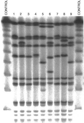
Gel electrophoresis is a technique used for the separation of nucleic acids and proteins (1). Separation of large (macro) molecules depends on two elements: charge and mass. When a biological sample (e.g., proteins or DNA) is mixed in a buffer solution and applied to a gel, these two factors act together. The electrical current from one electrode repels the molecules while the other electrode simultaneously attracts the molecules. The frictional force of the gel material acts as a molecular sieve, separating the molecules by size. During electrophoresis, macromolecules are forced to move through the pores when the electrical current is applied. Their rate of migration through the electric field depends on the strength of the field, size, and shape of the molecules, the relative hydrophobicity of the samples, and the ionic strength and temperature of the buffer in which the molecules are moving. After staining, the separated macromolecules in each lane can be seen in a series of bands spread from one end of the gel to the other, as seen, for example, in Fig. 1.
Some of the concepts of gel electrophoresis are used even in some of the most advanced commercially available
428 DNA SEQUENCING
Figure 1. A photograph of a gel from an electrophoresis experiment. Spots higher on columns represent lighter molecules. The lanes at the left and right end are control lanes where the DNA used had known length.
techniques for DNA sequencing. The role of gel electrophoresis will be made clear in describing later Sanger’s method.
TYPES OF DNA SEQUENCING
The inherent meaning of the word sequencing translates into finding the sequence of nucleotides of an unknown DNA strand. This type of DNA sequencing is usually referred to as de novo sequencing (de novo in Latin means from the beginning).
On the other hand DNA detection refers to the process of identifying known sequences of DNA within a sample. There are many laboratory techniques commonly used to perform such a task. Polymerase Chain Reaction (PCR) is used in amplifying (multiplying the concentration of) DNA sequences that contain certain primers (1). DNA microarrays is a technology used in gene expression profiling; it is a high throughput DNA detection mechanism where multiple DNA probes are simultaneously detected. These microarrays will be further analyzed in the following paragraphs. Biotin–streptavidin bead-based detection is a process that permits single-stranded DNA molecules containing a given subsequence to be filtered out from a heterogeneous pool of other DNA molecules (1). Strands
complementary to the subsequence are attached with biotin to streptavidin coated magnetic beads. The heterogeneous solution is passed over the beads and strands containing the subsequence anneal to the complementary sequence and are retained, while strands not containing it, pass through.
In many cases the DNA sequence under examination is largely known, but only small regions are of interest. This is the case in genotyping when it is desired to detect small variations in a whole genome (or gene) when compared with a known DNA sequence. Much of the variation in organisms originates from single-base changes in genes. These small changes can significantly affect the translation, and hence the role of the gene. This type of variation termed single nucleotide polymorphism (SNP, pronounced ‘‘snips’’) is of extreme interest in molecular biology. When a genome is examined for certain SNPs usually it is desired to detect subsequences of the form xxx. . .xYxxx. . .x, where xxx. . .x indicates known DNA bases and Y can be any base of A,T, G, or C. For a variation to be considered a SNP, it must occur in at least 1% of the population. The SNPs, which make up 90% of all human genetic variation, occur every 100–300 bases along the 3-billion-base human genome. There exist >100 techniques for detecting known forms of SNPs. Many SNPs have no effect on cell function, but scientists believe others could predispose people to disease or influence their response to a drug. For more information on SNPs their significance and detection methods interested readers are directed to (3).
DNA SEQUENCING PRINCIPLES: THE SANGER METHOD
The foundations of DNA sequencing were laid in 1974. Two groups, a British headed by Sanger et al. (4) and an American lead by Maxam and Gilbert (5), independently discovered a technique that enables to break a fragment of DNA into smaller nested subfragments. Both groups shared the 1980 Nobel Prize in chemistry for their discovery. The method from the American team was based on a chemical cleavage protocol and used toxic chemicals and large amounts of radioactivity, whereas Sanger’s method essentially mimics DNA replication as it takes place in cells. Sanger’s method was eventually adopted by the industry and a form of it is still used today since it was simpler to implement in large-scale production sequencing.
For the Sanger method the following items are needed: (1) the unknown DNA; (2) a primer; (3) DNA polymerase; (4) a mixture of dNTPs (deoxynucleotide triphospates) and ddNTPs (di-deoxynucleotide triphosphates).
The unknown DNA, termed here template, is the fragment of DNA that needs to be sequenced. The fragment needs to be in a single-stranded form in the 30-50 direction. If in double-stranded form a single-stranded sequence can be obtained by melting (denaturing) the duplex. We also assume that the unknown template contains a known subsequence usually 12–24 bases long. The complement of this subsequence in the 50-30 direction is called the primer. The primer is chemically synthesized. Once the primer is inserted in the solution containing the unknown DNA it will anneal (bind) to its complementary sequence
|
DNA SEQUENCING |
429 |
5'-GAATGTCCTTTCTCTAAG-3’ |
Figure 2. The primer (in red) is annealed |
|
3'-GGAGACTTACAGGAAAGAGATTCAGGATTCAGGAGGCCTACCATGAAGATCAAG-5' |
to the template at the primer binding site. |
|
with hydrogen bonds (Fig. 2). The primer needs to be long enough to ensure that the annealing site is unique, but not very long such that the annealing is unstable.
Once the primer and the template have annealed the DNA polymerase starts reacting and catalyzes the DNA and extending from the 30 end of the primer starts filling-in nucleotides (dNTPs) that are complementary to the template at each position. This serial addition of nucleotides is dependent on the bases of the template. The incoming nucleotide forms a covalent bond with the 30 end of the previous sugar using its 50 end. Under normal conditions, nucleotides are filled-in till the end of the template is reached, that is, the strand is fully extended. Sanger’s idea was to modify this process such that it ceases before it reaches the end of the template.
By using a simple chemical modification, nucleotides can be transformed such that they prohibit the addition of another nucleotide in their 30 end. The necessary chemical alteration is the substitution of the hydroxyl (OH) group on the 30 end of the nucleotide with a hydrogen (H). Such modified nucleotides are called ddNTPs or dideoxynucleotide triphosphates and are usually termed as terminators. When such terminators are incorporated in the extension the replication stops, as shown in Fig. 3.
The DNA polymerase will stop extending when a ddNTP is incorporated. Now, if in the solution a certain mixture of dNTPs and ddNTPs is present at the end, DNA polymerase will create a mixture of strands of various lengths terminated by ddNTPs. To distinguish between the different ddNTPs (A,T,G, or C) a unique fluorescent label is attached to each one of them. In some implementations the label is attached to the primer, but assumes that the reaction is run in parallel in four tubes where each tube contains only one type of ddNTP. The relative concentrations of the dNTPs and ddNTPs are adjusted in such a way that we end up with about the same number of copies of fragments between 100 bp and 500 bp long, and a smaller number of shorter and longer fragments.
At the end the solution contains a random mixture of partially terminated double-stranded sequences. If the ddNTPs were labeled, and hence only one test tube was used, the sequences are denatured into single-stranded DNA molecules and are run on a polyacrylamide–urea gel in a single lane. The gel is dried onto chromatography paper (to reduce its thickness and keep it from cracking) and exposed to X-ray film. Since the template strand is not radioactively labeled, it does not generate a band on the X-ray film.
The fragments will be ordered on the gel lane according to length. A laser (for stimulating the emission of radiation) and a detector (for collecting the stimulated radiation) are placed at a certain distance away from the initial position. When a fragment is scanned by the laser, the fluorescent label attached to the terminator is excited, and a signal at a certain wavelength depending on the label will be emitted and sensed by the detector, as shown in Fig. 4. Multiple copies of each fragment will ensure high signal strength, which will hopefully be strong enough to be detected. By examining the peaks of the time sequence of the fluorescence intensity at different wavelengths, the bases of the unknown sequence can be determined.
The above procedure is similar if the label was attached on the primer or in the dNTPs and four tubes and separate reactions were run. In this case, the results of each tube correspond to a specific ddNTP. Each tube’s contents are placed in a different lane (four in total) as seen in Fig. 5. With this setup only one fluorescent label is used, hence the excitation and detection mechanism is much more simplified. By examining the peaks of the intensity at each lane and working from bottom to top the base path or the ‘‘base ladder’’ can be determined.
It is evident that the resolution of the gel electrophoresis is rather critical and a single base resolution is usually a prerequisite. To improve the resolution of gel assays, the gels must be much large so that the molecules migrate further and are better resolved. They must contain a high concentration of urea (7–8 m) to prevent folding of the
5'-GAATGTCCTTTCTCTAAGTCCTAAG 3'-GGAGACTTACAGGAAAGAGATTCAGGATTCAGGAGGCCTACCATGAAGATCAAG-5'
5'-GAATGTCCTTTCTCTAAGTCCTAAGTCCTCCG 3'-GGAGACTTACAGGAAAGAGATTCAGGATTCAGGAGGCCTACCATGAAGATCAAG-5'
5'-GAATGTCCTTTCTCTAAGTCCTAAGTCCTCCGG 3'-GGAGACTTACAGGAAAGAGATTCAGGATTCAGGAGGCCTACCATGAAGATCAAG-5'
5'-GAATGTCCTTTCTCTAAGTCCTAAGTCCTCCGGATG 3'-GGAGACTTACAGGAAAGAGATTCAGGATTCAGGAGGCCTACCATGAAGATCAAG-5'
5'-GAATGTCCTTTCTCTAAGTCCTAAGTCCTCCGGATGG 3'-GGAGACTTACAGGAAAGAGATTCAGGATTCAGGAGGCCTACCATGAAGATCAAG-5'
5'-GAATGTCCTTTCTCTAAGTCCTAAGTCCTCCGGATGGTACTTCTAG 3'-GGAGACTTACAGGAAAGAGATTCAGGATTCAGGAGGCCTACCATGAAGATCAAG-5'
Figure 3. A mixture of the products of synthesis for the G ddNTP reaction.

430 DNA SEQUENCING
T ddNTP reaction
A ddNTP reaction
G ddNTP reaction
C ddNTP reaction
Intensity
Figure 4. An example of a gel where labels are attached on the ddNTPs, and hence a single-lane gel is only used.
Scan
Figure 6. A chromatogram example from an experiment with four dyes. The curves correspond to intensity measurements of fluorescent emission at different wavelengths corresponding to the dyes used for each ddNTP reaction.
G C A T
Figure 5. An example of a gel where labels are attached on the primer, and hence four lanes are used.
molecules and formation of DNA secondary structures by hydrogen bonding that would alter the mobility of the molecule. Similarly, the samples are denatured before they are loaded. The gels must run at higher temperature ( 50 8C), to prevent hydrogenbond formation.
From the above analysis, it is clear that in order to extract the bases from the gel reactions the peaks have to be identified. Traditionally the laser scanners output the fluorescence intensity into chromatograms (Fig. 6). Prior to the development of computers the chromatograms were interpreted by humans. The peaks were assigned to bases in a procedure known as base calling (Fig. 7). Ideally a periodic peak detection scheme would have been adequate if the signal was noiseless and perfect. There are certain
T T A A G A T T
Intensity
Scan
Figure 7. A decoded sequence from the chromatogram example of Fig. 6.
errors and limitations that make the base calling aspect of DNA sequencing a rather challenging task. Some of the sources of error are
1.Errors in fragment formation: (a) Abnormalities in primer extension (false stops, terminator is not incorporated, or conversely, several terminators accumulate at the same position). (b) Poor choice of relative concentrations of ddNTPs and dNTPs resulting in too many short or long fragments. (c) While DNA moves down the gel, secondary (e.g., hairpin) structures may form and change the mobility properties of the DNA fragments.
2.Convolution: Due to the stochastic nature of the DNA migration in the gel, the time scale of the chromatograms changes resulting in more elongated and less discreet peaks.
|
DNA SEQUENCING |
431 |
Table 1. Common Parameters Used when Evaluating DNA Sequencing Equipment |
|
|
|
|
|
Parameter |
Explanation |
|
|
|
|
Technology used |
Electrophoretic or nonelectrophoretic, capillary, and so on affects many of the other parameters |
|
|
in a DNA sequencing system |
|
Length of the gel |
Applies only to electrophoretic systems and refers to the length of the gel material. The longer |
|
|
the better since it increases resolution |
|
Throughput |
Measured in bases per cycle (or per day), illustrates the processing capability of the system |
|
Cycle speed |
The equipment may need replenishing of reagents after a run. The number of runs per day |
|
|
define the cycle speed |
|
Read length |
Maximum length of the DNA template that can be sequenced |
|
Capacity |
Number of different DNA templates that can be sequenced simultaneously |
|
Sample volume |
Defined as the volume of the template needed for a certain outcome quality (the less the better) |
|
Error rates |
Number of bases in error out of 1000 usually defines the error rate. Error rates are tightly bound |
|
|
to base calling quality assessment |
|
Maintenance and operation cost |
Number of dyes and labeling method used, have a direct impact on maintenance and operation cost |
|
|
|
|
3.Intensity cross-talk: Due to the overlapping of the fluorescent response spectra of the fluorophores employed in the four-dye sequencing strategy there is a need for a transformation to recover the relative concentrations of the four dyes from the fluorescence intensities measured at four different wavelengths.
4.Measurement errors: White noise can originate from several sources, including background, detector, and other noise from the operating environment. Another type of noise encountered is low frequency variation due to slow changes in the background light level during collection. Such variations may be caused by deformation of the gel due to heating, the formation of bubbles in the path of the laser, variations in laser output power and other systematic changes in the environment.
Nowadays advances in statistics, signal processing, electronics, laser optics and software have lead to automated DNA sequencing and base calling capable of sequencing many different DNA templates.
EVALUATING DNA SEQUENCING TECHNIQUES
When deciding on DNA sequencing equipment, a prospective buyer has to evaluate certain aspects of the DNA sequencing scheme offered by the vendor. The buyer has to consider the traits shown in Table 1. All these parameters are critical, but their importance is weighted differently according to the application sought after by the buyer.
CURRENT COMMERCIAL STATE OF THE ART
Since the development of the early DNA sequencing methods the capabilities of the DNA sequencing equipment have improved dramatically. This change can be attributed to the radical advances in the fields of DNA chemistry, laser and optics, statistics, robotics, automation, and software. In many of the laboratories involved in the human genome
project, the high throughput DNA sequencing machines that were employed used robotic arms to move samples in and out of the machines and heavy automation to perform those tasks with minimal human intervention. Advances in laser optics led to even finer scanning and detection resolution with lower error rates. As seen in the previous section, one of the most critical aspect in DNA sequencing is the analysis of the chromatograms to determine the bases. Nowadays this task is performed by sophisticated software packages that employ statistics, digital signal processing, and adaptive algorithms that can identify the bases from the fluorescence graphs. A comparison of some of the most commonly used packages can be found in Table 2.
In the following section, sequencing devices are first presented that rely on electrophoretic principles followed by those that do not.
Electrophoretic-Based Methods
Slab-Gel. It was expected that the first automated DNA sequencers would be based on the Sanger method. Acrylamide slab gel electrophoresis until recently was the most widespread method of de novo sequencing (6). The Prism 373 by Applied Biosystems (ABI) Prism (Foster City, CA) was the first sequencer that could scan and detect such gels using a procedure very similar to the one described in the previous sections. Some of the drawbacks of slab gel instruments are gel casting (preparing the gel), gel loading (loading the gel into the device), and lane tracking (detecting lanes on the gel).
The Prism 373 underwent many changes in order to increase throughput and read length before it was replaced by ABI PRISM 377. The PRISM 377 is based on a four dye chemistry coupled with a CCD (charged couple device) imaging detector and can process up to 96 samples per cycle (9–11 h) with read lengths of 650–750 bases. Despite their drawbacks, slab gel systems are still preferred for applications with low throughput requirements but large read lengths. Reviews of experimental and commercial systems based on slab gels can be found in (7,8). Of such systems the following are worth noting since they are still used due to their unique properties.
432 |
DNA SEQUENCING |
|
|
|
Table 2. A Comparison of Base Calling and Sequence Analysis Softwarea |
|
|||
Name |
|
Publisher |
License |
Short Description |
|
|
|
|
|
Phred |
|
University of Washington, |
Free, Open Source |
The phred software reads DNA |
|
|
Phil Green Laboratory |
|
sequencing trace files, calls bases, |
|
|
|
|
and assigns a quality value to |
|
|
|
|
each called base (1,3,4) |
Autoseq |
|
Reece Hart |
Free, Open Source |
Autoseq is a small package of |
|
|
|
|
base-calling software for ABI |
|
|
|
|
automated DNA sequencers (5) |
Sequence Analyzer |
GE Healthcare |
Commercial |
Usually bundled with MegaBASE |
|
|
|
|
|
sequencers (6) |
Lasergene |
|
DNAstar |
Commercial |
Comprehensive suite of easy-to-use |
|
|
|
|
sequence analysis software (7) |
Sequencher |
Gene Codes Corp |
Commercial |
Allows SNP detection (8) |
|
Staden |
|
R. Staden and other |
Free, Open Source |
A suite of sequence assembly, |
|
|
contributors |
|
editing, etc. (9) |
Sequencing Analysis Software |
Applied Biosystems |
Commercial |
Usually accompanies ABI Prism |
|
|
|
|
|
sequencers (10) |
TraceTuner |
Paracel |
Discontinued |
Another base calling application |
|
|
|
|
|
|
The DNA 4300 System by LI-COR Inc (Lincoln, NE) uses two dyes at near-(IR) frequencies. Based on this innovation, the ability to operate and sequence from both ends of the template in parallel and coupled with an excellent software suite, the higher end version of the 4300 has read length of up to 1250 bases thus making it ideal for applications with large read length requirements.
The BaseStation from MJ Research Inc (Waltham, MA) uses a 75 mm thick polyacrylamide gel to improve heat dissipation in the gel thus reducing significantly the run time. Armed with robotic gel loading, a four-color photomultiplier with high sensitivity and a 100 sample capacity, the instrument offers a nice alternative to capillary systems when long read lengths are needed.
Capillary Systems. The persistent drawbacks of slab gel electrophoresis and the desire for faster sequencing runs and higher throughput led to the development of capillary array electrophoresis (CAE). Electrophoresis works in a way similar to slab gels, except that each capillary contains a single sample, and therefore tracking problems are eliminated. Furthermore, the high surface/volume ratio of a capillary allows for more rapid heat dissipation than is possible in slab gels, thus allowing higher operating voltages and faster run times.
Capillary electrophoresis uses capillaries usually 50 mm in diameter. Capillaries are very narrow tubes that based on the capillary action can draw liquid against gravity. Similarly to the technique used for manufacturing fiber optics, the capillaries are made from highly pure fused silica.
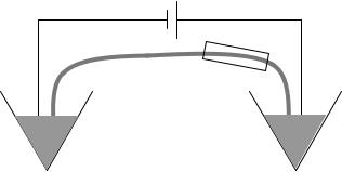
As seen in Fig. 8 the instrumentation is rather simple. The sample is injected into the capillary and high electrical field is applied to advance the sample into the capillary. Subsequently, the sample is replaced by a buffer solution and the field is reapplied to migrate the samples through the capillary. Since the capillary is filled with a sieving medium it allows for the separation of the DNA sequences according to length. The fragments pass through a laser-induced
Detector
Sample |
Buffer |
Figure 8. A single capillary electrophoresis device where a fusedsilica capillary is used for the separation. The left end of the capillary is submerged into the sample solution while the other is dipped into a buffer-filled holding tank. At some point the capillary goes through a detection apparatus. High voltage is applied at each end using platinum electrodes.
fluorescence detector, which can record at different frequencies the fluorescence response of the four dyes similar to the slab gel techniques.
High voltage (double compared to slab gel techniques) allows for rapid separation, but certain phenomena limit the resolution at high read lengths (9). Although the ability to use high voltages is rather attractive, the most interesting aspect of capillaries is their flexibility, allowing them to be incorporated easily into automated systems.
Capillary array electrophoresis uses a collection of capillaries each of which is injected with a different sample. In some instruments the detector sequentially moves from capillary to capillary while in most advanced ones each capillary is scanned simultaneously using detectors attached to each one (10,20–22).
Some of the sequencers currently available in the market are Applied Biosystems PRISM 310 and 3730, Hitachi, BioRad/MJ Research (Hercules, CA) BaseStation, GE
Healthcare (Piscataway, NJ) MegaBASE 4000, Beckman Coulter (Fullerton, CA) CEQ 8000, SpectruMedix (State College, PA) Aurora, and RIKEN (Tsukuba and Wako, Japan) RISA.
Nonelectrophoretic-Based Methods
During the last 20 years, several techniques for sequencing have been discovered that do not rely on electrophoretic principles. Although these techniques have now reached the performance of CAEs for de novo sequencing they are mostly used for other types of sequencing described in a previous section.
Pyrosequencing. Pyrosequencing is a sequencing technique developed around real-time monitoring of the release of pyrophosphate (PPi) during polymerase assisted DNA synthesis (23). Similarly to electrophoretic methods a sequencing primer is hybridized to a single-stranded DNA template, and incubated with the enzymes, DNA polymerase, ATP sulfurylase, luciferase, and apyrase, and the substrates, adenosine 50-phosphosulfate (APS) and luciferin. One of the four dNTPs is added to the solution. Assisted by polymerase the correct dNTP will be incorporated in the chain resulting in the release of PPi at a concentration analogous to the amount of incorporated nucleotides. The amount of PPi is constantly monitored by a coupled enzymatic reaction where PPi is converted to ATP by ATP sulfurylase. The ATP subsequently assists in the conversion of luciferin to oxyluciferin by firefly luciferase, which results in light emission. The process is repeated iteratively for the other dNTPs. A critical component for the success of the method is the removal of excess dNTP and ATP prior to a new dNTP addition. These can be achieved by attaching the template sequence on solid support that is washed prior to a new dNTP addition or by solution enzymatic reaction where apyrase is added to catalyze the remaining dNTPs.
The read length of pyrosequencing is smaller when compared to electrophoretic methods thus making pyrosequencing less advantageous for de novo sequencing. This is not true, however, for applications, such as genotyping of SNPs, resequencing, tag sequencing, microbial typing, and many others where pyrosequencing shines.
The technique, although in its infancy, can claim the first automated sequencer, the PSQ HS 96 from Pyrosequencing/Biotage (Uppsala, Sweden). It uses a disposable inkjet cartridge for precise delivery of small volume (200 nL) of six different reagents into a temperaturecontrolled microtiter plate and is widely used for SNP detection with a throughput of 96 samples per hour.
Sequencing by Hybridization: DNA Arrays, Microarrays.
Hybridization arrays or microarrays or DNA arrays were originally developed for de novo sequencing. Sequencing by Hybridization (SBH) requires annealing a labeled unknown DNA fragment to a complete array of short oligonucleotides (e.g., all 65,336 combinations of 8-mers) and decoding the unknown sequence from the annealing pattern (24). The array could be imaged using laser scanners and CCD devices or photomultiplier tubes. The
DNA SEQUENCING |
433 |
computational complexity of decoding the annealed pattern limited the popularity of such systems for de novo sequencing. Nowadays, the key applications of DNA arrays are SNP and expression analysis (25).
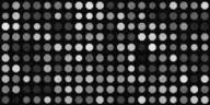
The DNA microarrays are small, solid supports onto which the sequences from thousands of different genes are immobilized, or attached, at fixed locations. The supports themselves are usually glass microscope slides, of various sizes, but can also be silicon chips or nylon membranes. The DNA can be printed, spotted, or actually synthesized directly onto the support. It is important that the gene sequences in a microarray are attached to their support in an orderly or fixed way, because the location of each spot in the array identifies a particular gene sequence. The spots themselves can be DNA, cDNA, or oligonucleotides. In each microarray experiment two samples are tested simultaneously, each labeled with a different fluorescent dye. The control sample is labeled by the Cy5 dye and the test sample by the Cy3. Both samples are introduced in the microarray simultaneously and when excited by different laser frequencies each dye returns a distinct response, which can be recorded as intensity measurements. This process produces two images; the green image, which corresponds to intensity measurements of the Cy5 dye, and the red image, which corresponds to intensity measurements of the Cy3. These fluorescence intensities correspond to the levels of hybridization of the two samples to the DNA sequences spotted on the slide. An example of a microarray image is shown in Fig. 9.
The list of manufacturers of DNA arrays is rather exhaustive with >30 entries. Of those, the pioneer and first in market Affymetrix (Santa Clara, CA), Agilent (Palo Alto, CA), and Nimblegen (Madison, WI) should be mentioned. The war on density and throughput of microarrays is everlasting. Nimblegen, for example, can produce microarrays with >40,000 genes, with each spot being 80 mm in diameter.
GENOME SEQUENCING
As of now, the maximum read length permitted by today’s commercial and experimental techniques does not exceed 2000 bases. The human genome is currently estimated to be >3 billion bases with 20,000–25,000 genes, while organisms such as the Escheuchia coli bacterium has4.6 million bases. It is clear that in order to sequence whole genomes of organisms with the currently available techniques a method for combining sequencing results of smaller reads is needed (26).
Most of the techniques rely on shotgun sequencing, which is based on the idea of sequencing overlapping
Figure 9. An example of a microarray image.
434 DNA SEQUENCING
fragments of DNA (27). The genomic segment is sheared into overlapping fragments of DNA 500 bases long and each fragment is then sequenced. The fragments are assembled into continuous sequences called contigs using complicated computer algorithms where they examine the overlappingsequencesandtry toorderthefragments. There aretwoissueswiththistechnique:gapsanderrors. Sincethe shearing technique is random, and to avoid laboratory errors usually multiple shearing experiments are performed and the fragments are sequenced to assemble contigs. Another source of errors are repeats. If sequences appear in multiple positions throughout the genomic segment it will lead to errors when the fragments are overlapped. In some cases during contig assembly nonoverlapping sequences are formed that create gaps. Gaps are resolved with directed sequencing experiments with primers derived from the contigs that surround the gap.
The difference between the Human Genome Project (HGP) and Celera Corp was the origin of the target sequence. Human Genome Project used a directed sequencing method (also seen as hierarchical shotgun sequencing) for which the whole genome is first broken into long fragments. The fragments are then mapped into the genome, which is equivalent to finding their order (location) within the genome. Each fragment then is sequenced using shotgun sequencing. The advantage of this approach is the relatively easy assembly, while the disadvantages are the difficulty of building the library, mapping the long fragments, and the need for redundant sequencing.
Celera Corp relied on whole genome shotgun sequencing, which is essentially shotgun sequencing applied directly on the whole genome. The challenge is to assemble the whole genome from small 500 base fragments. While this technique overcomes the shortcomings of hierarchical sequencing and is faster and less expensive, the assembly is rather complicated and resolving the repeats requires sequencing of many clones.
THE FUTURE OF DNA SEQUENCING
Microscale Systems
Micro capillary Systems are microfabricated systems that in principle work similarly to CAE systems. Such systems have the potential of reducing cost while increasing speed and throughput. Due to their small size, lab-on-chip solutions are even considered where most of the sample preparation, amplification, and sequencing is all taking place on a single glass surface (chip). Their unique manufacturing methods allow for a large number of capillaries at low cost with arbitrary geometries, which are not possible with standard capillaries.
The Mathies group at University of California at Berkeley (28) published a method for fabricating capillary systems with complex architecture and layout on glass substrates using photolithography. One of their systems can achieve a read length of 500 bases with 99% accuracy and a cycle speed of 20 min (29).
A rather interesting technique is Massive Parallel Signature Sequencing (MPSS) (30). Using a microarray structure and beads, targets are sequenced iteratively at each
cycle using a type IIs restriction enzyme that cleaves (cuts) within a target sequence, exposing a four-base-pair overhang. The overhang is identified using a sequencespecific ligation of a fluorescent linker. The method can read up to 20 bases (in 4–5 cycles) making it well suited for expression analysis.
Mass Spectrometry Based DNA Sequencing
Matrix Assisted Laser Desorption Ionization Time-of- Flight Mass Spectrometry (MALDITOF–MS) is the first MS based DNA sequencing technique (31). The method can replace the electrophoretic molecule separation step in DNA sequencing by a MS component. The mass of the molecule is estimated by measuring the time to travel of gas-phase DNA molecules within a flight tube that connects an excitation source (ultraviolet, UV laser) and an ion-to-electron conversion detector. The molecules collide at the detector thus registering the time to travel, which is analogous to molecular mass. The technique has the advantages of allowing the fast and parallel separation of a heterogeneous mixture of molecules without being affected by possible secondary structures that the DNA molecules have fallen into. Although the read lengths remain small, the availability of fast and autonomous MALDITOF–MS DNA sequencers (Sequenom Corporation in San Diego, CA) makes them good candidates for precise resequencing of small fragments useful in SNP detection.
DNA Sequencing at the Nanoscale
Most of the methods described below are based on manipulating properties of DNA at the nanoscale or utilizing properties of other materials at the nanoscale. Since such methods work with very low concentrations they can also be viewed as single-molecule sequencing methods and are suitable for applications when the template DNA is in very low concentration and amplification techniques could not be applied efficiently.
DNA Detection with Nanoparticles. Nanosphere Inc. (Northbrook, IL) has developed a method for rapid and low concentration detection of proteins and nucleic acids (32). Their technology is based on attaching oligonucleotide probes on nanoparticles. The probes attach to the target DNA and due to the unique properties of the used nanoparticles the event can be detected electrically, optically, or magnetically without amplification of the target sequence. The concentrations needed are below the operational threshold of PCR reactions. Although the techniques have not been extended to de novo sequencing the unique detection characteristics are proving very useful in detection scenarios.
In another effort from the founder of Nanosphere, Dr. Mirkin at Northwestern University (Evanston, IL), the electrical detection of DNA was first proposed (33). With this protocol the imaging aspect of microarray applications can be eliminated using gold nanoparticles that once hybridized onto the DNA probes and deposited with silver can close an electric circuit thus enabling detection of the hybridization event with electrical signals.
Sequencing with Atomic Force Microscopy. Atomic Force Microscopy (AFM) was invented at IBM Zurich Labs in 1986 and has completely revolutionized research at the nanoscale (34). A nanoscopic tip that is attached at the end of the cantilever interacts with the surface of the target material and records the tips deflections to create a topographic map of the surface. The AFM can be used to study the surface of duplex DNA and detect mismatches or it can be used as force measuring tool to study the mechanical properties of DNA. One application of particular interest is the AFM assisted unzipping of the DNA duplex, where a DNA duplex is suspended between a solid support and the AFM tip. Pulling the AFM tip further causes the duplex to unzip. The force needed to unzip depends on the percentage of the GasChromatography (GC)content, andhence canbeused to estimate the GC content of an unknown target if needed in a more large-scale sequencing function (35). A very similar idea was proposed in Ref. 36 where optical traps are used to stretch DNA molecules and to measure force.
Nanopore Sequencing. Another interesting technique that uses features at the nanoscale is nanopore sequencing. As DNA passes through an 1.5 nm nanopore, different base pairs hinder the pore to different degrees, altering the electric conductivity of the pore (37). The pore conductance can be measured and monitored to identify the DNA sequence. The accuracy of base calling ranges from 60% for single events to 99.9% for 15 events. The technique has only been shown to work experimentally on certain sequences but exhibits a big potential for super fast sequencing without amplification of the target. It is evident that the evolution of this technique depends on nanopore engineering. To break apart from this restriction Visigen (Houston, TX) and Li-cor (Lincoln, NE: U.S. Patent 6,306,607) are in the process of engineering DNA polymerases or fluorescent labeled nucleotides that can provide real-time, base-dependent signals during the natural DNA synthesis process.
Sequencing by Fluorescence Microscopy. This is a new class of DNA sequencing methodologies, for which the fluorescence emitted during single molecule interactions is detected (38). The interactions most commonly referred to are single nucleotide incorporation during DNA polymerase replication or nucleotide digestion from an exonuclease. The change in fluorescence emission is detected using microscopes and CCDs. An enabling technology is fluorescence resonance electron transfer (FRET), where the fluorescence emission of two molecular dyes can be affected by their proximity. The research in the area is vast and already three companies, Nanofluidics (Menlo Park, California), Solexa (Essex, UK), and GenoVoxx (Lubeck, Germany), are developing products based on this technology for high throughput DNA detection and genotyping.
DNA Computing Based DNA Sequencing
Up to this point instruments and electronic computers were assigned the task of analyzing and processing DNA sequences. In 1994, the roles were reversed by the first proof of concept experiment by Adleman of using DNA to
DNA SEQUENCING |
435 |
perform computations (39). This development led to the birth of the field of DNA computing [for a short introduction see (40)].
Landweber and Lipton were the first to suggest that DNA computing can be used to improve the performance of DNA sequencing (41). Their approach is based on DNA2DNA computations, where nucleotides of an unknown sequence are translated into a new DNA sequence using a unique mapping transformation. A library of DNA oligonucleotides is synthesized and mixed in the solution containing the template DNA. The oligonucleotides then anneal to complementary parts. The partially double-stranded sequences are ligated and hybridized on a DNA chip. The reconstruction of the encoded sequence is achieved by analyzing the DNA array image. Although the technique was never implemented in large scale it points to potential future applications where instruments can be assisted by DNA computers.
The first proof of such development came a few years later in an announcement by Dr. Suyama from the University of Tokyo and Olympus Corp. (Japan), where they developed the first DNA-computer-assisted gene expression instrument (42). The instrument is a hybrid of a molecular computer and an electronic–digital computer. The molecular computer is in charge of DNA input– output, DNA reactions, capture of DNA results, and DNA detection while the electronic is responsible for information processing by means of DNA reaction calculations and result analysis.
In Ref. 43, a new laboratory protocol is proposed that assists in the faster sequencing of genomes. The distance between primers (probes) that have annealed on a target sequence can be estimated by measuring the intensity and color of light emission of specialized hybridization array. Although the method is not intended for de novo sequencing it is proposed as an alternative method of comparing genomes.
Bio-informatics, a subfield of computational biology, refers to processing, analyzing or storing DNA sequencing data with computers. The field of analyzing DNA sequences using digital signal processing theory has been known as genomic signal processing (44). The idea is to process the sequence of DNA as a digital signal and find certain characteristics. Recently, the application of DNA computing in digital signal processing, termed as DNA-based Digital Signal Processing, has been suggested (45). A future is envisioned where a DNA based digital signal processor can process DNA sequences and output certain characteristics in the form of DNA sequences that can be subsequently detected (or sequenced). This will allow researchers to process a vast amount of DNA sequences without prior sequencing.
BIBLIOGRAPHY
1.Watson JD, et al. Molecular biology of the gene. 5th ed. San Francisco: Pearson/Benjamin Cummings; 2004.
2.Shopsin B, Kreiswirth BN, Molecular Epidemiology of Methicillin-Resistant Staphylococcus aureus, [serial on the Internet], 2001; 7(2) Accessed 2005 July 14. Available at http://www.cdc.gov/ncidod/eid/vol7no2/shopsin.htm.
436 DNA SEQUENCING
3.Weiner MP, Hudson TJ. Introduction to SNPs: discovery of markers for disease. Biotechniques 2002;32(Suppl.) S4–S13.
4.Sanger F, Nicklen S, Coulson AR. DNA sequencing with chainterminating inhibitors. Proc Natl Acad Sci USA 1977;74:5463– 5467.
5.Maxam AM, Gilbert W. A new method of sequencing DNA. Proc Natl Acad Sci USA 1977;74:560–564.
6.Studier FW. Slab-gel electrophoresis. Trends Biochem Sci 2000;25(12):588–590.
7.Meldrum D. Automation for genomics. Part Two: Sequencers, microarrays, and future trends. Genome Res 2000;10:1288– 1303.
8.Huang GM. High-throughput DNA sequencing: a genomic data manufacturing process. DNA Seq 1999;10:149–153.
9.Viovy JL, Duke T. DNA electrophoresis in polymer solutions: Ogston sieving, reptation and constraint release. Electrophoresis 1993;14(4):322–329.
10.Zagursky RJ, McCormick RM. DNA sequencing separations in capillary gels on a modified commercial DNA sequencing instrument. Biotechniques 1990;9(1):74–79.
11.Green P (No date). Phrep, Phrap and Consed [Online]. University of Washington. Available at http://www.phrap.org/ phredphrapconsed.html. Accessed 2005, June 29.
12.Ewing B, Green P. Basecalling of automated sequencer traces usingphred.II.Errorprobabilities.GenomeRes1998;8:186–194.
13.Ewing B, Hillier L, Wendl M, Green P. Basecalling of automated sequencer traces using phred. I. Accuracy assessment. Genome Res 1998;8:175–185.
14.Hart C. (1997, August 1). Autoseq home page. [Online]. InMachina. Availabel at http://www.in-machina.com/~reece/ autoseq/ Accessed 2005, June 29.
15.Software Sequencing (No date). GE Healthcare - formerly Amersham Biosciences - Sequencing [Online]. GE Healthcare. Available at http://www5.amershambiosciences.com/aptrix/ upp01077.nsf/Content/autodna_software_sequencing. Accessed 2005, June 29.
16.Lasergene (No date). DNASTAR. [Online]. DNASTAR, Inc. Available at http://www.dnastar.com/web/index.php. Accessed 2005, June 29.
17.Sequencher (No date). Gene Codes Corporation: Sequencher. [Online]. Gene Codes Corporation. Available at http://www. genecodes.com/sequencher/. Accessed 2005, June 29.
18.Staden Package (No date). Staden Package Home Page [Online]. SourceForge. Available at http://staden.sourceforge. net/ Accessed 2005, June 29.
19.Applied Biosystems Product Information Page (No date). Sequence Analysis Software [Online]. Applied Biosystems. Available at http://www.appliedbiosystems.com/. Accessed 2005, June 29.
20.Huang XC, Quesada MA, Mathies RA. DNA sequencing using capillary array electrophoresis. Anal Chem 1992;64(18):2149– 2154.
21.Kambara H, Takahashi S. Multiple-sheathflow capillary array DNA analyzer. Nature(London) 1993;361(6412):565–566.
22.Crabtree HJ. Capillary array DNA sequencer based on a micromachined sheath-flow cuvette. Electrophoresis 2000; 21:1329–1335.
23.Ronaghi M. Pyrosequencing Sheds Light on DNA Sequencing. Genome Res 2001;11:3–11.
24.Drmanac R, et al. DNA sequence determination by hybridization: a strategy for efficient large-scale sequencing. Science 1993;260:1649–1652; Erratum, Science 1994;163(5147):596.
25.Schena M, Shalon D, Davis RW, Brown PO. Quantitative monitoring of gene expression patterns with a complementary DNA microarray. Science 1995;270(5235):467–470.
26.Venter JC, et al. The Sequence of the Human Genome. Science 2001;291:1304–1351.
27.Sanger F, et al. Nucleotide sequence of bacteriophage lambda DNA. J Mol Biol 1982;162(4):729–773.
28.Woolley AT, Mathies RA. Ultra-High-Speed DNA Fragment Separations Using Microfabricated Capillary Array Electrophoresis Chips. Proc Natl Acad Sci USA 1994;91:11348–11352.
29.Simpson PC. High-throughput genetic analysis using microfabricated 96-sample capillary array electrophoresis microplates. Proc Natl Acad Sci USA 1998;95:2256–2261.
30.Brenner S, et al. In vitro cloning of complex mixtures of DNA on microbeads: physical separation of differentially expressed cDNAs. Proc Natl Acad Sci USA 2000;97:1665–1670.
31.Cantor CR, et al. DNA sequencing after the Human Genome Project. Nucleosides Nucleotides 1997;16:591–598.
32.Nam J-M, Park S-J, Mirkin CA. Bio-barcodes based on oligo- nucleotide-modified nanoparticles. J Am Chem Soc 2002;124: 3820–3821.
33.Park SJ, Taton TA, Mirkin CA. Array-Based Electrical Detection of DNA Using Nanoparticle Probes. Science Feb. 2002; 295(5559):1503–1506.
34.Binnig G, Quate CF, Gerber C. Atomic force microscope. Phys Rev Lett 1986;56:930–933.
35.Essevaz-Roulet B, Bockelmann U, Heslot F. Mechanical separation of the complementary strands of DNA. Proc Natl Acad Sci USA 1997;94:11935–11940.
36.Wang MD, et al. Stretching DNA with optical tweezers. Biophy J 1997;72:1335–1346.
37.Deamer DW, Branton D. Characterization of nucleic acids by nanopore analysis. Acc Chem Res 2002;35:817–825.
38.Braslavsky I, Hebert B, Kartalov E, Quake SR. Sequence information can be obtained from single DNA molecules. Proc Natl Acad Sci USA 2003;100:3960–3964.
39.Adleman L. Molecular computation of solutions to combinatorial problems. Science Nov. 1994;266:1021–1024.
40.Tsaftaris SA, Katsaggelos AK, Pappas TN, Papoutsakis ET. DNA computing from a signal processing viewpoint. IEEE Sig Proc Mag 2004;21(5):100–106.
41.Landweber LF, Lipton RJ. DNA2DNA Computations: A potential ‘killer app’? Proceedings of the 24th International Colloquium on Automata, Languages and Programming (ICALP). Lecture Notes in Computer Science. New York: SpringerVerlag; 1997. 672–683.
42.Normile D. DNA-Based Computer Takes Aim at Genes. Science 2002;295(5557):951.
43.Mishra B. Comparing Genomes. Comp Sci Eng 2002;4(1):42–29.
44.Anastassiou D. Genomic Signal Processing. IEEE Sig Proc Mag 2001;18(4):8–20.
45.Tsaftaris SA, Katsaggelos AK, Pappas TN, Papoutsakis ET. How can DNA-Computing be applied in Digital Signal Processing?. IEEE Sig Proc Mag 2004;21(6):57–61.
Further Reading
The following two articles provide a well-rounded review of commercially available and experimental sequencing techniques.
Marziali A, Akeson M. New DNA sequencing methods. Annu Rev Biomed Eng 2001;3:195–223.
and
Shendure J, Mitra RD, Varma C, Church GM. Advanced sequencing technologies: methods and goals. Nature Rev Genet 2004;5(5):335–344.
The reader is suggested to study the projections into the future of sequencing technology of the first article and compare it to the presentation of the current status of the second article. The advancement in technology is rather interesting given that the papers are only three years apart.