

- •VOLUME 2
- •CONTRIBUTOR LIST
- •PREFACE
- •LIST OF ARTICLES
- •ABBREVIATIONS AND ACRONYMS
- •CONVERSION FACTORS AND UNIT SYMBOLS
- •CARBON.
- •CARDIAC CATHETERIZATION.
- •CARDIAC LIFE SUPPORT.
- •CARDIAC OUTPUT, FICK TECHNIQUE FOR
- •CARDIAC OUTPUT, INDICATOR DILUTION MEASUREMENT OF
- •CARDIAC PACEMAKER.
- •CARDIAC OUTPUT, THERMODILUTION MEASUREMENT OF
- •CARDIOPULMONARY BYPASS.
- •CARDIOPULMONARY RESUSCITATION
- •CARTILAGE AND MENISCUS, PROPERTIES OF
- •CATARACT EXTRACTION.
- •CELL COUNTER, BLOOD
- •CELLULAR IMAGING
- •CEREBROSPINAL FLUID.
- •CHEMICAL ANALYZERS.
- •CHEMICAL SHIFT IMAGING.
- •CHROMATOGRAPHY
- •CO2 ELECTRODES
- •COBALT-60 UNITS FOR RADIOTHERAPY
- •COCHLEAR PROSTHESES
- •CODES AND REGULATIONS: MEDICAL DEVICES
- •CODES AND REGULATIONS: RADIATION
- •COGNITIVE REHABILITATION.
- •COLORIMETRY
- •COMPUTERS IN CARDIOGRAPHY.
- •COLPOSCOPY
- •COMMUNICATION AIDS FOR THE BLIND.
- •COMMUNICATION DEVICES
- •COMMUNICATION DISORDERS, COMPUTER APPLICATIONS FOR
- •COMPOSITES, RESIN-BASED.
- •COMPUTED RADIOGRAPHY.
- •COMPUTED TOMOGRAPHY
- •COMPUTED TOMOGRAPHY SCREENING
- •COMPUTED TOMOGRAPHY SIMULATOR
- •COMPUTED TOMOGRAPHY, SINGLE PHOTON EMISSION
- •COMPUTER-ASSISTED DETECTION AND DIAGNOSIS
- •COMPUTERS IN CARDIOGRAPHY.
- •COMPUTERS IN THE BIOMEDICAL LABORATORY
- •COMPUTERS IN MEDICAL EDUCATION.
- •COMPUTERS IN MEDICAL RECORDS.
- •COMPUTERS IN NUCLEAR MEDICINE.
- •CONFOCAL MICROSCOPY.
- •CONFORMAL RADIOTHERAPY.
- •CONTACT LENSES
- •CONTINUOUS POSITIVE AIRWAY PRESSURE
- •CONTRACEPTIVE DEVICES
- •CORONARY ANGIOPLASTY AND GUIDEWIRE DIAGNOSTICS
- •CRYOSURGERY
- •CRYOTHERAPY.
- •CT SCAN.
- •CUTANEOUS BLOOD FLOW, DOPPLER MEASUREMENT OF
- •CYSTIC FIBROSIS SWEAT TEST
- •CYTOLOGY, AUTOMATED
- •DECAY, RADIOACTIVE.
- •DECOMPRESSION SICKNESS, TREATMENT.
- •DEFIBRILLATORS
- •DENTISTRY, BIOMATERIALS FOR.
- •DIATHERMY, SURGICAL.
- •DIFFERENTIAL COUNTS, AUTOMATED
- •DIFFERENTIAL TRANSFORMERS.
- •DIGITAL ANGIOGRAPHY
- •DIVING PHYSIOLOGY.
- •DNA SEQUENCING
- •DOPPLER ECHOCARDIOGRAPHY.
- •DOPPLER ULTRASOUND.
- •DOPPLER VELOCIMETRY.
- •DOSIMETRY, RADIOPHARMACEUTICAL.
- •DRUG DELIVERY SYSTEMS
- •DRUG INFUSION SYSTEMS
378 CUTANEOUS BLOOD FLOW, DOPPLER MEASUREMENT OF
potentiated by two consecutive freeze–thaw cycles. Cryobiology 2003;46:99–102.
31.Otten DM, Onik G, Rubinsky B. Distributed network imaging and electrical impedance tomography of minimally invasive surgery. Tech Cancer Res Treatment 2004;3:125–133.
32.Maiwand MO, Asimakopoulos G. Cryosurgery for lung cancer: Clinical results and technical aspects. Tech Cancer Res Treatment 2004;3:143–150.
33.Bahn DK, Lee F, Bodalament R, Kumar A, Greski J, Chernick M. Targeted cryoablation of the prostate: 7-year outcomes in the primary treatment of prostate cancer. Urology 2002;60:3–11.
34.Seifert JK, Junginger T. Cryotherapy for liver tumors: current status perspectives, clinical results, and review of the literature. Tech Cancer Res Treatment 2004; 3:151–163.
35.Spaliviero M, Moinzadeh A, Gill I. Laparoscopic cryotherapy for renal tumors. Tech Cancer Res Treatment 2004;3:177–180.
36.Bickels J, Meller I, Shmookler BM, Malawer MM. The role and biology of cryosurgery in the treatment of bone tumors. A review Acta Orthop Scand 1999;70:308–315.
37.Rodriguez LM, Timmermans C. Transvenous cryoablation of cardiac arrythmias. Tech Cancer Res Treatment 2004;3: 515–524.
38.Gage AA. Selective cryotherapy. Cell Pres Tech 2004; 2:3–14.
39.Gage AA, Baust JG. Cryosurgery for tumors—a clinical overview. Tech Cancer Res Treatment 2004;3:187–199.
40.Baust JG, Gage AA. Progress toward optimization of cryosurgery. Tech Cancer Res Treatment 2004;3:95–101.
See also MINIMALLY INVASIVE SURGERY; TISSUE ABLATION.
CRYOTHERAPY. See HEAT AND COLD, THERAPEUTIC.
CT SCAN. See COMPUTED TOMOGRAPHY.
CUTANEOUS BLOOD FLOW, DOPPLER MEASUREMENT OF
LALITA KHAODHIAR
ARISTIDIS VEVES
Harvard Medical School
Boston, Massachusetts
INTRODUCTION
The necessity for measuring the skin blood flows occurs in many areas of physiology, pharmacology, and clinical medicine. Although the measurement of blood flow in the large blood vessels in the human body has been performed for centuries, the use of techniques to explore microcirculation have just evolved over the past 30 years. Available tests for assessing the skin microcirculation include tissue pH measurement, radioactive isotope clearance, capillary microscopy, plethysmography, transcutaneous oxygen tension, ultrasonic Doppler flowmetry, and laser Doppler flowmetry (1–5). Each method relies on different physiology principles and has its own advantages and disadvantages (Table 1). Currently, there is no gold standard test for the evaluation of skin blood flow and clinical observation remains the most acceptable method for assessing blood flow in the skin in clinical practice (6,7). The ideal blood flow measurement
technique should be simple, noninvasive, reproducible, and able to provide a continuous measurement of skin blood flow.
LASER DOPPLER FLOWMETRY
Laser Doppler flowmetry is the most widely accepted technique currently used for evaluating blood flow in the skin microcirculation. The basic technology underlying laser Doppler was introduced in 1975 by Stern, who demonstrated that the use of laser Doppler shifted light to measure the moving blood cell in the skin microcirculation. This technique has been in clinical use since 1977 (8,9) and since then it has been extensively studied, particularly in the field of vascular surgery, rheumatology, and dermatology. Although the laser Doppler flowmetry technology and data processing have continued to evolved, it has yet to gain the widespread acceptance for clinical applications. In this article, the principle, instrumentation (laser probe and laser scanning), and the clinical applications are discussed.
PRINCIPLES
This technique depends on the Doppler principle, which is the alteration in the frequency of light that is emitted or reflected by a moving object. The Doppler frequency shift can be calculated using the following equation:
df ¼ v=c f
where df is magnitude of the frequency shift; v is the velocity of the moving object with respect to the observer; c is the velocity of light, and f is the frequency of unshifted light. This means when light hits a moving object, it undergoes a frequency shift that is proportional to the velocity of the moving object (10).
Because of the movement of red blood cells in the skin microvascular network, low power light from a monochromatic stable laser is scattered and as a result is frequency shifted. Since the velocity of the red blood cells is 10 orders of magnitude smaller than the speed of light, it is impossible to measure this frequency-shifted light directly. The laser Doppler flowmetry, however, provides an indirect measurement of red cell velocity as follows: when the coherent laser light hits a surface, the light scattered from the red blood cells undergoes a frequency shift, but the light from the surrounding area remains at the same frequency as the transmitted light. The mixing of these two different light frequencies produces a beat frequency, that is, an oscillation of the measured light intensity. This beat frequency can be detected by the laser Doppler machine, and then analyzed to provide a skin blood flow measurement (4,11,12).
The term commonly used to describe blood flow measured by the Doppler techniques is flux, which is the amount relative to the product of the number of moving red cells in a given volume and their mean net velocity. The flux can be calculated using analogue circuitry or high speed digital processing. All laser measurements are usually expressed in volts and depend on the voltage difference created by the returned light to the computer. Higher blood flow at the skin
|
CUTANEOUS BLOOD FLOW, DOPPLER MEASUREMENT OF |
379 |
|
Table 1. Techniques for Assessing Skin Microcirculation |
|
|
|
|
|
|
|
Technique |
Advantages |
Disadvantages |
|
|
|
|
|
Photoplethysmography |
Noninvasive, accurate, reproducible |
Estimate based on changes in blood volume, not blood flow. |
|
|
|
Not useful in darkly pigmented skin |
|
Transcutaneous oxygen |
Noninvasive, provides |
Indirect estimate of blood flow, the measurement |
|
tension |
physiologic and nutritional |
is affected by the affinity of blood for oxygen and |
|
|
microcirculation assessment |
the change in skin temperature |
|
Thermometry |
Noninvasive, correlated |
Flow estimated based on skin temperature, which can be |
|
|
closely with capillary density |
influenced by ambient temperature, pain, anxiety |
|
Capillary microscopy |
Noninvasive, provides |
Provides a relative estimate of blood flow based |
|
|
information on capillary |
on visual characteristics |
|
|
size and number |
|
|
Ultrasonic doppler flowmetry |
Noninvasive, measure |
Probe is highly sensitive to motion, |
|
|
nutritive perfusion |
|
|
Laser doppler flowmetry |
Noninvasive, provides |
Probe-poor reproducibility |
|
|
continuous real-time |
|
|
|
measurement of skin perfusion |
|
|
|
|
|
|
level results in a higher amount of light picked by the laser Doppler and a higher voltage recorded by the computer.
Generally, the laser Doppler flowmetry measures blood flow in the very small blood vessels of the microvasculature, such as flow in the underlying arterioles and venules that regulate skin temperature and the low speed flows associated with nutritional blood flow in capillaries close to the skin surface. Thus, this technique does not differentiate between nutritional and non-nutritional skin perfusion (13).
INSTRUMENTATION
There are two types of laser Doppler flowmetry devices; a single-point laser probe, which evaluates the microvascular blood flow at one point of the skin, or a real-time laser scanner, which evaluates the blood flow in an area of skin.
Single-Point Laser Probe
In a single-point laser probe, laser light is transmitted to the tissue surface via optic fiber. The optic fiber terminates in an optic probe, which can be attached to the tissue surface. One or more light collecting fibers also terminate in the probe head and these fibers transmit a proportion of the scattered light to a photodetector and the signal processing electronics. Normal fiber separations in the probe head are a few tenths of a millimeter, so consequently blood flow is measured in a tissue volume of typically 1 mm3 or smaller. The measuring volume (depth) of laser penetration is generally 1–1.5 mm, but it is dependent on many factors, such as probe configuration (14), laser light wavelength (3), and skin pigmentation. A light source with wavelength 543 nm has less penetration depth than 633 nm, which has less penetration depth than 780 nm (15). In clinical medicine, a wavelength of 633 nm, is generally used. The distance between the transmitting and receiving fibers (fiber separation) also influences the penetration depth, with the increasing depth with greater fiber separation.
The single-point laser probe is mainly used for evaluating the hyperemic response to a heat stimulus, or for evaluating the nerve-axon-related hyperemic response.
Heat-Related Hyperemic Response. To assess heatrelated hyperemic response, the baseline blood flow is first measured. The skin is then heated to 44 8C for 20 min using a small brass heater. The measurement of the maximum blood flow is subsequently repeated to evaluate the magnitude of change from baseline.
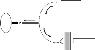
Nerve-Axon-Related Hyperemic Response. The nerveaxon related-hyperemic response evaluates the integrity of the neurovascular function. In healthy subjects, the ability to increase blood flow depends on the existence of normal neurogenic vascular response, which is conducted through the C nociceptive nerve fibers. Stimulation of these nerve fibers leads to antidromic stimulation of adjacent C fibers, which secrete substance P, calcitonin gene-related peptide (CGRP) and histamine, causing vasodilatation and increased blood flow to the injured tissues, thereby promoting wound healing (Lewis’ triple flare response) (Fig. 1). For this measurement, two single-point laser probes are applied (Fig. 2). One probe measures the blood flow to an area of skin, which is exposed directly to
Neurogenic vascular response

 Acetylcholine
Nerve cell
Vasodilatation
Substance P CGRP histamine
Figure 1. Stimulation of the C-nociceptive nerve fibers leads to antidromic stimulation of the adjacent C fibers, which secrete substance P, calcitonin gene related peptide (CGRP), and histamine that cause vasodilatation and increased blood flow.

380 CUTANEOUS BLOOD FLOW, DOPPLER MEASUREMENT OF
Figure 2. Measurements of direct and indirect effect of vasoactive substance using single-point laser probes: One probe is used in direct contact with the iontophoresis solution chamber (colored ring) and measures the direct response. The center probe measures the indirect response (nerve axon-related effect). A small quantity (<1 mL) of 1% acetylcholine chloride solution or 1% sodium nitroprusside solution is placed in the iontophoresis. A constant current of 200 mA is applied for 60 s achieving a dose of 6 mC cm 2 between the iontophoresis chamber and a second nonactive electrode placed 10–15 cm proximal to the chamber (black strap around the wrist). This current causes a movement of solution to be iontophorized toward the skin.
acetylcholine (Ach). The second probe, placed in close proximity (5 mm), measures the indirect effect of applied Ach. This indirect effect results from stimulation of C-nociceptive nerve fibers of the adjacent area and reflects the stability of the nerve-axon-related reactive hyperemia.
Our lab has examined the contribution of the nerveaxon reflex-related vasodilation to the total endotheliumdependent vasodilation at the forearm and the foot level in healthy adults and patients with diabetes mellitus (16). In healthy adults, the nerve-axon reflex-related response is approximately equal to one-third of the total response to Ach at both the forearm and the foot level. In diabetic patients with microvascular complications including diabetic neuropathy, Charcot arthropathy, and peripheral vascular disease, this contribution was significantly diminished. Another study demonstrated that the nerve-axon reflex-related vasodilatation is directly related to the function of the C-nociceptive fibers and is significantly associated with other nerve function measurements (17). As this method is an objective measurement, it is potentially useful as an alternative to currently employed techniques to evaluate small nerve fiber function.
Single-point measurements give a high temporal resolution (40 Hz data rates are typical) enabling rapid blood flow changes to be recorded. However, there are several limitations of the conventional laser Doppler probe. Because the probe is directly contacted to the skin, it can only measure the restricted area 1 mm2 at one time. Its pressure on the skin itself may also alter the skin blood flow (18). In addition, the probe is very sensitive to motion and vibration while it has poor reproducibility (3).
The Laser Scanning Method
The laser Doppler scanner–imager has been developed in response to the limitation of the laser Doppler probe. In the laser scanning method, a larger area of the skin can be studied while avoiding the contact between the scanner and the tissue being assessed. This technique is based on the same principle of measuring blood flow as the laser probe, but instead of the fiber optic probes, a system of mirrors and light-collecting lenses are used (14). This technique, the low intensity laser beam, is scanned across tissue surface in a raster fashion using a moving mirror. The scanner can scan the area from 5 5 cm to up to 50 50 cm. Light reflected back from the skin is then detected by a photodetector, which is connected to the computer enabling a mapping and a display of color-coded images of the blood flow. Regions of interest can then be defined and statistical data are calculated and recorded. This technique is also useful for the study of the skin microcirculation in response to various vasoactive substances.
To evaluate the endothelium-dependent and the endothelium-independent microvascular reactivity, the laser scanning method is used through the iontophoresis technique. The conditions associated with endothelial dysfunction are listed in Table 2.
The term iontophoresis denotes the introduction of soluble ions into the human skin by applying electric current. Using this technique, vasoactive substances can be applied to a localized area of the skin. The delivered dose depends on the current flowing and its duration. The test is noninvasive and avoids any systemic effects of the used drugs. By applying Ach chloride, the endotheliumdependent vasodilatation can be measured, while the use of sodium nitroprusside (SNP) measures the endotheliumindependent vasodilatation.
In this technique, a delivery vehicle device is attached firmly to the skin with double-sided adhesive tape. The device contains two chambers that accommodate two single-point laser probes. A small quantity of (< 1 mL) of 1% Ach solution or 1% of SNP solution is placed in the iontophoresis chamber and a constant current of 200 mA is applied for 60 s, achieving a dose of 6 mC cm 2 between the
Table 2. Conditions Associated with Impaired Endothelial Function
Atherosclerosis
Hypertension
Dyslipidemia; high LDL-C, low HDL-C, small dense LDL-C Diabetes mellitus and impaired glucose tolerance Metabolic syndrome
Obesity
Congestive heart failure Preeclampsia Vasculitis
Renal failure Menopause
Family history of coronary heart disease Family history of diabetes
Smoking
Inactivity
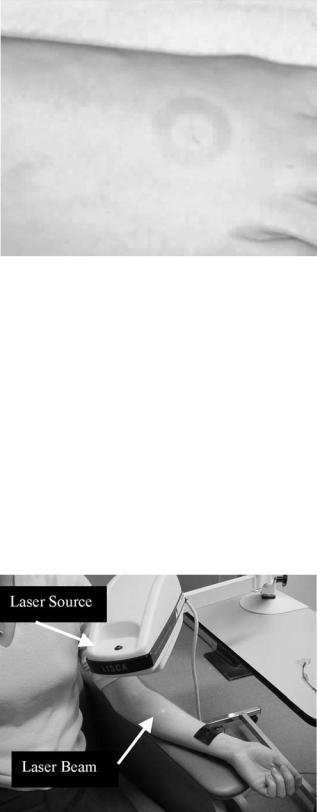
Figure 3. A normal response of blood flow in a skin to iontophoresis technique. Vasodilatation occurs in both the area that contact with the iontophoresis solution and area adjacent to, but not in direct contact with, the solution.
iontophoresis chamber and a second nonactive electrode placed 10–15 cm proximal to the chamber. This current causes a movement of solution to be iontophoresed toward the skin, resulting in vasodilatation (Fig. 3).
After the adhesive device has been removed, the localized area exposed to the vasoactive substances is scanned. The laser Doppler perfusion imager employs a 1 mW helium–neon laser beam of 633 nm wavelength, which sequentially scans an area of skin (Fig. 4). The maximum number of measured spots is 4096, and the apparatus produces a color-coded image of skin erythrocyte flux on a computer monitor. The scanner is set up to scan up to 32 32 measurement points over an area 4 4 cm.
Figure 4. Laser doppler flowmetry: A helium–neon laser beam is emitted from the laser source to sequentially scan the circular hyperemic area (seen surrounding the laser beam) produced by the iontophorized vasoactive substance to a small area on the volar surface of the forearm.
CUTANEOUS BLOOD FLOW, DOPPLER MEASUREMENT OF |
381 |
Generally, the laser Doppler scanner is not a reliable method for a quantification of the nerve-axon reflex related vasodilatation. However, Krishnan and Rayman recently published a technique assessing the axon-reflex related vasodilatation using the laser Doppler imager (LDI) called LDIflare. This method involved skin heating to 44 8C to evoke flare followed by scanning the site using a laser Doppler imager. The LDIflare was markedly reduced in healthy control subjects after topical administration of anesthesia, confirming its neurogenic nature. Similarly, LDIflare was decreased in patients with diabetic neuropathy when compared to diabetic patients with no neuropathy or healthy controls. The authors suggested that the LDIflare may be a simple objective method to detect early neuropathy and may be useful in assessing therapeutic interventions aimed at preventing or reversing C-fiber function.
In summary, the laser Doppler scanning offers a more global assessment of skin perfusion than the laser Doppler probe, while avoiding a direct contact to the skin surface. The scanning technique is best suited for studying the relative changes in flow induced by variety of physiological maneuvers or pharmaceutical intervention procedures.
VALIDATION
Single-Point Laser Probe
The technique has been validated against direct measurements of the capillary flow velocity (19). The day-to-day reproducibility of the technique was evaluated in healthy subjects who were repeatedly tested at their foot and arm for 10 consecutive working days in our lab. The coefficient of variation (CV) for the baseline blood flow measurement obtained with the laser probe evaluating the response to heat was 44.0%, while that for the maximal response to heat was 27.9%. The indirect response to Ach, measured by a single-point laser probe, had a CV of 60.6% for the baseline measurements and 35.2% for the maximal hyperemic response after the iontophoresis. Note that the variability of the technique is mostly a spatial one, that is, the variability is mainly related to the high heterogeneity of the skin microcirculation and not to the technique itself. The reproducibility of the single-point laser Doppler can be improved when one pays attention to place the laser probe approximately in the same skin area for repeated measurement.
The Laser Scanner Method
The laser scanner method has a significantly better reproducibility than a single-point laser probe. The CV at the foot and forearm level is ranged between 14 and 19%. For the laser Doppler perfusion imaging measurements before and after the iontophoresis of Ach, the reproducibility of the technique was evaluated in our lab in five healthy subjects (four males and one female, age 23–39 years) who were repeatedly tested at the forearm for 10 consecutive working days. The coefficient of variation of the baseline measurement before the iontophoresis of Ach was 14.1% and during maximal hyperemic response after the iontophoresis it was 13.7% (16,20).
382 CUTANEOUS BLOOD FLOW, DOPPLER MEASUREMENT OF
CLINICAL APPLICATIONS
Laser Doppler flowmetry have several clinical applications. It has been used to monitor to ischemic tissue (2), to follow the progress of atherosclerotic disease, to evaluate therapeutic effects of drugs or operations (21), and to study skin under both normal physiologic states and the pathogenetic mechanisms underlying pathologic skin disorders (22,23).
Atherosclerosis. Peripheral arterial disease (PAD) of the lower extremity is a common manifestation of the atherosclerotic process. As many as 10 million people in the United States have PAD with a prevalence > 10% in people aged > 60 years (24). Up to three-quarters of patients with PAD are asymptomatic (25,26). Those with symptoms usually present with intermittent claudication, resting ischemia or foot ulceration. Kvernebo et al. reported laser Doppler flowmetry values decreased progressively from healthy controls to patients with intermittent claudication to those with critical ischemia (21). The test was reproducible on a given population particularly with the local heating technique, but the reproducibility was poor for individual subjects. The possible explanations include changes in sympathetic vascular tone and different vascular architecture in the measuring volumes, which are only some few cubic millimeters (mm3). These patients with PAD also demonstrate a reduced skin hyperemic response to iontophoresis of ACh and to SNP (27), suggesting both endothelium and smooth muscle dysfunction. In addition, when they exercise, a reduction of the leg skin hyperemic response to ACh delivery is more pronounced at peak of claudication than prior exercise. The authors suggested acute endothelial dysfunction occurs with exercise-induced leg muscle ischemia and threshold of claudication should not be exceeded during rehabilitation programs for PAD patients.
Laser Doppler flowmetry may also be useful for followup after therapeutic intervention in patients with PAD. Ray et al. (28) studied 41 patients who underwent technically successful revascularization for severe leg ischemia. Laser Doppler flowmetry performed before the procedure was better than toe and ankle systolic pressures in identifying patients who continued to have ischemic symptoms or required amputation after surgery. Another study reported the prevalence of high frequency flux waves, which increased in peripheral ischemia decreased after successful percutaneous angioplasty (29). One randomized, doubleblind, placebo-controlled trial examining the effect of glutathione in patients with peripheral artery disease reported improvement in pain-free walking distance (PFWD) and microciculatory (laser Doppler flowmetry) test (30). Another study, the START-trial: STimulation of ARTeriogenesis using subcutaneous application of GMCSF as a new treatment for peripheral vascular disease (31), is underway. In this study, the primary endpoint will be the change walking distance from day 0 to day 14 as assessed by an exercise treadmill test, while the cutaneous microcirculation alterations assessed by laser Doppler flowmetry will be one of the secondary endpoints.
Diabetes Mellitus. Over the last two decades, it has become apparent that metabolic alterations in diabetes
cause both structural and functional changes in multiple areas within the arteriolar and capillary systems. The most characteristic structural changes of the capillary circulation in diabetic patients are a reduction in the capillary size and thickening of basement membranes, while the major functional change includes a marked limitation of microvascular vasodilation (32–34). Studies have shown that the structural changes are essentially responsible for the impairment of microvascular function. Microcirculatory test in patients with diabetes mellitus demonstrated a reduction in hyperemic response to heat stimulus and minor skin trauma, suggesting the role of endothelial dysfunction as the cause of the impaired vascular reactivity at microcirculatory level (35). Such dysfunction occurs early in the course of diabetes and may even predict diabetic microand macrovascular complications (36,37).
Several therapeutic interventions have been tested both in vitro and/or in vivo aiming to improve endothelial function in patients with diabetes mellitus. This includes insulin sensitizers (thiazolidenediones), angiotensin-con- verting enzyme (ACE) inhibitor, lipid lowering medications (statins), antioxidant, and exercise. To date, the studies have shown these interventions had no effects on the skin microcirculation in patients with diabetes, including both resting skin blood flow and blood flow after the iontophoresis of acetylcholine and sodium nitroprusside (38–41).
Skin Disease. Several skin diseases are associated with abnormal cutaneous blood flow, for example, psoriasis, skin inflammatory reaction, skin cancers, and scleroderma. Laser Doppler flowmetry has been used to determine differences in blood flow within psoriatic plaques, to identify the location of their leading edge, and to monitor the treatment (42,43). Laser Doppler perfusion imaging may also allow differentiation between different types of skin tumor. Stucker et al. found that malignant melanomas were significantly more perfused than basal cell carcinomas and tended also to be more so than melanocytic naevi (44,45).
Skin Burns. Laser Doppler flowmetry has been used to determine burn depth. Niazi et al. (46) demonstrated the Laser Doppler flowmetry flux values at 24 h after burn correlated with clinical assessment and histology reports, with low flux seen in deep dermal or full-thickness wound but high flux in superficial dermal wound (46). Park et al. (47) showed that laser Doppler flow measurements obtained within 72 h of burn injury correlated well with the depth of burn wound. The accuracy of laser Doppler in the assessment of burn depth is as high as 97%, compared with 60–80% for standard clinical methods (48). In addition, this technique has been used in concurrence with clinical judgment to objectively determine the need for excision of burns of indeterminate depth (49).
Skin Ulcer. Both ischemic and venous ulcers and their adjacent skin are associated with disorders in microcirculation. Together the deeper, subpapillary (thermoregulatory) network and the superficial (nutritive) network are affected. These network can be studied with laser Doppler flowmetry (subpapillary area) and capillary microscopy
(superficial area). Gschwandtner et al. (50–53) examined the microcirculatory characteristics of ulcers. They described the remarkable local differences in subpapillary and nutritive perfusion in ischemic and venous ulcers and their surrounding skin (51,52). Ulcer areas without granulation tissue (sign of ulcer without healing) demonstrated low laser Doppler area flux and the very low capillary density. The lack of capillary in this area suggested the breakdown of nutritive microcirculation as an underlying cause of ulcers. Ulcer with granulation tissue (sign of wound healing) had high laser Doppler area flux and an intermediate capillary density, implying both thermoregulatory, and nutritive networks are essential for wound healing of ulcers. The skin area adjacent ulcers where healing process nearly completed had an intermediate laser Doppler area flux and highest capillary density, indicating an adequate thermoregulatory and nutritive perfusion (53). Bornmyr et al. (54) reported the use of laser Doppler perfusion imaging and digital photograph for postoperative evaluation of vascularized grafts and monitoring of treatment of chronic skin ulcers.
CONCLUSION
Laser Doppler flowmetry has already been an important tool for studying blood flow in the skin. The technique provides continuous, noninvasive real-time assessment of skin perfusion. It has been successfully used in many studies of the cutaneous blood flow in patients with atherosclerosis, diabetes, skin diseases, or skin ulcers.
BIBLIOGRAPHY
1.Chittenden SJ, Shami SK. Microvascular investigations in diabetes mellitus. Postgrad Med J 1993;69:419–428.
2.Furnas H, Rosen JM. Monitoring in microvascular surgery. Ann Plast Surg 1991;26:265–272.
3.Schabauer AM, Rooke TW. Cutaneous laser Doppler flowmetry: applications and findings. Mayo Clin Proc 1994;69:564–574.
4.Choi CM, Bennett RG. Laser Dopplers to determine cutaneous blood flow. Dermatol Surg 2003;29:272–280.
5.Fagrell B. Microcirculatory methods for the clinical assessment of hypertension, hypotension, and ischemia. Ann Biomed Eng 1986;14:163–173.
6.Hellner D, Schmelzle R. Laser Doppler monitoring of free microvascular flaps in maxillofacial surgery. J Craniomaxillofac Surg 1993;21:25–29.
7.Hirigoyen MB, Urken ML, Weinberg H. Free flap monitoring: a review of current practice. Microsurgery 1995;16:723–726; discussion 727.
8.Stern MD, Lappe DL, Bowen PD, Chimosky JE, Holloway GA Jr, Keiser HR, Bowman RL. Continuous measurement of tissue blood flow by laser-Doppler spectroscopy. Am J Physiol 1977;232:H441–H448.
9.Holloway GA Jr, Watkins DW. Laser Doppler measurement of cutaneous blood flow. J Invest Dermatol 1977;69:306–309.
10.Fischer JC, Parker PM, Shaw WW. Laser Doppler flowmeter measurements of skin perfusion changes associated with arterial and venous compromise in the cutaneous island flap. Microsurgery 1985;6:238–243.
11.Rendell M, Bergman T, O’Donnell G, Drobny E, Borgos J, Bonner RF. Microvascular blood flow, volume, and velocity
CUTANEOUS BLOOD FLOW, DOPPLER MEASUREMENT OF |
383 |
measured by laser Doppler techniques in IDDM. Diabetes 1989;38:819–824.
12.Zinser G. Scanning laser Doppler flowmetry. In: Pillunat L, Harris A, Anderson D, Greve E, editors. Current concepts on ocularblood flow in glaucoma. The Hague: Kugler Publications; 1999: 197–204.
13.Rossi M, Carpi A. Skin microcirculation in peripheral arterial obliterative disease. Biomed Pharmacother 2004;58:427–431.
14.Essex TJ, Byrne PO. A laser Doppler scanner for imaging blood flow in skin. J Biomed Eng 1991;13:189–194.
15.Bonner RF, Nossal R. Principles of laser Doppler flowmetry. In: Shepherd AP, Oberg PA, editors. Laser Doppler Blood Flowmetry. Kluwer Academic Publishers; 1990.
16.Hamdy O, Abou-Elenin K, LoGerfo FW, Horton ES, Veves A. Contribution of nerve-axon reflex-related vasodilation to the total skin vasodilation in diabetic patients with and without neuropathy. Diabetes Care 2001;24:344–349.
17.Caselli A, Rich J, Hanane T, Uccioli L, Veves A. Role of C- nociceptive fibers in the nerve axon reflex-related vasodilation in diabetes. Neurology 2003;60:297–300.
18.Obeid AN, Barnett NJ, Dougherty G, Ward G. A critical review of laser Doppler flowmetry. J Med Eng Technol 1990;14:178–181.
19.Tooke JE, Ostergren J, Fagrell B. Synchronous assessment of human skin microcirculation by laser Doppler flowmetry and dynamic capillaroscopy. Int J Microcirc Clin Exp 1983;2:277–284.
20.Veves A, Akbari CM, Primavera J, Donaghue VM, Zacharoulis D, Chrzan JS, DeGirolami U, LoGerfo FW, Freeman R. Endothelial dysfunction and the expression of endothelial nitric oxide synthetase in diabetic neuropathy, vascular disease, and foot ulceration. Diabetes 1998;47:457–463.
21.Kvernebo K, Slagsvold CE, Stranden E, Kroese A, Larsen S. Laser Doppler flowmetry in evaluation of lower limb resting skin circulation. A study in healthy controls and atherosclerotic patients. Scand J Clin Lab Invest 1988;48:621–626.
22.Braverman IM. The cutaneous microcirculation. J Investig Dermatol Symp Proc 2000;5:3–9.
23.Braverman IM. The cutaneous microcirculation: ultrastructure and microanatomical organization. Microcirculation 1997;4: 329–340.
24.Criqui MH. Peripheral arterial disease—epidemiological aspects. Vasc Med 2001;6:3–7.
25.Hooi JD, Kester AD, Stoffers HE, Overdijk MM, van Ree JW, Knottnerus JA. Incidence of and risk factors for asymptomatic peripheral arterial occlusive disease: a longitudinal study. Am J Epidemiol 2001;153:666–672.
26.Stoffers HE, Rinkens PE, Kester AD, Kaiser V, Knottnerus JA. The prevalence of asymptomatic and unrecognized peripheral arterial occlusive disease. Int J Epidemiol 1996;25: 282–290.
27.Rossi M, Cupisti A, Perrone L, Mariani S, Santoro G. Acute effect of exercise-induced leg ischemia on cutaneous vasoreactivity in patients with stage II peripheral artery disease. Microvasc Res 2002;64:14–20.
28.Ray SA, Buckenham TM, Belli AM, Taylor RS, Dormandy JA. The predictive value of laser Doppler fluxmetry and transcutaneous oximetry for clinical outcome in patients undergoing revascularisation for severe leg ischaemia. Eur J Vasc Endovasc Surg 1997;13:54–59.
29.Bollinger A, Hoffmann U, Franzeck UK. Evaluation of flux motion in man by the laser Doppler technique. Blood Vessels 1991;28(1 Suppl): 21–26.
30.Arosio E, De Marchi S, Zannoni M, Prior M, Lechi A. Effect of glutathione infusion on leg arterial circulation, cutaneous microcirculation, and pain-free walking distance in patients with peripheral obstructive arterial disease: a randomized, double-blind, placebo-controlled trial. Mayo Clin Proc 2002; 77:754–759.