


Nervous System
I. OVERVIEW-NERVOUS SYSTEM
The nervous system can be organized by anatomical or functional divisions.
A. Nervous system is divided anatomically into the central nervous system (CNS), which includes the brain and spinal cord encased in the skull and vertebral column, and the peripheral nervous system (PNS), which includes the nerves outside the CNS and their associated ganglia as well as connections to the CNS, including the receptors and motor components of the peripheral nerves of the body.
B.Nervous system is divided functionally into a sensory component, which transmits electrical impulses (signals) to the CNS, and a motor component, which transmits impulses from the CNS to various structures of the body. The motor component is further divided into the somatic and autonomic systems. The somatic nervous system (SNS), located within both the CNS and PNS, controls sensory and motor functions of all conscious voluntary actions. However, it does not control functions related to smooth or cardiac muscles, reflex arcs, or glands.
The autonomic nervous system (ANS) functions to regulate the internal organs. It is further subdivided into the sympathetic, parasympathetic, and enteric systems. The sympathetic nervous system readies the body for action against hostile situations, resulting in "flight or fight or freeze;' whereas the parasympathetic nervous system is "calming" and supplies secretomotor innervation to most exocrine glands. The enteric nervous system, a very large component of the autonomic system, serves the digestive system. Although it operates independently, both the sympathetic and the parasympathetic components are utilized in modulating the functions of the enteric system.
C.Nervous tissue contains two types of cells: nerve cells (neurons), which conduct electrical impulses, and supporting cells, which support, nurture, and protect the neurons within the CNS
(glial [neuroglial] cells), as well as cells located in the PNS (satellite cells, capsule cells, and Schwann cells).
A. The neuroepithelium thickens and differentiates to form the neural plate.
B.The neural plate invaginates and thickens to form the neural groove.
C.The neural tube, a cylindrical structure that results from fusion of the edges of the neural groove, enlarges at its cranial end to form the brain. The remaining portion gives rise to the spinal cord.
1 43

1 44 |
BRS Cell Biology and Histology |
FIGURE 9.1. Early histogenesis of the nervous system. Notice how the neural crest forms and then fragments to migrate to its destinations.
D. Neural crest cells stream off the edges of the neural groove before formation of the neural tube. These cells migrate throughout the body and give rise to the following structures:
1 . Sensory neurons of cranial and spinal sensory ganglia
2.Most sensory neurons and Schwann cells of the PNS
3.Enteric and autonomic ganglia and their postganglionic neurons and associated glia
4.Most ofthe mesenchymal (ectomesenchymal) cells ofthe head and anterior portion ofthe neck
5.Melanocytes of the skin and oral mucosa
6.Odontoblasts (cells responsible for the production of dentin)
7.Cells of the arachnoid and pia mater (rostral to the mesencephalon)
8.Chromaffin cells of the adrenal medulla
CLINICAL
1 . Abnormal histogenesis of the CNS results in congenital
CONSIDERATIONS malformations.
2.Congenital malformations
a.Spina bifida is a defective closure of the spinal column. In severe cases, the spinal cord and meninges may protrude through the unfused areas. Very severe cases may be associated with defective development of the viscera of the thorax and abdomen.
b.Anencephaly is failure of the developmental anterior neuropore to close. This produces a poorly formed brain without a cranial vault. It is usually not compatible with life.
c.Hirschsprung disease (congenital megacolon) is the result of abnormal organogenesis in
which neural crest cells fail to migrate into the wall of the gut. The disease is characterized by the absence of Auerbach plexus, a part of the parasympathetic system innervating the distal segment of the colon. This loss of motor function leads to dilation of the colon.
d.Neuroglial tumors constitute 50% of intracranial tumors. CNS tumors are rarely associated with neurons; they are mostly derived from neuroglial cells (e.g., astrocytes, oligodendro cytes, and ependymocytes). These tumors range in severity from slowly growing benign oligodendrogliomas to rapidly growing fatal malignant astrocytomas.
e.Brains of patients with AIDS and HIV-1 possess large populations of microglial cells. Although these microglia do not attack neurons, they produce cytokines that are toxic to neurons.
i!1llll'1ttDllNervous System 145
A.Neurons consist of a cell body (soma, perikaryon), and its processes, which usually include multiple dendrites and a single axon. Neurons comprise the smallest and largest cells of the body, ranging
from 5 to 150 J.!m in diameter. |
|
|
|
|
|
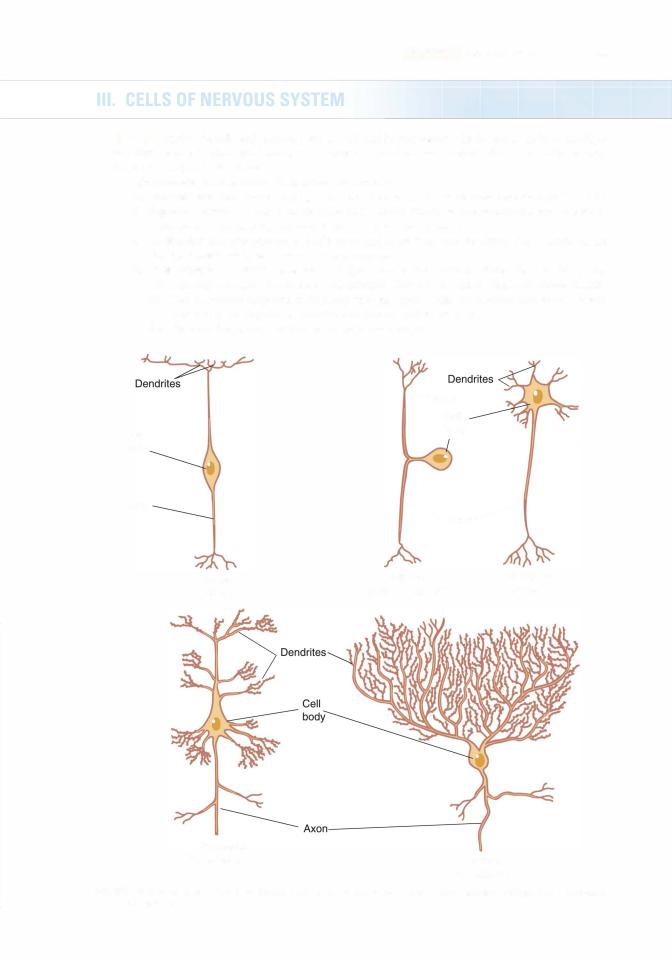
1 . Morphologic classification of neurons Figure 9.2) |
|
see Section III A l d |
|
. |
|
a. Unipolar neurons possess a single process( |
but are rare in vertebrates |
( |
) |
||
|
|
|
|
||
b.Bipolar neurons possess( a single axon and a single dendrite.)These neurons are present in some sense organs e.g., the vestibular-cochlear mechanism .
c.Multipolar neurons possess a single axon and more than one dendrite. These neurons are the most common type of neuron in vertebrates.
d.Pseudounipolar neurons possess a single process that extends from the cell body and subsequently branches into an axon and dendrite. They are present in spinal and cranial ganglia. (1 ) These neurons originate embryologically as bipolar cells whose axon and dendrite later
fuse into a single process functionally categorized as an axon.
(2)They are frequently referred to as unipolar neurons.
-Axon
|
Cell |
|
Cell |
body |
|
|
|
|
body |
|
|
Axon |
Axon ---------- |
|
|
||
Bipolar |
Unipolar |
Multipolar |
(retina) |
(pseudounipolar) |
(motor) |
Pyramidal
(hippocampus) Purkinje
(cerebellum)
FIGURE 9.2. Various types of neurons. (Reprinted with permission from Gartner LP, Hiatt JL. Color Textbook ofHistology. 2nd ed. Philadelphia, PA: Saunders; 2001 : 1 87.)
1 46 |
BRS Cell Biology and Histology |
2.Functional classification of neurons
a.Sensory neurons receive stimuli from the internal and external environments and conduct impulses to the CNS for processing and analysis.
b.lnterneurons (intercalated neurons) connect other neurons in a chain or sequence. They commonly connect sensory and motor neurons; they also regulate signals transmitted to neurons.
c.Motor neurons conduct impulses from the CNS to other neurons, muscles, and glands.
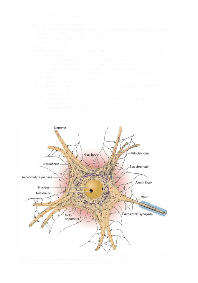
3.Neuron structure (Figure 9.3)
a. Neuronal cell body (soma, perikaryon) is the region of a neuron containing the nucleus, various cytoplasmic organelles and inclusions, and cytoskeletal elements.
(1 ) The nucleus is large, spherical, and pale staining and is centrally located in the soma of most neurons. It contains abundant euchromatin and a large nucleolus (owl-eye nucleus).
(2)Cytoplasmic organelles and inclusions
(a)Nissl bodies are composed of polysomes and rough endoplasmic reticulum (RER). They appear as clumps under light microscopy and are most abundant in large motor neurons.
(b)The Golgi complex is near the nucleus, and mitochondria are scattered throughout the cytoplasm.
(c)Melanin-containing granules are present in some neurons in the CNS and in the dorsal root and sympathetic ganglia.
Axodendritic synapses
FIGURE 9.3. A typical neuron with its constituents and syna pses&. (Reprinted with permission from Kiernan JA. Barr's TheHuman Nervous
8th ed. Baltimore, MD: Lippincott Williams Wilkins; 2005:19.)

i!1llll'1ttDllNervous System 147
FIGURE 9.4. Light micrograph ofthe spinal cord in cross section ( X 540l. Observe the multipolar neurons in the ventral horn of the spinal cord. D, dendrites; AH, axon hillock; A, axon.
(d)Lipofuscin-containing granules are present in some neurons and increase in number with age.
(e)Lipid droplets are occasionally present.
(3)Cytoskeletal components (Figures 9.3 and 9.4)
(a)Neurofilaments (10 nm in diameter) are abundant and run throughout the soma cytoplasm. They are intermediate filaments composed of three intertwining polypeptide chains.
(b)Microtubules (24 nm in diameter) are also present in the soma cytoplasm.
(c)Microfilaments (actin filaments 6 nm in diameter) are associated with the plasma membrane.
b.Dendrites receive stimuli (signals) from sensory cells, axons, or otherneurons and convert these signals into small electrical impulses (action potentials) that are transmitted toward the soma.
(1 ) Dendrites possess arborized terminals (except in bipolar neurons), which permit a
neuron to receive stimuli simultaneously from many other neurons.
(2)The dendrite cytoplasm is similar to that ofthe soma except that itlacks a Golgi complex.
(3)Organelles are reduced in number or absent near the terminals exceptformitochondria, which are abundant.
(4)Spines on the surface of dendrites increase the area available for synapse formation with other neurons. These diminish with age and poor nutrition and exhibit altered configurations in individuals with trisomy 21 or trisomy 13.
c.Axons conduct impulses away from the soma to the axon terminals without any dimin ution in their strength.
(1 ) The diameter and length of axons in different types of neurons vary. Some axons are as long as 100 em.
(2)Axons originate from the axon hillock, a specialized region of the soma that lacks RER, ribosomes, Golgi cisternae, and Nissl bodies but contains many microtubules and neurofilaments; abundance of the latter may regulate neuron diameter. Further, it permits passage of mitochondria and vesicles into the axon.

1 48 |
BRS Cell Biology and Histology |
(3)Axons may have collaterals, branching at right angles from the main trunk.
(4)Axon cytoplasm (axoplasm) lacks a Golgi complex but contains smooth endoplasmic reticulum (SER), RER, and elongated mitochondria.
(5)A plasma membrane surrounding the axon is called the axolemma.
(6)Axons terminate in many small branches (axon terminals) from which impulses are passed to another neuron or other types of cells.
Alzheimer disease is the most common cause of dementia. It is the sixth leading cause of death in the United States and is considered to be a
disease of the aged. Currently, about 5.2 million persons in the United States have been diagnosed with Alzheimer disease, of whom five million are aged 65 and older and 200,000 are younger than 65 years of age. It is estimated that in 201 3 Alzheimer disease is expected to cost the United States $203 billion, which is expected to rise to $1 .2 trillion by 2050. Alzheimer disease starts with memory loss and confusion, loss of finding words, loss of abstract thinking, disorientation, and depression, followed by irritability and mood swings with aggression. Finally, there is withdrawal and decline of senses and the loss of bodily functions. It appears that a combination of genetics, lifestyle, and the environment plays a role in triggering the onset. The disease is characterized by the loss of neurons and synapses mainly within the cerebral cortex followed by atrophy of the individual cerebral lobes. Patients with Alzheimer disease develop -amyloid plaques and neurofibrillary tangles that render the neurons nonfunctional. Recent studies have shown that -amyloid protein can act as a prion and be transferred from infected neurons to noninfected neurons by traveling down the axon of the
infected cell and entering the uninfected cell via synapses. Still other current investigations suggest that Alzheimer disease is a neuroendocrine disorder, specifically type Ill diabetes mellitus, result ing from specific deficiencies in insulin, insulin-like growth factor- 1 , and insulin-like growth factor-2 production, as well as in the scarcity of receptors for these three molecules confined to certain regions of the brain.
B. The neuroglial cells, astrocytes and oligodendroglia (as well as microglia and ependymal cells) are located only in the CNS. Schwann cells and capsule cells are their PNS equivalent cells.
1 . General characteristics. Neuroglial cells comprise several cell types and outnumber neurons by approximately a factor of 10. These cells are embedded in a web of tissue composed of modified ectodermal elements; the entire supporting structure is termed the neuroglia. They function to support and protect neurons, but they do not conduct impulses or form synapses with other cells; however, they do appear to provide some regulation of neurons in the process of neural transmissions. Neuroglia do that by actively monitoring synapses, removing neurotransmitter molecules from synapses, and delivering molecules known as gliotransmitter substances, such as glutamate and ATP into the synaptic vicinity. These gliotransmitters appear to suppress synaptic transmission in cultured hippocampal neurons. Additionally, neuroglial cells possess the capacity to undergo cell division. Neuroglia are revealed in histologic sections ofthe CNS only with special gold and silver stains.
2.Types of neuroglial cells (Figure 9.5). Note that Schwann cells are also discussed in this section, but they are present only in the PNS.
a. Astrocytes
(1)Astrocytes are the largest of the neuroglial cells that reside in the CNS. They have many processes, some of which possess expanded pedicles (vascular feet) that surround blood vessels, whereas others exhibit processes that contact the pia mater.
(2)Function
(a)Astrocytes scavenge ions and debris from neuron metabolism and supply energy for metabolism.
(b)Along with other components of the neuroglia, astroglia form a protective sealed barrierbetween the pia mater and the nervous tissue ofthe brain and the spinal cord.
(c)They provide structural supportfor nervous tissue.
(d)They proliferate to form scar tissue (glial scar) after injury to the CNS.

i!1llll'1ttDllNervous System 149
Protoplasmic |
|
astrocyte |
astrocyte |
|
Microglia |
Oligodendrocyte |
FIGURE 9.5. Various types of neuroglial cells. !Adapted with permission from Gartner LP. Hiatt JL. Color Textbook of Histology. 2nd ed. Philadelphia. PA: Saunders; 2001 : 1 92.1
(3)Types of astrocytes
(a)Protoplasmic astrocytes reside mostly in gray matter and have branched processes that envelop blood vessels, neurons, and synaptic areas. They contain some intermediate filaments composed of glial fibrillar acidic protein (GFAP). These astrocytes help establish the blood-brain barrier and may contribute to its maintenance.
(b)Fibrous astrocytes reside mostly in white matter and have long, slender processes with few branches. They contain many intermediate filaments composed of GFAP.
b.Oligodendrocytes
(1 ) Oligodendrocytes are neuroglial cells that live symbiotically with neurons (i.e., each
cell type is affected by the metabolic activities of the other). These cells are necessary for the survival of neurons in the CNS.
(2)They are located in both gray matter and white matter.
(3)They possess a small, round, condensed nucleus and only a few short processes.
(4)Their electron-dense cytoplasm contains ribosomes, numerous microtubules, many mitochondria, RER, and a large Golgi complex.
(5)Oligodendrocytes produce myelin, a lipoprotein material organized into a sheath that insulates and protects axons in the CNS. Each oligodendrocyte forms several processes, and each process produces myelin for a single internode for a single axon. In this fashion, an oligodendroglion can myelinate an internode for several axons.
c.Schwann cells
(1 ) Schwann cells, located in the PNS, are flat cells with only a few mitochondria and a small Golgi region.
(2)Although Schwann cells are derived from neural crest cells, they are still considered neuroglial cells.
(3)These cells perform the same function in the PNS as oligodendrocytes in the CNS: they protect and insulate neurons. Schwann cells form either unmyelinated or myelinated coverings over neurons. However, a single Schwann cell can only insulate a single axon, whereas a single oligodendrocyte may insulate several axons. A myelin sheath consists of plasmalemmae of copious numbers of Schwann cells wrapped around a single axon (see Section V).
1 50 |
BRS Cell Biology and Histology |
A
8
FIGUREB-D. 9.6. Myelin sheath formation. EA. Myelin sheathF. and the Schwann cell as they are seen (ideally) by light microscopy. Successive stages in the development of the myelin sheath from plasma membrane of the Schwann cell& . E. Ultrastruc ture of a node of Ranvier, sectioned longitudinally. Relation ofthe Schwann cell to several unmyelinated axons. IReprinted with permission from Kiernan JA. Barr's The Human Nervous System: An Anatomical Viewpoint. 8th ed. Baltimore, MD: Lippincott Williams Wilkins; 2005:22.)
(4)Unmyelinated peripheral nerve fibers are surrounded by a large number of Schwann cells where each segment ofthe axon is enveloped by a single Schwann cell. Several axon segments may be embedded in each Schwann cell (see Figure 9.6F). Since impulses in myelinated axons jump from one node of Ranvier to another and in unmyelinated axons they move sequentially from ion channel to ion channel, propagation of the impulse is much faster in myelinated axons. It is interesting to note that, frequently, in the CNS unmyelinated axons, unlike in the PNS, are not enveloped by neuroglial cells.
d.Microglia are small, phagocytic neuroglial cells that are derived from the mononuclear phagocytic cell population in the bone marrow. Theyhave a condensed, elongated nucleus and many short, branching processes. Normally they are inactive but during injury or pathogen invasion they release interferon-y which activates neighboring microglia. Activated microglial cells remove residues of cellular injury and secrete cytokines that attract T cells to the site of injury. Microglia also possess the ability to become antigen presenting cells and present the antigens to the newly arrived T cells.
e.Ependymal cells, derived from the neuroepithelium, are the epithelial cells that line the neural tube and ventricles of the brain. In certain regions of the brain, they possess cilia, which aid in moving the cerebrospinal fluid (CSF). Modified ependymal cells contribute to the formation of the choroids plexus.


1 52 |
BRS Cell Biology and Histology |
(2)Electrical synapses are much less numerous than chemical synapses.
(3)Signal transmission across these synapses is nearly instantaneous.
B.Synaptic morphology
1.Axon terminals mayvary morphologically, depending on the site of synaptic contact.
a.Boutons terminaux are bulbous expansions that occur singly at the end of axon terminals.
b.Boutons en passage are swellings along the axon terminal; synapses may occur at each swelling.
2.The presynaptic membrane is the thickened axolemma of the neuron that is transmitting the impulse. It contains voltage-gated Ca2+ channels, which regulate the entry of calcium ions into the axon terminal. Synaptic vesicles fuse with and become incorporated into the presynaptic membrane before releasing their neurotransmitter substances.
3.The postsynaptic membrane is the thickened plasma membrane of the neuron or other target cell that is receiving the impulse.
4.The synaptic cleft is the narrow space (20-30 nm wide) between the presynaptic and postsynaptic membranes. Neurotransmitters diffuse across the synaptic cleft.
5.Synaptic vesicles are small, membrane-bound structures (40-60 nm in diameter) in the axoplasm ofthe transmitting neuron. They discharge neurotransmitters into the synaptic cleft by exocytosis.
C.Neurotransmitters (Table 9.1) are produced, stored, and released by presynaptic neurons. They diffuse across the synaptic cleft and bind to receptors in the postsynaptic membrane, leading to generation of an action potential.
V. NERVE FIBERS
Nerve fibers are individual axons enveloped by a myelin sheath, Schwann cells in the PNS, or oligodendrocytes in the CNS.
A. Myelin sheath (Figure 9.6)
1 . The myelin sheath is produced by oligodendrocytes in the CNS and by Schwann cells in
the PNS. |
9.1 |
|
|
t a b I e |
Common Neurotransmitters |
|
|
Neurotransmitter |
|
Location |
Function |
Acetylcholine |
|
Myoneural junctions; all parasympathetic |
Activates skeletal muscle, autonomic nerves, |
|
|
synapses; preganglionic sympathetic syna pses |
brain functions |
Norepinephrine |
|
Postganglionic sympathetic synapses |
Increases cardiac output |
Glutamate |
|
CNS; presyna ptic sensory and cortex |
Most common excitatory neurotransmitter |
|
|
|
of CNS |
GABA |
|
CNS |
Most common inhibitory neurotransmitter |
|
|
|
of CNS |
Dopamine |
|
CNS |
Inhibitory and excitatory, depending on |
|
|
|
receptor |
Glycine |
|
Brainstem and spinal cord |
Inhibitory |
Serotonin |
|
CNS |
Pain inhibitor; mood control; sleep |
Aspartate |
|
CNS |
Excitatory |
Enkephalins |
|
CNS |
Analgesic; pain suppression |
Endorphins |
|
CNS |
Analgesic; pain suppression |
|
|
|
|
CNS . central nervous system; GABA. y-aminobutyric acid.

i!1llll'1ttDllNervous System 1 53
2.It consists of several spiral layers of the plasma membrane of an oligodendrocyte or Schwann cell wrapping around the axon.
3.It is not continuous along the length of the axon but is interrupted by gaps called nodes of Ranvier.
4.Its thickness is constant along the length of an axon; however, thickness usually increases as the axonal diameter increases.
5.The myelin sheath can be extracted by standard histological techniques. Methods using osmium tetroxide preserve the myelin sheath and stain it black.
6.Under electron microscopy, the myelin sheath displays the following features:
a.Major dense lines represent fusions between the cytoplasmic surfaces of the plasma membranes of Schwann cells (or oligodendrocytes).
b.lntraperiod lines represent close contact, but not fusion, of the extracellular surfaces of adjacent Schwann cell (or oligodendrocyte) plasma membranes.
c.Clefts (incisures) of Schmidt-Lanterman (observed in both electron and light microscopy) are cone-shaped oblique discontinuities of the myelin sheath due to the presence of Schwann cell (or oligodendrocyte) cytoplasm within the myelin.
B.Nodes of Ranvier are regions along the axon that lack myelin and represent discontinuities between adjacent Schwann cells or adjacent oligodendrocytes.
1 . In the PNS, the axon at the nodes of Ranvier is covered by interdigitated cytoplasmic processes ofadjacent Schwann cells that protect the myelin-free surface ofthe axon. In the CNS, however, the axon is not covered by cytoplasmic processes of oligodendrocytes. Instead, the myelin-free surface of the axon at the node ofRanvier is covered by a foot plate of an astrocyte.
2.The axolemma at the nodes contains many Na+ pumps, and in electron micrographs, it displays a characteristic electron density.
C. Internodes are the segments of a nerve fiber between adjacent nodes of Ranvier. They vary in length from 0.08 to 1 mm, depending on the size of the Schwann cells or oligodendrocytes associated with the fiber.
Multiple Sclerosis
Multiple sclerosis (MS) is an immune-mediated disease characterized by chronic and progressive dysfunction of the nervous system due to demyelination of the CNS, espe cially in the brain, spinal cord, and optic nerves. MS afflicts about 1 in 700 in this country, most com monly in the 20to 40-year age group, affecting 2 females to 1 male. There are random episodes of inflammation, edema, and demyelination of axons followed by periods of remission that may last for months to years. Each episode may reduce the vitality of the patient and be sufficient to cause death within months. It is believed that, in the CNS, T lymphocytes mount an attack on myelin sheaths, de myelinate axons, and interfere with normal propagation of action potentials. However, recent studies have suggested that the true causative factor of MS is oligodendrogliopathy and the T-cell response is merely a secondary reaction. Present treatments include immunosuppression with corticosteroids.
VI. NERVES
Nerves are cordlike bundles of nerve fibers surrounded by connective tissue sheaths (Figure 9.7). They are visible to the unaided eye and usually appear whitish because ofthe presence of myelin.
A. Connective tissue investments
1 . Epineurium is the layer of dense fibrous connective tissue (fascia) that forms the external coat of nerve bundles and is often embedded in adipose tissue.

1 54 |
BRS Cell Biology and Histology |
Epineurium
Endoneurium
FIGURE 9.7. A peripheral nerve in cross section showing the various& connective tissue sheaths. Each bundle of nerve fibers, or fascicle (one is extended in drawing), is covered by perineurium. (Reprinted with permission from Kelly DE. Wood RL. Enders AC. Bailey's Textbook ofMicroscopic Anatomy. 1 8th ed. Baltimore. MD: Williams Wilkins; 1 984:353.)
2.Perineurium surrounds each bundle of nerve fibers (fascicle). Its inner surface consists of layers of flattened cells joined by tight junctions (zonulae occludentes) that prohibit passage of most macromolecules, thus assisting in the formation of the blood-nerve barrier (Figure 9.8).
3.Endoneurium is a thin layer of reticular fibers, produced mainly by Schwann cells, that surrounds individual nerve fibers. This layer, difficult to observe without specialized stains, contains occasional mast cells and macrophages.
Meningitis
Meningitis results from an inflammation of the meninges caused by viral or bacterial infection of the CSF. Viral meningitis is not severe, but bacterial meningitis is contagious and dangerous, leading to hearing loss, learning disability, brain damage, and death, sometimes within 24 hours if untreated. In the United States, children 4 years of age or younger have been vaccinated for the most prevalent form of the bacterium. Recently, because of so many outbreaks of meningitis on college campuses, several schools have chosen to vaccinate all students and those in close contact with them. Major symptoms include fever, headache, stiff neck, and alteration of
consciousness with rapid onset and progression. Spinal tap and culture of CSF to determine the bac terial species is the only diagnosis. This is followed by treatment with a specific antibiotic. Bacterial meningitis can be spread by respiratory and throat secretions (i.e., coughing, sneezing, kissing).
B.Functional classification of nerves
1.Sensory nerves contain afferent fibers and carry sensory signals only from the internal and external environments to the CNS.
2.Motor nerves contain efferent fibers and carry signals only from the CNS to effector organs.
3.Mixed nerves are the most common type ofnerve, containing both afferent and efferent fibers and thus carry both sensory and motor signals.

i!1llll'1ttDllNervous System 1 55
FIGURE 9.8. Light micrograph of peripheral nerve in cross section I x 1 32). Note that an axon (tip of arrow) is located in the center of the myelin sheath IM). P, perineurium.
VII. GANGLIA
Ganglia are encapsulated aggregations of neuronal cell bodies (soma) outside the CNS.
A.Autonomic ganglia are motor ganglia in which axons of preganglionic neurons synapse on postganglionic neurons (see Section IX B 1).
B.Craniospinal ganglia are sensory ganglia associated with most cranial nerves and the dorsal roots of spinal nerves (dorsal root ganglia). Unlike autonomic ganglia, craniospinalganglia do not possess synapses. These ganglia contain the cell bodies of sensory neurons, which are pseudounipolar (unipolar) and transmit sensory signals fromreceptors to the CNS.
VIII. HISTOPHYSIOLOGY OF NERVOUS SYSTEM
A.Resting membrane potential
1. The resting membrane potential exists across the plasma membrane of all cells. The resting potential of most neuron plasmalemmae is negative -70 mV inside the cell compared to outside the cell.

1 56 |
BRS Cell Biology and Histology |
2.It is established and maintained mostly by K+ leak channels and to a lesser extent by the Na+-K+ pump, which actively transports three Na+ ions out of the cell in exchange for two K+ ions. The resting potential exists when there is no net movement of K+ (i.e., when outward diffusion ofK+ is just balanced by the external positive charge acting against further diffusion).
B.An action potential is the electrical activity that occurs in a neuron as an impulse is propagated along the axon and is observed as a movement of negative charges along the outside of an axon. It is an all-or-nothing event with a constant amplitude and duration.
1.Generation of the action potential
a.An excitatory stimulus on a postsynaptic neuron partially depolarizes a portion of the plasma membrane (the potential difference is less negative).
b.Once the membrane potential reaches a critical threshold, voltage-gated Na+ channels in the membrane open, permitting Na+ to enter the cell (Figure 9l.9).
c.The influx of Na+ leads to reversal of the resting potential in the immediate area (i.e., the external side becomes negative).
d.The Na+ channels close spontaneously and are inactivated for to 2 ms (refractory period).
e.Opening of voltage-gated K+ channels is also triggered by depolarization. Because these channels remain open longerthanthe Na+ channels, exit ofK+ during the refractory period repolarizes the membrane to its resting potential.
f.The ion channels then return to their normal states. The cell is now ready to respond to another stimulus.
2.Propagation of the action potential
a.Propagation results from longitudinal diffusion of Na+ (which enters the cell at the initial site of excitation) toward the axon terminals (orthodromic spread). The longitudinal diffusion of Na+ depolarizes the adjacent region of membrane, giving rise to a new action potential at this site.
b.Propagation does not result from diffusion of Na+ toward the soma (antidromic spread), because the Na+ channels are inactivated in this region.
c.Action potentials are propagated most rapidly in myelinated fibers, which exhibit saltatory conduction. In this type of conduction, the action potential jumps from one node ofRanvier to the next.
C.Axonal transport of proteins, organelles, and vesicles occurs at high, intermediate, and low velocities, depending on the nature of the transported materials.
1 . Anterograde transport carries material away from the soma.
2.Retrograde transport carries material toward the soma for reuse, recycling, or degradation. However, some viruses (e.g., herpes simplex and rabies) spread in this fashion. Also, some toxins (e.g., tetanus) move from the periphery to the CNS in this manner.
|
Extracellular space |
|
++++Charge++++ |
- - - -Charge- - - - |
++++Charge++++ |
- - - -Charge- - - |
++++Charge++++ |
- - - -Charge- - - - |
Closed |
Open |
Inactivated |
FIGURE 9.9. Model of the voltage-gated Na+ channel showing the transition between its closed, open, and inactivated states. In the resting state, the channel-blocking segment and gating keep the channel closed to entry of extracellular Na+. Depolarization of the membrane causes a conformational change that opens the channel to influx of Na+ The channel closes spontaneously and becomes inactive within a millisecond after opening.

i!1llll'1ttDllNervous System 1 57
D.Trophic function of nervous tissue
1.Denervation of a muscle or gland leads to its atrophy.
2.Reinnervation of a muscle or gland restores its structure and function.
IX. SOMATIC NERVOUS SYSTEM AND AUTONOMIC NERVOUS SYSTEM
Somatic and autonomic are functional concepts relating to all the neural elements involved in the transmission of impulses from the CNS to the somatic and visceral components of the body, respec tively. Neural crest cells give rise to neurons of both the parasympathetic and sympathetic divisions of the ANS whose preganglionic cell bodies are located in the CNS and postganglionic cell bodies are located in autonomic ganglia located outside the CNS.
A.The somatic nervous system (SNS) contains sensory fibers that bring information to the CNS and the motor fibers that originate in the CNS that innervate voluntary skeletal muscles.
B.The autonomic nervous system (ANS) is generally considered to be purely motor in function as it contains motor fibers that control and regulate smooth muscle, cardiac muscle, and some glands. It establishes and maintains homeostasis of the body's visceral functions. Anatomically and functionally, the ANS is divided into three parts: the sympathetic, parasympathetic, and enteric nervous systems. The sympathetic and parasympathetic nervous systems generally function antagonistically in a given organ (i.e., when the sympathetic system stimulates an organ, the parasympathetic inhibits it, and vice versa). The enteric nervous system is confined to the digestive system, where it controls digestive processes; however, it is modulated by the sympathetic and parasympathetic nervous systems.
1.Autonomic nerve chains
a.Cell bodies of preganglionic neurons are located in the CNS and extend their preganglionic fibers (axons) to an autonomic ganglion located outside of the CNS.
b.In the ganglion, the preganglionic fibers synapse with postganglionic neurons, which typically are multipolar and are surrounded by satellite cells.
c.Postganglionic fibers leave the ganglion and terminate in the effector organ (smooth muscle, cardiac muscle, and glands).
2.Sympathetic system (thoracolumbar outflow)
a.Preganglionic cell bodies ofthe sympathetic nervous system are located in the thoracic and the firsttwo lumbar segments ofthe spinal cord.
b.Function. The sympathetic system effects vasoconstriction. In general, it functions to prepare the bodyforflight-or-fight-or-freeze responses by increasing heart rate, respiration, blood pressure, and blood flow to skeletal muscles; dilating pupils; and decreasing visceral function.
3.Parasympathetic system (craniosacral outflow)
a.Preganglionic cell bodies of the parasympathetic nervous system are located in certain cranial nerve nuclei within the brain and in some sacral segments of the spinal cord.
b.Function. The parasympathetic system stimulates secretion (secretomotor function). In general, it functions to prepare the body for rest-or-digest functions by decreasing heart rate, respiration, and blood pressure; constricting pupils; and increasing visceral function.
4.Enteric nervous system
a.Enteric nervous system consists of two divisions of neurons located in the wall of the digestive tract: the myenteric plexus of Auerbach and the submucosal plexus of Meissner.
b.Function. The enteric nervous system is a stand-alone system that is generally responsible for the proper course of digestion, including the control of digestive secretions and blood flow to and from the gut. Although the enteric system is considered to be an independent system, its functions are modulated by the sympathetic and parasympathetic components of the ANS.

1 58 |
BRS Cell Biology and Histology |
The CNS consists of the brain, located in the skull, and the spinal cord housed in the bony vertebral column.
A.White matter and gray matter are both present in the CNS.
1 . White matter contains mostly myelinated nerve fibers but also some unmyelinated fibers and neuroglial cells.
2.Gray matter contains neuronal cell bodies, manyunmyelinated fibers, some myelinated fibers, and neuroglial cells.
3.Spinal cord gray matter appears in the shape of an H in cross sections of the spinal cord (Figure 9. 10).
a. A small central canal, lined by ependymal cells, is at the center of the crossbar in the H.
This canal is a remnant of the embryonic neural tube.
b.The dorsal vertical bars of the H (dorsal horns) consist of sensory fibers extending from the dorsal root ganglia and cell bodies of interneurons.
c.The ventral vertical bars of the H (ventral horns) consist of cell bodies and fibers of large multipolar motor neurons.
4.Brain gray matter is located at the periphery (cortex) of the cerebrum and cerebellum. White matter lies beneath the gray matter in these structures.
a. The Purkinje cell layer (cerebellar cortex only) consists offlask-shaped Purkinje cells. These
cells have a central nucleus, highlybranched (arborized) dendrites, and a single myelinated axon. These cells may receive several hundred thousand excitatory and inhibitory impulses to sort and integrate (Figure 9.1 1).
b.Brain gray matter also forms the basal nucleus (previously called basal ganglia) which are deep within the cerebrum and are surrounded by white matter.
B.Meninges are membranous coverings of the brain and spinal cord. They are formed from connective tissue. There are three layers of meninges: the outermost dura mater, lining the bony skull and spinal cord; the intermediate arachnoid mater, abutting the dura; and the innermost, highly vascular pia mater lying directly on the surface of the brain and spinal cord.
|
|
Ventral primary ramus |
horn |
root |
Lateral cutaneous nerve |
|
Anterior cutaneous nerve / & LP
FIGURE 9.10. Typical thoracic spinal cord segment with spinal nerve. (Reprinted with permission from Hiatt JL. Gartner Textbook of Head and Neck Anatomy. 4th ed. Baltimore, MD: Lippincott Williams Wilkins; 201 0:25.)

i!1llll'1ttDllNervous System 1 59
FIGURE 9.1 1 . Light micrograph of the cerebellum ( X 132). Observe the Purkinje cells (P) with their dendritic trees (arrows) protruding into the molecular layer (ML). The heavily populated and deeply stained region is the granular layer (GL) of the cerebellum.
C.Cerebrospinal fluid
1.CSF is a clear fluid produced primarily by cells of the choroid plexus in the ventricles of the brain. The choroid plexus is composed offolds ofpia mater and capillaries that are surrounded by cuboidal ependymal cells.
2.CSF circulates through the ventricles, subarachnoid space, and central canal, bathing and nourishing the brain and spinal cord; it also acts as a shock-absorbing cushion to protect these structures.
3.CSF is about 90% water and ions; it contains little protein, occasional white blood cells, and infrequent desquamated cells.
4.CSF is continuously produced and is reabsorbed by arachnoid granulations that transport it into the superior sagittal sinus. If reabsorption is blocked, hydrocephalus may occur.
Lumbar puncture: A small amount of CSF can be extracted from the spinal cord for analysis via a needle inserted into the vertebral canal between
the third and fourth lumbar vertebrae.
5.Blood-brain barrier functions to isolate the nervous tissue of the CNS from products carried by the blood vascular system. Tight junctions formed by adjacent endothelial cells lining the continuous capillaries supplying the neural tissues perform this function. Although this
barrier permits the passage of certain molecules (i.e., 02, H20, C02, lipids, and drugs), other substances such as vitamins, some drugs, and glucose are afforded access only via diffusion or receptor-mediated transport. Additionally, certain ions can also pass through the blood-brain barrier by the use of active transport. Protoplasmic astrocytes assist in the maintenance of the blood-brain barrier.

i!1llll'1ttDllNervous System 1 61
However, there is now evidence that neuronal stem cells within certain regions of the brain exhibit multipotential capability and can be stimulated to differentiate into glial cells and neurons, replacing those that were lost or injured in the damaged tissue.
2.In the CNS, neuronal death may be followed by proliferation of the neuroglia, which fills in areas left by dead neurons.
B.Transection ofperipheral axons induces changes in the soma, including chromatolysis (disruption of Nissl bodies with a concomitant loss of cytoplasmic basophilia), increase in soma volume, and movement of the nucleus to a peripheral position.
1.Degeneration of distal axonal segment (anterograde changes)
a.The axon and its myelin sheath, which are separated from the soma, degenerate completely (Wallerian degeneration), and the remnants are removed by macrophages.
b.Schwann cells proliferate, forming a solid cellular column that is distal to the injury and that remains attached to the effector cell.
2.Regeneration of proximal axonal segment (retrograde changes) (Figure 9.12)
a.The distal end, closest to the wound, initially degenerates, and the remnants are removed by macrophages.
b.Growth atthe distal end then begins (0.5-3 mm/day) and progresses toward the columns of Schwann cells.
c.Regeneration is successful if the sprouting axon penetrates a Schwann cell column and reestablishes contact with the effector cell.


i!1llll'1ttDllNervous System 1 65
dementia, and death. The cause is apparently the loss of neurons that produce the neurotrans mitter GABA. Dementia symptoms are thought to be related to the loss of cells secreting acetyl choline (see Chapter 9 IV A Clinical Considerations).
14. A. Meningitis results from an inflammation ofthe meninges caused by viral or bacterial infec tion in the CSF. Although viral meningitis is not severe, bacterial meningitis is contagious and dangerous, leading to hearing loss, learning disability, brain damage, and death if untreated, sometimes within 24 hours. Major symptoms include fever, headache, stiff neck, and alteration of consciousness with rapid onset and progression. Spinal tap and culture of CPS to determine the bacterial species is the only diagnosis. Treatment is by species-specific antibiotic. Bacterial meningitis can be spread by respiratory and throat secretions (i.e., coughing, sneezing, kissing) (see Chapter 9 VI A Clinical Considerations).
15.D. Parkinson disease is a progressive degenerative disease characterized by tremors, muscular rigidity, difficulty in initiating movements, slow voluntary shuffling movement, and masklike face. It is caused by the loss of dopaminergic neurons from the substantia nigra of the brain (see Chapter 9 IV A Clinical Considerations).