


Cartilage and Bone
I. OVERVIEW-CARTILAGE
Cartilage is an avascular specialized fibrous connective tissue. It has a firm extracellular matrix that is less pliable than that of connective tissue proper, and it contains chondrocytes embedded in matrix. Surrounding the cartilage is the perichondrium housing chondroblasts and chondrogenic cells. Cartilage functions primarily to support soft tissues and assists in the development and growth of long bones. The three types of cartilage- hyaline cartilage, elastic cartilage, and fibrocartilage vary in certain matrix components (Table 7.1).
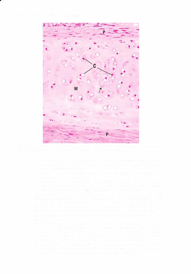
A.Hyaline cartilage (Figures 7. 1 and 7.2; Table 7.1) is the most abundant cartilage in the body, and it also serves as a temporary skeleton in the fetus until it is replaced by bone.
1.Structure
a.Matrix
(1 ) The matrix is composed of an amorphous ground substance containing proteoglycan aggregates and chondronectin, in which type II collagen is embedded (see Tables 4.2
and 7.1). |
|
|
|
|
t a b I e |
7.1 |
Cartilage Types, Characteristics, and Locations |
|
|
Type of Cartilage |
Identifying Characteristics |
Perichondrium |
Location |
|
Hyaline |
Type II collagen, basophilic |
Perichondrium usually |
Articular ends of long bones, |
|
|
matrix, chondrocytes usually |
present except on articular |
nose, larynx, trachea, bronchi, |
|
|
arranged in groups (isogenous |
surfaces |
ventral ends of ribs, template for |
|
|
groups) |
|
endochondral bone formation |
|
Elastic |
Type I I collagen; elastic fibers |
Perichondrium present |
Pinna of ear, auditory canal and |
|
|
|
|
|
tube, epiglottis, some laryngeal |
|
|
|
|
cartilages |
Fibrocartilage |
Type I collagen, acidophilic |
Perichondrium absent |
Intervertebral discs, articular |
|
|
matrix, chondrocytes arranged |
|
discs, pubic symphysis, insertion of |
|
|
in parallel rows between |
|
tendons, meniscus of knee |
|
|
bundles of collagen, always |
|
|
|
|
associated with dense |
|
|
|
collagenous connective tissue and/or hyaline cartilage
Adapted with permission from Gartner LP, Hiatt JL. Color Textbook of Histology. Philadelphia, PA: Saunders; 1 997;133.
106


|
7.2 |
|
l!iitJ'!1tiiiCartilage and Bone |
1 09 |
t a b I e |
Effects of Hormones and Vitamins on Hyaline Cartilage |
|
||
Hormones |
|
|
Function |
|
Thyroxine, testosterone, somatotropin |
Stimulate cartilage histogenesis |
|
||
Cortisone, hydrocortisone, estradiol |
Inhibit cartilage histogenesis |
|
||
Vitamins |
|
|
|
|
Hypovitaminosis A |
|
|
Diminishes thickness of epiphyseal plates |
|
Hypervitaminosis A |
|
|
Accelerates ossification of epiphyseal plates |
|
Hypovitaminosis C |
|
|
Stops matrix production, distorts cartilage columns in epiphyseal plates; |
|
|
|
|
scurvy develops |
|
Hypovitaminosis D |
|
|
Deficient absorption of calcium and phosphorus: epiphyseal cartilage cells |
|
|
|
|
proliferate, but matrix fails to calcify, and growing bones become deformed; |
|
|
|
|
rickets develops |
|
|
|
|
|
|
2.Histogenesis of hyaline cartilage is similar to that of elastic cartilage and fibrocartilage and is affected by certain hormones and vitamins (Table 7.2). It occurs by the following two processes:
a.Interstitial growth results from cell division of preexisting chondrocytes. This type of growth occurs only during the early stages of cartilage formation and in articular cartilage and the epiphyseal plates of long bones.
b.AppositionaI growth results from differentiation ofchondrogenic cells intheperichondrium. This type of growth results in the formation of chondroblasts and/or new chondrocytes,
which elaborate a new layer of cartilage matrix at the periphery.
3. Degeneration of hyaline cartilage occurs when chondrocytes undergo hypertrophy and die and the matrix becomes calcified, a process that becomes more frequent with age. Degeneration of hyaline cartilage is a normal part of endochondral bone formation (discussed later).
Arthritis, one of the processes of aging, is the degeneration of hyaline cartilage, especially as it covers the articulating surfaces of the mem
bers of bony joints. Over time, this causes joint pain, redness, swelling, stiffness, and restricts joint mobility.
Osteoarthritis is usually caused by wear and tear on the joint where the hyaline cartilage is worn away, resulting in bone grinding on bone. Other causes include joint injury or infection within the joint.
Rheumatoid arthritis is a very severe form of arthritis, where the immune system attacks the joint, including the cartilage, bone, and the synovial membrane. If left untreated, it may destroy the joint, including the cartilage and the bone.
B.Elastic cartilage (Figure 7.1; Table 7.1) possesses a perichondrium and is nearly identical to hyaline cartilage except for a network of elastic fibers, which impart a yellowish color. Although it contains type II collagen, it is less prone to degeneration than hyaline cartilage and does not calcifyin aging. It is located in areas where flexible supportis required. Elastic cartilage exists as in the cartilage of the auditory tube, external ear, and epiglottis.
C.Fibrocartilage (Figure 7.1; Table 7.1) lacks an identifiable perichondrium. It is characterized by alternating rows of fibroblast-derived chondrocytes surrounded by scant matrix and thick parallel bundles of type I collagen fibers. Fibrocartilage is located in areas where support and tensile strength are required and where tissues are exposed to compressive and shear forces. It is located in the intervertebral disks, menisci of the knees, sternoclavicular joints, and the pubic symphysis.
1 1 0 |
BRS Cell Biology and Histology |
|
|
Bone is the primary constituent of the adult skeleton. It is a specialized type of connective tissue with a calcified extracellular matrix in which characteristic cells are embedded. Bone functions to support and protect vital organs, and fleshy structures, as a hemopoiesis organ, and provides a storage site for phosphate and calcium (bone contains about 99% of the body's calcium). Secondarily, it functions to regulate blood calcium levels via input from two separate nonbony tissues. Parathyroid hormone (PTH) from the parathyroid gland acts to increase calcium levels in the blood, whereas calcitonin secreted by the parafollicular cells (C cells) of the thyroid gland decreases calcium levels in the blood. Thus, the activities of these two substances on the calcium levels in bone are responsible for maintaining the proper blood calcium level. Bone is also a dynamic tissue that constantly undergoes changes in shape, such that applied pressure results in bone resorption, whereas applied tension results in bone formation.
A.Structure
1 . Bone matrix
a.The inorganic (calcified) portion of the bone matrix (about 65% of the dry weight) is composed of calcium, phosphate, bicarbonate, citrate, magnesium, potassium, and sodium. It consists primarily of hydroxyapatite crystals, which have the composition
Ca10(P04)s(OH)2•
b. The organic portion of the bone matrix (about 35% of the dry weight) consists primarily of type I collagen (95%) and a minor contribution of type V collagen. It has a ground substance that contains chondroitin sulfate, keratan sulfate, and hyaluronic acid.
(1 ) Osteocalcin, stimulated by vitamin D, inhibits osteoblast function, and osteopontin, both glycoproteins, binds to hydroxyapatite and to integrins on osteoblasts and osteoclasts. Osteonectin, produced mostly by osteoblasts, is located in bone undergoing remodeling. Cytokines, growth hormones, and bone morphogenic proteins (BMPs), among others contribute to the bone matrix.
(2)Bone sialoprotein is a matrix protein that also binds to integrins of the osteoblasts and osteocytes and is thus related to adherence of bone cells to bone matrix.
2.The periosteum is a layer of noncalcified connective tissue covering bone on its external surfaces, except at synovial articulations and muscle attachments.
a.It is composed of an outer dense fibrous collagenous layer and an inner cellular osteoprogenitor (osteogenic) layer.
b.Sharpey fibers (type I collagen) attach the periosteum to the bone surface.
c.The periosteum functions to distribute blood vessels to bone.
3.The endosteum is a thin specialized connective tissue that lines the marrow cavities and supplies osteoprogenitor cells and osteoblasts for bone growth and repair.
B. Bone cells
1.Osteoprogenitor cells
a.These spindle-shaped cells are derived from embryonic mesenchyme and are located in the periosteum and the endosteum, and persist throughout life as stem cells that line bone. They can be activated later for bone repair of fractures or other repair.
b.They are capable of differentiating into osteoblasts. However, at low oxygen tensions, they may change into chondrogenic cells.
2.Osteoblasts
a.Osteoblasts are derived from osteoprogenitor cells under the influence of members of the BMP family and also transforming growth factor- . They possess receptors for PTH (see Chapter 13 V). Osteoblasts are responsible for the synthesis of organic protein components of bone matrix, including type I collagen, proteoglycans, and glycoproteins, which they secrete as osteoid (uncalcified bone matrix), which is mineralized under the influence and control by the osteoblasts. Additionally, they produce macrophage colony stimulating factor (M-CSF), a receptor for the activation of nuclear factor kappa B (RANKL),

l!iitJ'!1tiiiCartilage and Bone |
1 1 1 |
osteoprotegerin, osteocalcin (for bone mineralization), osteopontin (for formation of sealing zone between osteoclasts and the subosteoclastic compartment), osteonectin (related to bone mineralization), and bone sialoprotein (binding osteoblasts to extracellular matrix).
b.On bony surfaces, they resemble a layer of cuboidal, basophilic cells as they secrete organic matrix (see Figure 6.1).
c.They possess cytoplasmic processes with which they contact the processes of other osteoblasts and osteocytes and form gap junctions.
d.When synthetically active, they have a well-developed RER and Golgi complex.
e.These cells become entrapped in lacunae, but maintain contact with other cells via their cytoplasmic processes. Entrapped osteoblasts are known as osteocytes.
3.Osteocytes (Figure 7.3)
a.Osteocytes are mature bone cells housed in their own lacunae.
b.They have narrow cytoplasmic processes that extend through canaliculi in the calcified matrix (see Figures 6.1 and 7.3).
c.They maintain communication with each other via gap junctions between their processes.
d.They are nourished and maintained by nutrients, metabolites, and signal molecules carried by the extracellular fluid that flows through the lacunae and canaliculi. In addition, calcium released from bone enters the extracellular fluid located within these spaces.
e.They contain abundant heterochromatin, a paucity ofRER, and a small Golgi complex.
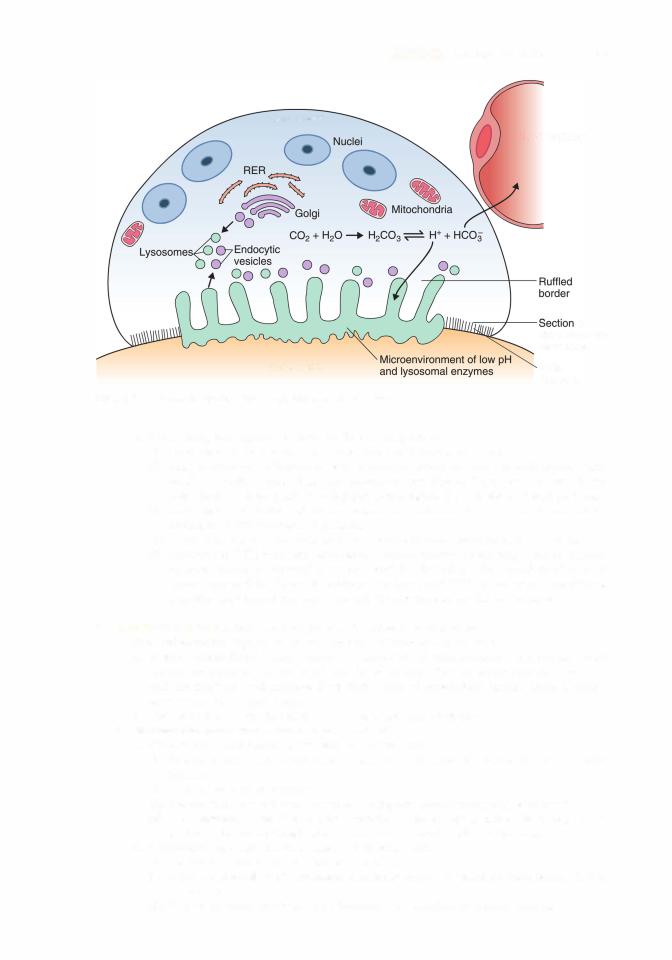
4.Osteoclasts
a.Overview. Osteoclasts are large, motile, multinucleated cells (up to 50 nuclei) that resorb bone. They are derived from cells of the mononuclear-phagocyte system, comprising blood-borne monocytes that enter the connective tissue spaces where they differentiate into various types of macrophages and osteoclasts (see Section (3)).
(1 ) Osteoclasts possess cell surface receptors: colony-stimulating factor- 1 receptor, calcitonin receptor, and RANK (nuclear factor kappa B).
(2)Osteoblasts that have been stimulated by PTH promote osteoclast formation, whereas osteoblasts that have been stimulated by calcitonin inhibit osteoclast formation by stimulating osteoid synthesis and calcium deposition.
(3)Via a series of three osteoblast signals, osteoclast precursors (macrophages) are stimulated by M-CSF to undergo mitosis. Another signaling molecule, RANKL, binds to the precursor, inducing it to differentiate into the multinucleated osteoclast, thus activating it to commence bone resorption. A third signal, osteoprotegerin (OPG), a member of the tumor necrosis factor receptor (TNFR) family produced by osteoblasts and other cells, can prohibit RANKL from binding to the macrophage, thus prohibiting osteoclast formation.
b.Osteoclast cytoplasm is acidophilic. Osteoclasts function in the resorption of bone (osteolysis). They form and reside in depressions known as Howship lacunae, which represent areas of bone resorption.
c.Morphology. Osteoclasts activated by osteoclast-stimulating factor produced and released by osteoblasts display four regions in electron micrographs.
(1 ) Basal zone is that part of the osteoclast housing most of the organelles and is the
farthest from the subosteoclastic compartment.
(2)The ruffled border is the site of active bone resorption. It is composed of irregular fingerlike cytoplasmic projections extending into the subosteoclastic compartment, a slight depression that deepens as the osteoclast resorbs bone and then that depression is referred to as Howship lacuna. The ruffledborder ofan inactive osteoclast is collapsed as the cell is in a resting stage.
(3)The clear zone surrounds the ruffled border. It contains actin filaments at the periphery that help osteoclasts maintain contact with the bony surface. This isolates and seals the region of osteolytic activity. Further, osteopontin, secreted by osteoblasts, is used to seal the zone between osteoclasts and the subosteoclastic compartment.
(4)The vesicular zone contains exocytotic vesicles that transfer lysosomal enzymes to Howship lacunae and endocytotic vesicles that transfer degraded bone products from Howship lacunae to the interior of the cell.
1 1 2 |
BRS Cell Biology and Histology |
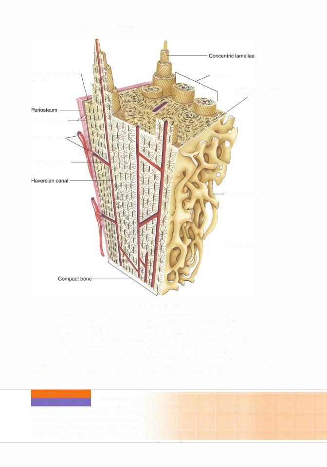
Outer circumferential lamellae
Sharpey fibers
Blood vessels
Volkmann canal
Osteons
Inner circumferential lamellae
Cancellous bone
Marrow cavity
Compact Bone
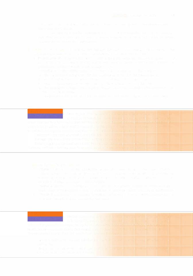
Compact bone is surrounded by dense irregular collagenous connective tissue, the periosteum, which is attached to the outer circumferential lamellae by Sharpey fibers. Blood vessels of the periosteum enter the bone via larger nutrient canals or small Volkmann canals, which not only convey blood vessels to the Haversian canals of osteons but also interconnect adjacent Haversian canals. Each osteon is composed of concentric lamellae of bone whose collagen fibers are arranged so that they are perpendicular to those of contiguous lamellae. The inner circumferential lamellae are lined by endosteal lined cancellous bone that protrudes into the marrow cavity.
FIGURE 7.3. H istological aspects of a long bone. Inset: cross section of an osteon. (Reprinted& with permission from Gartner LP. Hiatt JL. ColorAtlas and Text ofHistology. 6th ed. Baltimore, M D : Wolters Kluwer Health/Lippincott, Williams Wilkins; 201 3:88.1
CLINICAL |
Osteopetrosis, unlike osteoporosis, is a genetic disorder affecting os |
|
CONSIDERATIONS |
||
teoclasts so thatthey do not possess ruffled borders; therefore, these |
||
|
osteoclasts cannot resorb bone, which creates an imbalance between bone formation and bone resorption. Thus, persons with osteopetrosis display increased bone density. This condition leads to anemia because of decreased marrow space, blindness, deafness, and damage to the cranial nerves as the foramina of the skull become narrow and impinge on the nerves.

l!iitJ'!1tiiiCartilage and Bone |
1 1 5 |
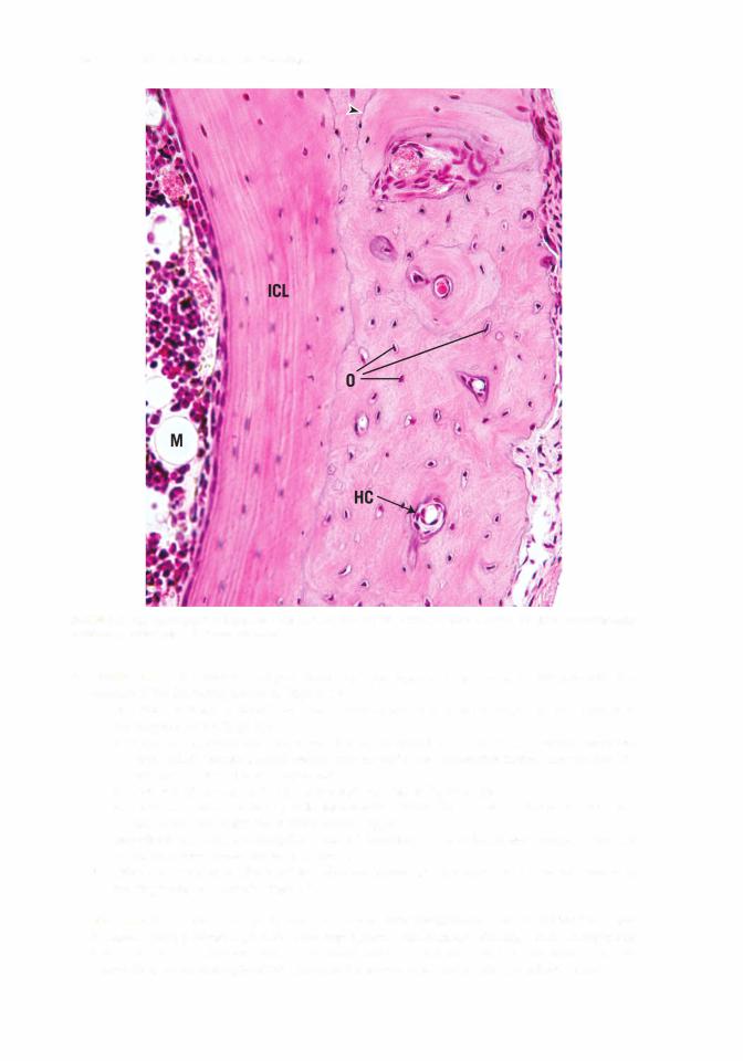
FIGURE 7.6. Light micrograph of ground bone (x 1 32). Observe the haversian canal (HC), cementing line (CL). interstitial lamellae (IL), Volkmann canal (VC). and osteocytes within lamellae (arrows).
1. Intramembranous bone formation (Figure 7.7) is the process by which most of the flat bones
(e.g., parietal bones of the skull) are formed. It involves the following events:
a.Mesenchymal cells, in the presence of a vascular zone, condense into primary ossification centers, differentiate into osteoblasts, and begin secreting osteoids in a rather haphazard form, known as woven bone.
b.As appositional bone growth continues and calcification occurs, osteoblasts become trapped in their own matrix and become osteocytes. These centers of developing bone are called trabeculae (fused spicules).
c.Fusion of the bony trabeculae produces spongy bone as blood vessels invade the area and other undifferentiated mesenchymal cells become hematopoietic cells forming blood cells of the bone marrow.
d.The periosteum and endosteum develop from portions of the mesenchymal layer that do not undergo ossification.
e.Mitotic activity ofthe mesenchymal cells gives rise to osteoprogenitor cells, which undergo cell division and form more osteoprogenitor cells or differentiate into osteoblasts within the inner layer of the developing periosteum.
f. Finally, intramembranous bone may then be converted to lamellar bone.
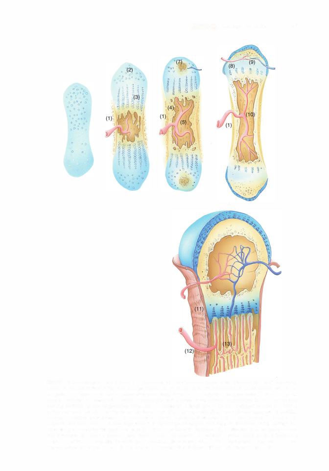
2.Endochondral bone formation (Figure 7.8) is the process by which long bones are formed. It begins in a segment of hyaline cartilage that serves as a small model for the bone. The two

1 1 6 |
BRS Cell Biology and Histology |
FIGURE 7.7. Light micrograph of membranous bone formation (X 1 321. Observe the forming haversian canals (fHCl. osteo blasts (Obi. and osteocytes (arrows).
stages of endochondral bone formation involve the development of primary and secondary centers of ossification.
a. The primary center of ossification develops at the midriff of the diaphysis of the hyaline cartilage model, containing type II collagen, by the following sequence of events:
(1 ) Vascularization of the perichondrium at this site causes the transformation of chondrogenic cells to osteoprogenitor cells, which differentiate into osteoblasts. This region of the perichondrium is now called the periosteum.
(2)Osteoblasts elaborate matrix deep to the periosteum, and via intramembranous bone formation, form the subperiosteal bone collar.
(3)Chondrocytes within the core of the cartilaginous model undergo hypertrophy and degenerate, and their lacunae become confluent, forming large cavities that eventually become marrow spaces.
(4)Osteoclasts create perforations in the bone collarthat permit the periosteal bud (blood vessels, osteoprogenitor cells, and mesenchymal cells) to enterthe newlyformed spaces in the cartilaginous model. The cartilage that constitutes the walls of these spaces then becomes calcified.
(5)Newly developed osteoblasts elaborate bone matrix that becomes calcified on the surface ofthe calcified cartilage, forming a calcified cartilage-calcified bone complex.

1 1 8 |
BRS Cell Biology and Histology |
In histological sections, the calcified cartilage stains basophilic, whereas the calcified bone stains acidophilic.
(B)6) The subperiosteal bone collar becomes thicker and elongates toward the epiphysis.
7) Osteoclasts begin to resorb the calcified cartilage-calcified bone complex, thus enlarging the primitive marrow cavity.
Repetition of this sequence of events results in bone formation spreading toward the epiphyses.
b. Secondary centers of ossification develop at the epiphyses in a sequence of events similar to that described for the primary center, except a bone collar is not formed.
(1 ) Development of these centers begins when osteoprogenitor cells invade the epiphysis and differentiate into osteoblasts, which elaborate bone matrix to replace the disintegrating cartilage. When the epiphyses are filled with bone tissue, cartilage remains in two areas, the articular surfaces and the epiphyseal plates.
(2) Articular cartilage persists and does not contribute to bone formation.
(3) Epiphyseal plates continue to grow by adding new cartilage at the epiphyseal end while it is being replaced with bone at the diaphyseal end (lengthening the bone).
(4)Ossification of the epiphyseal plates and cessation of growth occur at about 20 years of age.
3.Zones of the epiphyseal plates are histologically distinctive and arranged in the following order:
a. The zone of reserve cartilage is at the epiphyseal side of the plate. It possesses small,
randomly arranged inactive chondrocytes.
b. The zone of proliferation (of chondrocytes) is a region ofrapid mitotic divisions giving rise to rows of isogenous cell groups.
c.The zone of cell hypertrophy and maturation is the region where the chondrocytes are greatly enlarged.
d.The zone of calcification is the region where hypertrophied chondrocytes die and the cartilage becomes calcified.
e.The zone of ossification is the area where newly formed osteoblasts elaborate bone matrix on the calcified cartilage, forming a calcified cartilage-calcified bone complex, which is resorbed and replaced by bone.
4.Calcification of bone is not clearly understood, however.
a.Osteonectin, proteoglycans, and bone sialoprotein are known to stimulate calcification.
b.Bone matrix contains high concentrations ofcalcium Ca2+ along with several other organic
compounds and enzymes. Osteocalcin and sialoproteins further concentrate the calcium, resulting in osteoblasts secreting alkaline phosphatase, thus concentrating P043- ions, which further concentrates the calcium ions. Small matrix vesicles are released into the bone matrix from the osteoblasts, resulting in crystallization of calcium phosphate within the matrix vesicles.
c.Calcium pumps in the matrix vesicle membranes bring in more calcium, concentrating it and forming calcium hydroxyapatite crystals that grow and eventually puncture the matrix vesicle expelling its contents.
d.Calcium hydroxyapatite crystals that become free in the matrix become nidi of crystallization.
e.Released enzymes free phosphate ions that unite with the calcium forming calcium phosphate.
f.Calcium phosphate then begins to calcify the matrix around the nidi of crystallization.
g.Water is removed from the matrix, permitting hydroxyapatite crystals to be deposited into gaps within the collagen fibrils.
h.Nidi of mineralization enlarge and fuse with neighboring nidi, eventually calcifying the entire matrix.
F.Bone remodeling. Bone is constantly being remodeled as necessary for growth and to alter its structural makeup to adapt to changing stresses in the environment throughout life.
1 . Early on, bone development outpaces bone resorption as new haversian systems are added and fewer are resorbed.
l!iitJ'!1tiiiCartilage and Bone |
1 1 9 |
2 . Later, when the epiphyseal plates are closed, ending bone growth, bone development and resorption are balanced.
Several factors, including calcitonin and PTH, are responsible for this phenomenon regarding compact bone (see Section II J). Remodeling ofcancellous bone is under the control of many factors within the bone marrow.
G. Repair of a bone fracture. A bone fracture damages the matrix, bone cells, and blood vessels in the region and is accompanied by localized hemorrhaging and blood clot formation.
1 . Proliferation of osteoprogenitor cells occurs in the periosteum and endosteum in the vicinity of the fracture. As a result of this proliferation, cellular tissue surrounds the fracture and penetrates between the ends of the damaged bone.
2.Formation of a bony callus occurs both internally and externally at a fracture site.
a.Fibrous connective tissue and hyaline cartilage are formed in the fracture zone.
b.Endochondral bone formation replaces the cartilage with primary bone.
c.Intramembranous bone formation also produces primary bone in the area.
d.The irregularly arranged trabeculae of primary bone join the ends of the fractured bone, forming a bony callus.
e.The primary bone is resorbed and replaced with secondary bone as the fracture heals.
Bone Repair. After severe injury where segments of bone have been lost or must be removed, the remaining bone is prevented from forming a bony union followed by a bony callus that over time would result in completed bone repair. When the bony
union is not possible, a bone graft is required. For this purpose, bone fragments that are stored frozen to maintain osteogenic potential may then may be utilized in bone grafts.
Autographs are most successful since the bone donor is the recipient. Homographs are of bone donated from a different individual.
Heterographs are the least successful because the donor bone comes from another species. However, calf bone that has been frozen can serve as a viable bone graft when necessary.
H.Role of vitamins in bone formation
1.Vitamin D is necessary for absorption of calcium from the small intestine. Vitamin D deficiency results in poorly calcified (soft) bone, a condition known as rickets in children and osteomalacia in adults. Vitamin D is also necessary for bone formation (ossification), whereas an excess ofvitamin D causes bone resorption.
2.Vitamin A deficiency inhibits proper bone formation and growth, whereas an excess accelerates ossification of the epiphyseal plates. Deficiency or excess ofvitamin A results in small stature.
3.Vitamin C is necessary for collagen formation. Deficiency results in scurvy, characterized by poor bone growth and inadequate fracture repair.
Rickets occurs in children deficient in vitamin D, which results in calcium deficiency. It is characterized by deficient calcification in newly formed
bone and is generally accompanied by deformation of the bone spicules in epiphyseal plates; as a result, bones grow more slowly than normal and are deformed by the stress of weight bearing. Osteomalacia (rickets of adults) results from calcium deficiency.
1 . It is characterized by deficient calcification in newly formed bone and decalcification of already calcified bone.
2. This disease may be severe during pregnancy because the calcium requirements of the fetus may lead to calcium loss from the mother.
1 20 |
BRS Cell Biology and Histology |
I. Role of hormones in bone formation |
|
1. |
PTH activates osteoblasts to secrete osteoclast-stimulating factor, which then |
|
activates osteoclasts to resorb bone, thus elevating blood calcium levels. Excess PTH |
|
(hyperparathyroidism) renders bone more susceptible to fracture and subsequent deposition |
|
of calcium in arterial walls and certain organs, such as the kidney. |
2.Calcitonin is produced byparafollicular cells (C cells) ofthe thyroid gland. It eliminates the ruffled border of osteoclasts and inhibits bone matrix resorption, preventing the release of calcium.
3.Pituitary growth hormone (somatotropin) is produced in the pars distalis of the pituitary gland. It stimulates overall growth, especially that of epiphyseal plates, and influences bone development via insulin-like growth factors (somatomedins), especially stimulating growth of the epiphyseal plates. Children deficient in this hormone exhibit dwarfism, whereas adults with an excess of somatotropin in their growing years display pituitary gigantism and acromegaly.
Osteoporosis is a disease characterized by low bone mass (low bone mineral density) and structural deterioration of bone tissue, making the
bone fragile and susceptible to fracture. Osteoporosis is associated with an abnormal ratio of mineral to matrix.
1. It results from increased bone resorption, decreased bone formation, or both.
2. Estrogen activates bone formation by osteoblasts, and in its absence an imbalance causes osteoclastic activity to render bones fragile and susceptible to fracture.
a. Osteoporosis is most common in postmenopausal women because of diminished estrogen secretion, and in immobile patients because of lack of physical stress on the bone.
b.Estrogen therapy was employed for decades to minimize the onset of osteoporosis. Recently, it was determined that estrogen replacement therapy increases the risk of heart disease, stroke, breast cancer, and blood clots. Now, instead of estrogen, a recent new group of drugs, the bisphosphonates, has been developed that reduces the incidence of osteoporosis.
c.Preventive measures include a balanced diet rich in calcium and vitamin D and weight bearing exercise.
Acromegaly
Acromegaly results from an excess of pituitary growth hormone in adults. It is characterized by very thick bones in the extremities and in portions of the facial skeleton.
A. Synarthroses are immovable joints composed of connective tissue, cartilage, or bone. These joints unite the first rib to the sternum and connect the skull bones to each other.
B.Diarthroses (synovial joints) permit maximum movement and generally unite long bones. These joints are surrounded by a two-layered capsule, enclosing and sealing the articular cavity. The articular cavity contains synovial fluid, a colorless, viscous fluid that is rich in hyaluronic acid and proteins.
1 . The external (fibrous) capsular layer is a tough, fibrous layer of dense connective tissue.
2.The internal (synovial) capsular layer is also called the synovial membrane. It is lined by a layer of squamous to cuboidal epithelial cells on its internal surface. Two cell types are displayed in electron micrographs of this epithelium.
a.Type A cells are intensely phagocytic and have a well-developed Golgi complex, many lysosomes, and sparse RER.
b.Type B cells resemble fibroblasts and have a well-developed RER; these cells probably secrete synovial fluid.

Review Test
Directions: Each of the numbered items or incomplete statements in this section is followed by answers or by completions of the statement. Select the ONE lettered answer or completion that is BEST in each case.
1 . Which of the following statements character izes osteoclasts?
(A)They are enucleated cells.
(B)They produce collagen.
(C)They secrete osteoid.
(D)They are derived from osteoprogenitor cells.
(E)They occupy Howship lacunae.
2.Which one of the following statements is correct concerning the periosteum?
(A)It is devoid of a blood supply.
(B)It produces osteoclasts.
(C)It is responsible for interstitial bone growth.
(D)Its inner layer contains osteoprogenitor cells.
(E)Its outer layer is devoid of fibers.
3.Which one of the following statements is characteristic of osteocytes?
(A)They communicate via gap junctions between their processes.
(B)They contain large amounts of RER.
(C)They are immature bone cells.
(D)They are housed as isogenous groups in lacunae.
(E)They give rise to osteoclasts.
4.Which one of the following statements con cerning hyaline cartilage is correct?
(A)It is vascular.
(B)It contains type IV collagen.
(C)It undergoes appositional growth only.
(D)It is located at the articular ends of long bones.
(E)Its chondrocytes are aligned in rows.
5.A 7-year-old boy is seen by his pediatrician because the child broke his humerus as he tripped and fell while walking. The pediatrician asked about the child's diet and learned that he might have a dietary deficiency. Which of the following may be lacking in his diet?
(A)Potassium
(B)Calcium
(C)Iron
(D)Carbohydrates
(E)Protein
6.A 22-year-old woman is seen for the first time by her new physician, who notes that she has verythick bones in her extremities and face. The physician suspects acromegaly, caused by which of the following?
(A)Hypervitaminosis A
(B)Excess growth hormone
(C)Hypovitaminosis A
(D)Hypervitaminosis D
(E)Hypovitaminosis D
7.Which of the following statements is characteristic of bone?
(A)Bone matrix contains primarily type II collagen.
(B)About 65% of the dryweight of bone is organic.
(C)Haversian canals are interconnected via Volkmann canals.
(D)Bone growth occurs via interstitial growth only.
(E)Bone growth occurs via appositional growth only.
1 21

1 22 |
BRS Cell Biology and Histology |
8.Which one ofthe following inhibits histogenesis of cartilage?
(A)Thyroxine
(B)Hypervitaminosis A
(C)Hypovitaminosis D
(D)Hydrocortisone
(E)Hypovitaminosis C
9.Which one of the following stimulates carti lage histogenesis?
(A)Thyroxine
(B)Hypervitaminosis A
(C)Hypovitaminosis D
(D)Hydrocortisone
(E)Hypovitaminosis C
10.Which one of the following accelerates epiphyseal ossification?
(A)Thyroxine
(B)Hypervitaminosis A
(C)Absence of vitamin D
(D)Hydrocortisone
(E)Hypovitaminosis C
11 . Which one of the following makes epiphy seal cartilage matrix fail to calcify?
(A)Thyroxine
(B)Hypervitaminosis A
(C)Hypovitaminosis D
(D)Hydrocortisone
(E)Hypovitaminosis C
12.A 25-year-old patient, anemic for several years, complains offailing eyesight and hearing loss. During a physical examination, it is deter mined that the patient has lost function of some of the cranial nerves. The diagnosis could be which one ofthe following?
(A)Osteoporosis
(B)Osteomalacia
(C)Rickets
(D)Acromegaly
(E)Osteopetrosis

Answers and Explanations
1 . E. Osteoclasts are multinucleated cells that produce proteolytic enzymes and occupy Howship lacunae. They are not derived from osteoprogenitor cells but from monocyte precursors (see Chapter 7 II C 4).
2.D. The inner layer ofthe periosteum possesses osteoprogenitor cells, whereas the outer layer of the periosteum is fibrous. The periosteum functions to distribute blood vessels to the bone; thus, appositional bone growth takes place here (see Chapter 7 II B 2).
3.A. Osteocytes communicate with each othervia gap junctions on narrow cytoplasmic pro cesses that extend through canaliculi. They are mature bone cells that occupy individual lacu nae as mature resting bone cells (see Chapter 7 II C 3).
4.D. Hyaline cartilage is avascular, contains type II collagen, and grows both interstitially and ap positionally. It is located at the articulating ends of long bones (see Chapter 7 I A).
5.B. Because calcium must be maintained at a constant level in the blood and the tissues, a diet deficient in calcium leads to calcium loss from the bones. As a result, the bones become fragile (see Chapter 7 II C 4 Clinical Considerations).
6.B. Excessive growth hormone causes acromegaly. Excessive vitamin D causes bone resorption. Both an excess and a deficiency ofvitamin A result in short stature (see Chapter 7 II J Clinical Considerations).
7.C. Haversian canals run longitudinally, parallel to the long axis of bone. They are connected to one another by Volkmann canals that run perpendicular (or obliquely) to them (see Chapter 7 II E 1).
8.D. Hydrocortisone inhibits cartilage growth and matrix formation (see Chapter 7 I A 2).
9.A. Thyroxine, testosterone, and somatotropin stimulate cartilage growth and matrix formation (see Chapter 7 I A 2).
10.B. Hypervitaminosis A accelerates ossification of epiphyseal plates, whereas hypovitaminosis A reduces the width of the epiphyseal plates (see Chapter 7 II I).
11 . C. In the absence ofvitamin D, epiphyseal chondrocytes continue to proliferate, but their ma trix does not calcify, which leads to rickets (see Chapter 7 II I Clinical Considerations).
12.E. Osteopetrosis is a genetic defect involving the osteoclasts. Persons with this defect possess osteoclasts without ruffled borders, which prohibit them from resorbing bone. Therefore, bone forms but is not resorbed. This leads to increased bone density, anemia, blindness, deafness, and cranial nerve involvement because the nerves are impinged upon as theyexit the cranium via their foramina (see Chapter 7 II B Clinical Considerations).
1 23