


Epithelia and Glands
I. OVERVIEW-EPITHELIA
A. Structure. Epithelia are specialized layers of tissue arising from allthree embryonic germ layers, namely, the ectoderm, mesoderm, and endoderm, that line the internal and cover theAnexternal surfaces of the body except in certain areas such as tooth surfaces and articular cartilages. epithelium con sists of a sheet of cells lying close together with little extracellular space. These cells have distinct bio chemical, functional, and structuraldomainsthat confer polarity, or sidedness; thus, thesecellsare said to have apical, lateral, and basal epithelial domains (or as some authors prefer: basolateral domain).
1.A basement membrane, composed of a basal lamina and a lamina reticularis, separates the epithelium from underlying connective tissue and blood vessels.
2.Epithelia are avascular and receive nourishment by diffusion of molecules through the basal lamina to which they are attached.
B. |
Classification (Table 5.1). Epithelia are classified into various types based on the number of cell |
|||
|
layers (one cell layer is simple; more than one is stratified) and the shape of the superficial cells |
|||
t |
a b I e |
5.1 |
Classification of Epithelia |
|
Type |
|
Shape of Superficial Cell Layer |
Typical Locations |
|
One cell layer |
|
|
|
|
Simple squamous |
|
Flattened |
Endothelium (lining of blood vessels). |
|
|
|
|
|
mesothelium (lining of peritoneum and pleura) |
Simple cuboidal |
|
Cuboidal |
Lining of distal tubule in kidney and ducts in |
|
|
|
|
|
some glands, surface of ovary |
Simple columnar |
|
Columnar |
Lining of intestine, stomach, and excretory |
|
|
|
|
|
ducts in some glands |
Pseudostratified |
All cells rest on basal lamina, but |
|
not all reach the lumen; thus the |
|
epithelium appears falsely stratified |
More than one cell layer |
|
Lining of trachea, primary bronchi, nasal cavity, and excretory ducts in parotid gland
Stratified squamous (not |
Flattened (nucleated) |
Lining of esophagus, vagina, mouth, and true |
keratinized! |
|
vocal cords |
Stratified squamous (keratinized) |
Flattened (without nuclei) |
Epidermis of skin |
Stratified cuboidal |
Cuboidal |
Lining of ducts in sweat glands |
Stratified columnar |
Columnar |
Lining of large excretory ducts in some glands |
|
|
and cavernous urethra |
Transitional |
Dome-shaped (when relaxed). |
Lining of urinary passages from renal calyces |
|
flattened (when stretched) |
to the urethra |
|
|
|
75


l!illlJttQijEpithelia and Glands 77
4.Selective permeability results from the presence of tight junctions between epithelial cells and permits fluids with different compositions and concentrations to exist on separate sides of an epithelial layer (e.g., intestinal epithelium).
5. Protection from abrasion and injury is provided by the epidermis, the epithelial layer ofthe skin.
CLINICAL
First-degree burns are lesions caused by heat, friction, or other
CONSIDERATIONS agents.
1 . |
Damage is limited to the superficial layers of the epithelium usually the epidermis of the skin). |
|
2. |
Redness and edema occur, but blisters do not form. |
|
|
a. |
Mitotically active cells remain viable in the deeper layers of the epidermis. |
|
b. |
They divide and replace the damaged or destroyed cells. |
These surfaces contain specialized molecules that form junctions that permit cells to adhere to each other and restrict movement of materials between adjacent cells (paracellular route) into and out of the lumen lined by the epithelium.
A.The junctional complex is an intricate arrangement of membrane-associated cell adhesion molecules that function in cell-to-cell attachment of columnar epithelial cells. It corresponds to the terminal bar observed in epithelia by light microscopy and consists of three distinct components that are visible by electron microscopy.
1 . The tight junction (zonula occludens; plural: zonulae occludentes) is a zone that surrounds the entire apical perimeter of adjacent cells and is formed by fusion of the outer leaflets ofthe
cells' plasma membranes (Figure 5.3).
a.In freeze-fracture preparations of this zone, the tight junction is visible as a branching anastomosing network of intramembrane strands ridges) on the plasma membrane inner leafletnext to the cytoplasm (P-face) and grooves on the corresponding external E-face, the inner aspect ofthe outer leaflet (Figure 5.3). The strands consist oftransmembrane proteins of each cell attached directly to one another, thus sealing offthe extracellular space.
b.The intramembrane strands that close off the paracellular route possess four groups of transmembrane proteins: claudins, occludins, nectins, and junctional adhesive molecules (JAMs).
1 ) Claud ins are believed to bear the greatest responsibility in closing offthe paracellular route by creating a physical barrier that prevents the movement ofmaterial between the cells. However, claudins do form small aqueous pores that allow water and small ions to penetrate this barrier.
2) Occludins are also present in most, but not all, epithelial cell tight junctions. Their role is not understood as yet.
3) Nectins are also present in the zonula occludens, and their extracellular domains form a part of the physical barrier.
4) JAMs are similar to nectins in that their extracellular domains probably impede the movement of molecules in the extracellular space of the tight junction.
These four proteins have to be reinforced so that they maintain their proper position, and this reinforcement is due to the presence of actin filaments (F-actin) of the cytoskeleton. However, there are intermediary proteins that are capable of binding both to F-actin as well as to the four proteins just described. These are the three zonula occludens proteins, Z0-1, Z0-2, and Z0-3, as well as a fourth protein, known as afadin.
These four intermediary proteins are located on the cytoplasmic aspect ofthe region of the cell membrane involved in the formation of the tight junction and, in that fashion, are interposed between the F-actin and the claudins, occludins, nectins, and JAMs, forming a strong bond that maintains the integrity of the tight junction.


l!illlJttQijEpithelia and Glands 79
c.The tight junction prevents not only the movement of substances into the extracellular space from the lumen but also intermingling of the transmembrane proteins of the apical with those ofthe lateral domains. This ability (its tightness) is directly related to the number and complexity of the intramembrane strands and to the function of the epithelia housing the particular tight junction.
d.The fascia occludens, a ribbon-like area of fusion between transmembrane proteins on adjacent endothelial cells lining capillaries, is analogous to the zonula occludens but does not encircle the perimeter of the entire cell.
2.Anchoring junctions of epithelial cells consist of four types, two on the cell's lateral domain, zonula adherens (plural: zonulae adherentes) and desmosome (macula adherens; plural: maculae adherentes) and two, discussed in the section below, on the cell's basal domain, namely hemidesmosomes and focal adhesions.
a.The adhering junction, zonula adherens, surrounds the entire perimeter of epithelial cell, and is located just basal to and reinforcing the zonula occludens (Figure 5.3).
(1 ) It is characterized by a 10-to 20-nm separation between the adjoining cell membranes where the extracellular portions of the transmembrane glycoprotein E-cadherin molecules occupy the intercellular space.
(2)A mat of actin filaments is located on each of the cytoplasmic surfaces of the zonulae adherentes. The actin filaments are linked to each other via the actin-binding proteins a-actinin, and are linked, via another actin-binding protein vinculin, to the protein catenin, which also binds to the intracellular portion of the E-cadherin molecules. It is in this fashion that the E-cadherin molecules are reinforced by the a-actinin filaments
of the cytoskeleton. The extracellular moieties of E-cadherins of adjacent cells face each other in the extracellular space and, in the presence of Ca2+ ions, bind to each other, promoting adhesion of adjacent cells to each other.
(3)Fasciae adherentes, ribbon-like adhesion zones in the intercalated disks of cardiac muscle, are analogous to zonulae adherentes, but they do not surround the entire perimeter of the cardiac muscle cells.
b.Desmosomes (maculae adherentes) are small, discrete, disk-shaped adhesive sites.
Desmosomes are commonly found at sites other than the junctional complex, where they join epithelial cells to each other.
(1 ) Desmosomes are characterized by having five regions, two intracellular regions in each cell, namely the outer dense plaque and inner dense plaque, and an extracellular region, known as the extracellular core, in the space between the two cells.
(2)The extracellular moieties of the transmembrane glycoproteins, desmogleins and desmocollins, members ofthe E-cadherin superfamily, of one cell contact desmogleins
and desmocollins of the other cell in the extracellular space. In order for these glycoproteins to form strong adhesive bonds with their counterparts, Ca2+ ions must be present. These extracellular moieties of desmogleins and desmocollins of each cell form the extracellular core and hold the two cells to each other.
(3)The intracellular moieties of desmogleins and desmocollins form bonds with the desmosomal plaque proteins plakoglobins and plakophilins. Additional large proteins, known as desmoplakins, contact both plakoglobins and plakophilins, and together they form the outer dense plaque that presses against the cytoplasmic aspect of each cell membrane.
(4)The desmoplakins are large molecules, and their tails extend farther into the cytoplasm where they contact and form bonds with the keratin intermediate filaments of the epithelial cell. This region of bonding between the desmoplakins and the intermediate filaments forms the inner dense plaque, and it is here that the cytoskeleton is affixed to the site of adhesion.
B.Gap junctions (communicating junctions; nexus) are not part of the junctional complex and are frequent components of certain tissues other than epithelia (e.g., central nervous system, cardiac muscle, and smooth muscle).
1.Gap junctions are small aqueous pores that are inserted into the plasma membranes that couple adjacent cells metabolically and electrically (see Figure 5.3).
80BRS Cell Biology and Histology
2.A gap junction is composed of subunits, called connexons (hemichannels), which extend beyond the cell surface into the gap (a 2-nm-wide intercellular space) (Figure 5.3). Two
connexons, one in the plasma membrane of each adjacent cell contacting each other in the intercellular space, form a single gap junction.
a. Connexons consist of six subunits (composed of proteins called connexins), which are arranged radially around a central channel with a diameter of 1.5 nm (see Figure 5.3).
b.Precise alignment of connexons on adjacent cells produces a junction where they form cell-to-cell channels permitting the passage of ions and small molecules with a molecular weight of less than 1 kDa (kilodaltons) but preventing these molecules from escaping into the extracellular space.
c.Since there are different connexons, depending on the amino acid sequence of their connexins, gap junctions may be homotypic or heterotypic.
d.Connexins may alter their conformation to shut off communication between cells, especially if one of the cells is dying.
e.Usually, a large number of gap junction channels are clustered together to form a gap junction plaque where exchange ofions and small second messenger molecules may occur.
f.Connexons are in abundant supply where intercellular communication and coordination is essential such as in smooth and cardiac muscles, nerves, and certain epithelia.
Deafness
M utations of certain connexins genes, which are abundant in the cochlea, are responsible for deafness.
Bone development and mineralization
Certain genes code for connexins located in gap junctions between osteoblasts and osteocytes in developing bone. When a particular gene (Cx43) is deleted, skeletal defects develop and bone mineralization is delayed.
Darier disease (also known as keratosis follicularis) is recognizable due to the pus-filled, dry, dark regions on the skin (although the pus may be absent). It is an inherited, autosomal dominant, noncontagious condition. Histologically, the keratinocytes of the skin (especially those of the stratum spinosum and stratum granulosum) are rounded, and because the desmosomal contacts are com promised, the intercellular connections are weakened and ineffective, leading to acantholysis. The distinguishing characteristics of the disease are a specific scent of the affected skin and the fragility of the fingernails. On the average, 1 in 1 00,000 people is afflicted by Darier disease worldwide. The condition appears to be due to a problem of intracellular trafficking of desmoplakin to the lateral cell membrane during desmosomal assembly.
C.Lateral interdigitations (plicae) are irregular fingerlike projections that interlock adjacent epithelial cells. These lateral interdigitations are most frequently present in cells that function in fluid and/ or electrolyte transport (e.g., epithelial lining of the intestines, proximal tubules of the kidney).
A.The basement membrane is a narrow, flexible, PAS-positive (i.e., it stains purplish with periodic acid-Schiff reagent) acellular supportive structure that is consistently interposed between the epithelium and the underlying connective tissue. The thickness of the basement membrane depends on its location, so that it is much thicker in thick skin than in the lining of the trachea, but on the average it is usually 0.3 Jlm wide. When viewed with the electron microscope, the basement membrane is resolved to be composed of two layers, the basal lamina, approximately 100 nm in thickness, and the lamina reticularis, which is at least 200 nm in thickness (Figure 5.4).
1.The basal lamina is manufactured bythe epithelial layer and is composed of two regions, the electron-lucent lamina Iucida, which is in direct contact with the basal plasma membrane of


82 |
BRS Cell Biology and Histology |
B.Two additional components of the basal domain of the cell membrane, the anchoring junctions focal adhesions and hemidesmosomes, also participate in the adhesion mechanism between the
epithelium and the connective tissue.
1 . Focal adhesionsare regions ofrelativelyweak anchoringjunctionsthatassist in the attachment ofthe epithelium to the basal lamina. The principal components are clusters oftransmembrane proteins, alpha and beta integrins, whose cytoplasmic moieties are attached to actin filaments of the cytoskeleton via the intracellular anchorage proteins alpha actinin, talin, paxillin, and vinculin, and their extracellular regions are attached to laminin and fibronectin of the basal lamina. Focal adhesions may be attachments of long duration, but mostly they are formed as cells that migrate along the basal lamina surface and continuously detach and reattach during their movement. Both the attachment and the detachment are dynamic processes that occur due to intracellular and/or extracellular signals that alter the three-dimensional conformation ofthe integrin molecules, causing them to form or break bondswith the intracellular anchorage proteins and with the proteoglycans of the basal lamina.
2.Hemidesmosomes are specialized anchoring junctions whose morphology resembles that of a half of a desmosome. However, instead of attaching cells to each other, hemidesmosomes mediate strong adhesion of epithelial cells to the underlying extracellular matrix (see Figure 5.3). Unlike focal adhesions, hemidesmosomes are mostly of longer duration and provide a firmer attachment of the cell to the basal lamina. Moreover, instead of binding to actin filaments of the cytoskeleton, they are bound to the more robust intermediate filaments.
a.There are two types of hemidesmosomes: the classical type (type I) located in the stratum basale of skin, in the lining of the esophagus, in the masticatory and lining mucosae of the oral cavity, as well as in the cells of pseudostratified epithelia of structures such as the trachea, and type II hemidesmosomes are present mostly in the simple columnar epithelia of the intestinal lining. Type I hemidesmosomes are more complex, whereas type II hemidesmosomes have much fewer components.
(1 ) Type I hemidesmosomes have several components, namely the dense cluster of transmembrane proteins a6 4 integrin, whose intracytoplasmic moieties bind to the protein erbin and the plakin proteins plectin and bullous pemphigoid antigen 230 (BP230). It is these two plakin proteins that connect a6 4 integrins to the intermediate
filaments (keratin-5 and keratin-14, also known as tonofilaments). Erbin assists in the binding of the integrin molecule to BP230. Two additional proteins are associated with the a6 component of the integrin, namely bullous pemphigoid antigen 180 (BP 180, also known as type XVII collagen) and cluster of differentiation protein 151 (CD151). BP 180 binds intracellularly to both the a6 component of the integrin and to plectin, and extracellularlyto the a6 component ofthe integrin and to laminin ofthe basal lamina. The extracellular moieties of a6 4 integrins also bind to the laminins and type IV collagens of the basal lamina. CD151 functions to ensure that enough integrin molecules are recruited to the area so that hemidesmosome formation can occur. The intracytoplasmic components of the hemidesmosome form an electron-dense, plaque-like structure that resembles but is different from the outer dense plaque of a desmosome.
(2)Type II hemidesmosomes are much simpler than the classical hemidesmosomes, in that they are composed only of the dense cluster of transmembrane proteins a6 4 integrin whose intracytoplasmic moieties bind to the plakin protein plectin, which in turn binds to the intermediate filaments keratin-S and keratin-18 (tonofilaments). As in type I hemidesmosomes, the intracytoplasmic components form an electron-dense, plaque-like structure that resembles but is different from the outer dense plaque of a desmosome.
b.Because of the large number of a6 4 integrins and their connection to the tougher intermediate filaments rather than to the less sturdy actin filaments, hemidesmosomes
form much stronger anchoring junctions than focal adhesions. However, similar to focal adhesions, the presence of Ca2+ ions are necessary for the formation and maintenance of these anchoring junctions, and the cell is able to form and disassemble hemidesmosomes as required. Both the formation and disassembly are regulated by a series of intracellular and extracellular signaling events, many ofwhich act during the formation ofnew cells that
l!illlJttQijEpithelia and Glands 83
either become anchored to the basal lamina or are moving toward the free surface of the epithelium.
c.Keratin filaments (tonofilaments) in the cell terminate in the hemidesmosome plaque, allowing these junctions to link the cytoskeleton with the components of the extracellular matrix.
CLINICAL |
Bullous pemphigoid is an autoimmune disease in which antibodies |
|
CONSIDERATIONS |
||
against hemidesmosomes are produced. |
||
|
1 . This disease is characterized by chronic generalized blisters in the skin.
2. These blisters cause the epithelium to separate from the underlying substratum.
C. Basal plasma membrane infoldings are commonin ion-transporting epithelia (e.g., distal convoluted tubule of the kidney, striated ducts in salivary glands).
1 . They form deep invaginations that compartmentalize mitochondria.
2.Function. They increase the surface area and bring ion pumps (Na+-K+ adenosine triphosphatase [ATPase]) in the plasma membrane close to their energy supply (ATP produced in mitochondria).
These surfaces may possess specialized structures such as microvilli, stereocilia, and cilia. Flagella, modified cilia that are present only in spermatozoa, are discussed inl Chapter 20, The Male Reproduc tive System.
A. Microvilli are fingerlike projections of epithelia approximately J..lmlong that extend into a lumen and increase the cell's surface area.
1 . A glycocalyx (sugar coat) is present on their surfaces (see Chapter I II C).
2.A bundle of approximately 25 to 30 actin filaments runs longitudinally through the core of each microvillus, extending from the tip of the microvillus into the terminal web, a zone of intersecting filaments in the apical cytoplasm.
a.The actin filaments within the microvillus are bound to each other by the actin-binding proteins villin, fimbrin, espin, and fascin, and the actin filaments at the perimeter of the actin bundle are affixed to the plasmalemma ofthe microvillus by calmodulin and myosin I.
b.The actin filaments are arranged in a specific orientation within the microvillus, so that their plus ends (barbed ends) extend to the tip ofthe microvillus, where they are embedded in an amorphous material known as viii in. The minus ends (pointed ends) ofthe bundle of actin filaments extend into the terminal web ofthe epithelial cell, where they are affixed to the spectrin and actin filaments of the terminal web.
3.The myosin II and tropomyosin molecules located at the terminal web can interact to contract the apical region of the cell, thereby causing the microvilli to diverge from each other, increasing the intermicrovillar spaces. These enlarged intermicrovillar spaces facilitate the increased transport of materials into the cell.
4.Microvilli constitute the brush border ofkidneyproximaltubule cells and the striated border of intestinal absorptive cells.
B.Stereocilia are very long (15-20 J..lm in length) microvilli (they are not cilia) in the hair cells of the inner ear, in the epididymis, and in the vas deferens of the male reproductive tract. The core of the stereocilia is composed of actin filaments that are attached to each other by fimbrin and to the plasmalemma of the stereocilia by villin-2 and ezrin (but not in the hair cells of the inner ear). As in microvilli, the barbed ends ofthe actin filament bundle extend to the tip ofthe stereocilia, where villin is absent, and their pointed ends reach and are anchored in the cell web.

l!illlJttQijEpithelia and Glands 85
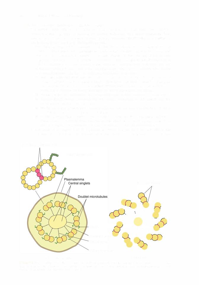
FIGURE 5.6. Electron micrograph of a cross-sectioned centriole. Notice the nine triplet microtubules arranged radially in the shape of a pinwheel. This is known as the 9 + 0 configuration (compare with the cilium). and little central organization is observed.
(9 + 0 configuration). It resembles a centriole (see Figure 5.6) buthas a less complex central organization. The inner two triplets of the basal body give rise to the doublet microtubules of the cilium axoneme. The outermost, third microtubule, referred to as microtubule C, of the triplet is also incomplete. It is composed of only 10 protofilaments and shares 3 of microtubule B's protofilaments so that microtubule C is also a completely closed structure. The region where the basal body and the axoneme merge with each other is frequently referred to as the transition zone. It is the basal body and structures coupled with it that not only secure the cilium to the cell but also function in ensuring that all cilia of the cell beat in the same direction. Structures that are associated with the basal body are the alar sheet, basal feet, and striated rootlets.
(1 ) A funnel-shaped fibrous membrane, known as the alar sheet, attaches to the microtubules C of the basal body at the transition zone and sweeps upward to merge with the cell membrane at the origin of the cilium. The alar sheet not only ensures a strong attachment of the basal body to the plasmalemma but also functions as a semipermeable membrane that limits access to the ciliary cytoplasm.
(2)Attached to the basal body is a structure known as the basal foot that is believed to ensure that all cilia ofthe same cell are facing in the same direction, thereby controlling the synchronous, unidirectional bending of all cilia of the same cell.
(3)Extending from the basal body, and securing itintothe cytoplasm, arethe protofilaments, known as the striated rootlet, which affixes the cilium relatively deeply into the apical cytoplasm.
c.Ciliary movement occurs as the dynein arms of a doublet grasp and "climb" along the "back" of the adjacent doublet, thereby bending the cilia in a preferred direction. This is an energy-requiring process and is fueled by the ATPase activity of the dynein arms. As the cilium bends, the elastic proteins of the axoneme become stretched. Once the cilium is bent sufficiently, the dynein arms relax their grasp on the adjacent doublet, and the cilium snaps back to its original position propelling materials located at its tip, thus moving material, such as mucus in the tracheal lumen, along the epithelial surface. The process of "snapping back" does not require energy because it is fueled by the stretched elastic proteins returning to their resting positions.
d.Transport of material occurs within the motile cilia, primary cilia, and flagella, referred to as axonemal transport. Within cilia it is known as intraciliary transport, and within flagella it is known as intraflagellar transport. The transport of tubulin dimers and other molecules required by the cilia occurs via carrier proteins, known as raft proteins that pick up cargo, such as tubulin dimers. The raft proteins then become attached to kinesin or to

86 |
BRS Cell Biology and Histology |
dynein, motor proteins that ferry cargo along microtubules in an anterograde or retrograde direction, respectively. Anterograde is from the basal body toward the tip of the cilium, and retrograde is in the opposite direction, from the tip of the cilium toward the basal body. Defects in the intraciliary transport result in various anomalies, some with even lethal consequences.
2.A single nonmotile cilium (primary cilium) is present on nearly every human cell that is in the G0 stage of the cell cycle. These structures were believed to be nonfunctional evolutionary remnants, but recently it has been demonstrated that they have essential functions in the organization of signaling pathways not only during embryonic development but also in the adult organism. It has been amply demonstrated that impaired primary cilia are responsible
for various anomalous conditions, known as ciliopathies.
a. The axoneme of primary cilia differs+ from those of motile cilia in that they possess no central singlets; thus, they have a 9 0 configuration; moreover, the central sheet, outer
and inner dynein arms, and most radial spokes are absent.
b.A set of proteins, known as BBSome (named after Bardet-Biedl syndrome), is located at the distal end of the basal body and is responsible for the formation as well as the proper functioning ofthe primary cilium. It is the BBSome protein complexthat determines which molecules are permitted entry into the cytoplasm of the primary cilium. Disruption of the BBSome is responsible for a number of ciliopathies, including Bardet-Biedl syndrome.
c.If a cell leaves the G0 stage of the cell cycle and enters the G1 phase, the primary cilium becomes resorbed, and the basal body returns to its previous function as a centriole.
d.Primary cilia of a particular region, such as the kidney tubule or fibroblasts, are oriented in the same direction, and this precise alignment is dictated by the basal foot. This precise alignment permits the primary cilia to perform their functions, whether in the monitoring of the flow of the ultrafiltrate in the kidney tubule or in the migration of fibroblasts in the proper direction during wound healing.
e.It has been demonstrated that a number of ion channels and select receptors are present only in the membranes of primary cilia. The specific reason for this exclusivity has not been elucidated, but it is believed that it may have some association with the intraciliary transport mechanism, and disturbances of this particular cellular event are responsible for the various ciliopathies.
1 . lmmotile cilia syndrome results from a genetic defect that causes an abnormal ciliary beat or the absence of a beat.
a. In this syndrome, cilia have axonemes that lack ciliary dynein arms and have other abnormalities.
b. The syndrome is associated with recurrent lower respiratory tract infections, reduced fertil ity in women, and sterility in men.
2. Polycystic kidney disease (PKD), a genetic disorder, is an autosomal dominant disease occur ring in about two per thousand births. There are three mutations responsible for this anomaly, namely PKD - 1, PKD-2, and PKHD-1 , but most cases are due to mutations in gene PKD-1 located on chromosome 1 6. For an unknown reason, individuals with these mutations develop abnormal primary centrioles, resulting in abnormal cell cycles as well as defective calcium transport within the cell. During embryonic development, cysts begin forming on the kidneys and increase in size and number. As these cysts continue to gain fluid and become larger, they apply pressure on the uriniferous tubules and prevent them from performing their function. In
most patients, the symptoms, such as pain in the back and sides as well as headaches, become evident by the 40th year of life, and eventually the kidney function of the patients is reduced until they have to be placed on renal dialysis and become eligible for kidney transplant.
3.Bardet-Biedl syndrome is a disorder due to disruptions in the normal functioning of BBSome located at the base of the basal body of the primary cilium. The manifestations of this syndrome
are varied and include night blindness, speech disorder, malformations of the rods and cones of the retina with a subsequent loss of vision, presence of extra digits on the hands and feet, kidney failure, urogenital defects, and obesity. Many patients succumb to kidney failure.


88BRS Cell Biology and Histology
(2)A connective tissue capsule may surround the gland, or septa of connective tissue may divide the gland into lobes and smaller lobules.
(3)Glands may have ducts between lobes (interlobar), within lobes (intralobar), between lobules (interlobular), or within lobules (intralobular), such as striated and intercalated ducts.
(4)Multicellular glands secrete various substances.
(a)Mucus is a viscous material that usually protects or lubricates cell surfaces.
(b)Serous secretions are watery and often rich in enzymes.
(c)Mixed secretions contain both mucous and serous components.
(5)Mechanisms of secretion vary.
(a)In merocrine glands (e.g., parotid gland), the secretory cells release their contents by exocytosis.
(b)In apocrine glands (e.g., lactating mammary gland), part of the apical cytoplasm of the secretory cell is released along with the contents.
(c)In holocrine glands (e.g., sebaceous gland), the entire secretory cell along with its contents is released.
2.Endocrine glands may be unicellular (e.g., individual endocrine cells in gastrointestinal and respiratory epithelia) or multicellular (e.g., adrenal gland), and they lack a duct system. In multicellular glands, secretory material is released into fenestrated capillaries, which are abundant just outside the basal lamina of the glandular epithelium.
1 . Epithelia sometimes undergo metaplasia in response to persistent in jury. Metaplasia is the conversion of one type of differentiated epithe
lium into another. Most commonly, a glandular epithelium is transformed into a squamous epithelium. However, in cases of chronic acid reflux from the stomach into the lower esopha gus, the stratified squamous nonkeratinized epithelium is replaced by a glandular mucus secreting epithelium (Barrett epithelium) similar to that found lining the cardia of the stomach. This helps to protect the esophagus against the injurious effects ofthe acid and pepsin, but is also a well-known precursor of esophageal adenocarcinoma.
2. Epithelial cell tumors occur when cells fail to respond to normal growth regulatory mechanisms.
a.These tumors are benign when they remain local.
b.They are malignant when they invade neighboring tissues. Then they may (or may not) metastasize to other parts of the body.
i. Carcinomas are malignant tumors that arise from surface epithelia.
ii. Adenocarcinomas are malignant tumors that arise from glands.

Review Test
Directions: Each of the numbered items or incomplete statements in this section is followed by answers or by completions of the statement. Select the ONE lettered answer or completion that is BEST in each case.
1 . Which one of the following statements about the desmosome is true?
(A)It is sometimes called a nexus.
(B)It permits the passage of large proteins
from one cell to an adjacent cell.
(C) It has a plaque made up of many connexons.
(D)It facilitates metabolic coupling between adjacent cells.
(E)It is a disk-shaped adhesion site between epithelial cells.
2.A medical student who has chronic lower respiratory infections seeks the advice of an ear, nose, and throat specialist. A biopsy of the stu dent's respiratory epithelium reveals alterations in certain epithelial structures. This patient is most likely to have abnormal
(A)microvilli.
(B)desmosomes.
(C)cilia.
(D)hemidesmosomes.
(E)basal plasmalemma infoldings.
3.Which one of the following statements about the gap junction is true?
(A)It extends as a zone around the apical perimeter of adjacent cells.
(B)It possesses dense plaques composed in part of desmoplakins.
(C)It permits the passage ofions from one cell to an adjacent cell.
(D)Its adhesion is dependent upon calcium ions.
(E)It possesses transmembrane linker glycoproteins.
4.Which one of the following statements about glands is true?
(A)Exocrine glands lack ducts.
(B)Simple glands have ducts that branch.
(C)Endocrine glands secrete into ducts.
(D)Serous secretions are watery.
(E)Holocrine glands release their contents by exocytosis.
5.Which one of the following statements about epithelia is true?
(A)They are polarized.
(B)Theyare vascular.
(C)They are completely surrounded by a basal lamina.
(D)They contain wide intercellular spaces.
(E)They are not part of the wall of blood vessels.
6.Which one of the following statements about cilia is true?
(A)They possess a 9 + 0 configuration of microtubules.
(B)They do not contain an axoneme.
(C)They contain ciliary dynein arms.
(D)They are nearly identical to centrioles.
(E)They play a major function in absorption.
7.Which one of the following statements about stratified squamous epithelium is true?
(A)The surface layer ofcells is always keratinized.
(B)The cells in its most superficial layer are flattened.
(C)Its basal cells rest on an elastic lamina.
(D)Its cells lack desmosomes.
(E)It lines the ducts of sweat glands.
8.Which one of the following is an autoim mune disease?
(A)Adenocarcinoma
(B)Bullous pemphigoid
(C)Carcinoma
(D)First-degree burn
(E)Immotile cilia syndrome
9.Which one of the following is a hereditary disease that may be associated with infertility?
(A)Adenocarcinoma
(B)Bullous pemphigoid
(C)Carcinoma
(D)Edema
(E)Immotile cilia syndrome
89
90BRS Cell Biology and Histology
10.Which one ofthe following is a tumor aris ing from glandular epithelium?
(A)Adenocarcinoma
(B)Bullous pemphigoid
(C)Carcinoma
(D)Edema
(E)Immotile cilia syndrome
11 . Which of the following is a condition affect ing the epidermis of the skin in which blisters do not form?
(A)Adenocarcinoma
(B)Bullous pemphigoid
(C)Carcinoma
(D)First-degree burn
(E)Immotile cilia syndrome

Answers and Explanations
1 . E. Desmosomes are sites of adhesion characterized by dense cytoplasmic plaques and associ ated keratin filaments. Only gap junctions permit cell-to-cell communication ofsmall mol ecules via their connexon channels (see Chapter 5 II A).
2.C. Individuals with abnormal respiratory cilia commonly have recurrent respiratory infections if the cilia are unable to clear the respiratory epithelium of microorganisms, debris, and so forth. The student may have immotile cilia syndrome, which is caused by a genetic defect, resulting in cilia with axonemes that lack ciliary dynein arms and thus are unable to beat (see Chapter 5 IV C 2 Clinical Considerations).
3.C. The gap junction channel regulates the passage of ions and small molecules from cell to cell, excluding those having a molecular weight greater than 1200 Da. The tight junction is the zone of adhesion around the apical perimeter of adjacent cells. The other statements are characteris tics of desmosomes (see Chapter 5 II B).
4.D. Serous secretions produced by glands are oftenrich in enzymes and watery in consistency. Exocrine glands secrete into ducts, and endocrine glands lack ducts. Merocrine glands use exo cytosis to release their products (see Chapter 5 V B).
5.A. Epithelia are polarized, meaning they show sidedness and have apical and basolateral sur faces with specific functions (see Chapter 5 I A).
6.C. Cilia contain an axoneme with ciliary dynein arms extending unidirectionally from one member of each doublet. Ciliary dynein has ATPase activity, and when it splits ATP, the adjacent doublets slide past one another and the cilium moves. Microvilli, not cilia, function in absorp tion (see Chapter 5 IV C).
7.B. Stratified squamous epithelium is characterized by flattened cells with or without nuclei in its superficial layer. It may or may not be keratinized, and it rests on a basal lamina pro duced by the epithelium. Stratified cuboidal epithelium lines the ducts in sweat glands (see Chapter 5 I B).
8.B. Bullous pemphigoid is an autoimmune disease. Affected individuals form antibodies against their own hemidesmosomes (see Chapter 5 III B Clinical Considerations).
9.E. Immotile cilia syndrome results from a genetic defect that prevents synthesis of ciliary dynein ATPase, resulting in cilia that cannot actively move. Men are sterile because their sperm are not motile (the flagella in their tails lack this enzyme). Women may be infertile because cilia along their oviducts may fail to move oocytes toward the uterus (see Chapter 5 IV C 2 Clinical Considerations).
10.A. Adenocarcinomas are epithelial tumors that originate in glandular epithelia. Carcinomas originate from surface epithelia (see Chapter 5 V B 2 Clinical Considerations).
1 1 . D. First-degree burns damage the upper layers ofthe epidermis only, and blisters do not form in the skin (see Chapter 5 I C Clinical Considerations).
91