

Cytoplasm and Organelles
I. OVERVIEW-THE CYTOPLASM
The cytoplasm contains three main structural components: organelles, inclusions, and the cytoskel eton. The fluid component is called the cytosol. The functional interactions among certain organelles result in the uptake and release of material by the cell, protein synthesis, and intracellular digestion.
A. Organelles (Figure 3.1) are metabolically active units of cellular matter.
1 . The plasma membrane, which envelops the cell and forms a boundary between it and adjacent structures, is discussed in Chapter 1 .
2. Ribosomes
a.Structure. Ribosomes are nonmembranous organelles that are 12 nanometers (nm) wide and 25 nm long and consist of a small and a large subunit. The two subunits are composed of several types of ribosomal ribonucleic acids (rRNAs) and numerous proteins (Table 3.1; Figure 2.5).
b.Ribosomes may be free in the cytosol or bound to membranes of the rough endoplasmic reticulum (RER) or the cytoplasmic surface of the outer nuclear membrane. Whether free or bound, the ribosomes constitute a single interchangeable population.
c.A polyribosome (polysome) is a cluster of ribosomes along a single strand of messenger ribonucleic acid (mRNA) where every ribosome is concurrently engaged in protein synthesis.
d.Function. Ribosomes are the sites where mRNA is translated into protein. Proteins destined for transport (secretory, membrane, and lysosomal) are synthesized on polyribosomes bound to the RER, whereas proteins not destined for transport are synthesized on polyribosomes in the cytosol. Protein synthesis that forms proteins not destined for transport occurs in the following manner (synthesis of proteins destined to be transported is discussed below in Section III b):
(1 ) The small ribosomal subunit binds both mRNA and activated transfer ribonucleic acids (tRNAs); the codons of the mRNA then base-pair with the corresponding anticodons of
the tRNAs.
(2) Next, an initiator tRNA recognizes the start codon (AUG) on the mRNA.
33

34 |
BRS Cell Biology and Histology |
Plasma membrane
•
•
•
Nuclear • envelope •
•
•
• • •
• •
FIGURE 3.1 . A eukaryotic cell and its major organelles and inclusions. (Reprinted&with permission from Chandar N. Viselli S. Cell and
North American ed. Baltimore. MD: Wolters Kluwer Health/Lippincott Williams Wilkins; 201 0:48 )
(3)The large ribosomal subunit then binds to the complex. The enzyme peptidyl transferase located in the large subunit catalyzes peptide bond formation, resulting in addition of amino acids to the growing polypeptide chain.
(4)A chain-terminating codon (UAA, UAG, or UGA) causes release of the polypeptide from the ribosome, and the ribosomal subunits dissociate from the mRNA.
3.Rough endoplasmic reticulum (Figures 3.1 and 3.2)
a. Structure. RER is a system of membrane-bounded sacs, or cavities. The outer surface of
|
RER is studded with ribosomes, which makes it appear rough. The interior region of RER is |
|||
|
called the cisterna, or the lumen. The outer nuclear membrane is continuous with the RER |
|||
|
membrane, which brings the perinuclear cisterna into continuity with the cisternae of the |
|||
|
RER. The RER membrane also has receptors (ribophorins) in its membrane to which the |
|||
|
large ribosomal subunit binds. |
|
||
t a b I |
e |
3.1 |
Ribosome Composition |
|
Subunit |
|
|
rRNA Types |
Number of Proteins |
Large (60S) |
|
|
5S |
49 |
|
|
|
5.8S |
|
|
|
|
28S |
|
Small (40S) |
|
|
1 8S |
33 |
rRNA. ribosomal ribonucleic acid.

l!1i!tttttiilCytoplasm and Organelles |
35 |
FIGURE 3.2. An electron micrograph showing rough endoplasmic reticulum (RERI. portions of several mitochondria and their cristae (C), matrix (MI. and matrix granules (MGI. and a peroxisome with a nucleoid (N).
b.RER is abundant in cells synthesizing secretory proteins; in such cells, the RER is organized into many parallel arrays.
c.The RER sac closest to the Golgi apparatus gives rise to buds free of ribosomes that form vesicles known as transfer vesicles. This sac is known as a transitional element and represents the region of exit from the RER.
d.Function. The RER is where membrane-packaged proteins are synthesized, including secretory, plasma membrane, and lysosomal proteins. In addition, the RER monitors the assembly, retention, and even degradation ofcertain proteins. Proteins that are to be retained in the RER cisternae are marked by the presence of a small peptide composed of a specific sequence offour amino acids at their C terminus; these amino acids form the KDEL sequence and are composed of lysine, asparagine, glutamine, and leucine. Proteins that do not sport the KDEL sequence at the C terminus are transported out of the cisternae of the RER.
4.Smooth endoplasmic reticulum (SER)
a.Structure. SER is an irregular network of membrane-bounded channels that lacks ribosomes on its surface, which makes it appear smooth.
b.It usually appears as branching, anastomosing tubules, or vesicles, whose membranes do not contain ribophorins.
c.SER is less common than RER but is prominent in cells synthesizing steroids, triglycerides, and cholesterol.
d.Function. SER has different functions in different cell types.
(1 ) Steroid hormone synthesis occurs in SER-rich cells such as the Leydig cells ofthe testis, which make testosterone.
(2)Cells synthesizing fatty acids and phospholipids are rich in SER.
(3)Drug detoxification occurs in hepatocytes following proliferation ofthe SER in response to the drug phenobarbital; the oxidases that metabolize this drug are located in the SER.
(4)Muscle contraction and relaxation involve the release and recapture of Ca2+ by the SER in skeletal muscle cells, called the sarcoplasmic reticulum.
5.Annulate lamellae
a.Structure. Annulate lamellae are parallel stacks of membranes (usually 6-10) that resemble the nuclear envelope, including its pore complexes. They are often arranged with their annuli (pores) in register and are frequently continuous with the RER.
b.Function. Annulate lamellae are found in rapidly growing cells (e.g., germ cells, embryonic cells, and tumor cells), but their function and significance remain unknown.

36BRS Cell Biology and Histology
6.Mitochondria (Figures 3.1 and 3.2)
a. Structure. Mitochondria are rod-shaped organelles that are 0.2 J.Lmwide and up to 7 J.Lm long. They possess an outer membrane, which surrounds the organelle, and an inner
membrane, which has a high concentration of cardiolipin, a phospholipid that does not allow ions to cross the membrane in either direction. The inner membrane invaginates to form cristae. Mitochondria are subdivided into an intermembrane compartment between the two membranes and an inner matrix compartment. Granules within the matrix bind the divalent cations Mg2+ and Ca2+ .
b.Enzymes and genetic apparatus. Mitochondria contain the following:
(1 ) All of the enzymes of the Krebs (tricarboxylic acid) cycle are in the matrix, except for
succinate dehydrogenase, which is located on the inner mitochondrial membrane.
(2)Elementary particles (visible on negatively stained cristae) represent adenosine triphosphate (ATP) synthase, a special enzyme embedded in the inner mitochondrial membrane. It consists of a head portion and a transmembrane H+ carrier and is involved in coupling oxidation to phosphorylation of adenosine diphosphate (ADP) to form ATP (Figure 3.3).
(3)A genetic apparatus in the matrix composed of circular deoxyribonucleic acid (DNA), mRNA, tRNA, and rRNA (with a limited coding capacity), although most mitochondrial proteins are encoded by nuclear DNA.
(4)Those mitochondrial proteins that are formed in the cytosol are transported into the mitochondria in a very specific fashion. The proteins must possess a terminal amino acid presequence thathas a positive charge and the protein has to be accompanied by cytosolic heat shock protein 70. These two signals are recognized by a carrier embedded in the outer mitochondrial membrane, known as the TOM (translocase of the outer mitochondrial membrane complexes). The recognition permits the translocation of the protein into the intermembrane compartment via the proteins known as the ITM (inner mitochondrial membrane complexes). Once in the matrix, mitochondrial heat shock protein 70 cleaves the amino acid-positive presequence.
Electron transport chain |
ADP + Pi |
ATP |
Matrix |
|
|
H+ |
|
FIGURE 3.3. Chemiosmotic coupling mechanism for generating adenosine triphosphate (ATP)W in mitochondria. As elec trons move (sequentially) along the enzyme complexes of the electron transport chain, ions (protons) are pumped from the matrix compartment across the inner mitochondrial membraneW into the intermembrane compartment generating a proton gradient. This electrochemical proton gradient drives the production of ATP as protons pass down their electro chemical gradient through ATP synthase and reenter the matrix. As passes through ATP synthase, this enzyme uses the energy of the proton flow to drive the production of ATP from adenosine diphosphate (ADP) and Pi.

l!1i!tttttiilCytoplasm and Organelles |
37 |
c.Origin and proliferation
(1 ) Mitochondria may have originated as symbionts (intracellular parasites). According
to this theory, anaerobic eukaryotic cells endocytosed aerobic microorganisms that evolved into mitochondria, which function in oxidative processes.
(2)Mitochondria proliferate by division (fission) ofpreexisting mitochondria and typically have a 10-day life span. Proteins needed to sustain mitochondria are imported into them from the cytosol.
d.Mitochondrial ATP synthesis
(1 ) Mitochondria synthesize ATP via the Krebs cycle, which traps chemical energy and produces ATP by oxidation of fatty acids, amino acids, and glucose.
(2)ATP is also synthesized via a chemiosmotic coupling mechanism involving enzyme complexes of the electron transport chain (composed of NADH dehydrogenase complex, cytochrome b1-c complex, and cytochrome oxidase complex through which energy-rich protons enter the intermembrane compartment) and ATP synthase present in elementary particles of cristae (Figure 3.3). It is through the ATP synthase that the energy-rich protons leave the intermembrane compartment to reenter the matrix compartment where they yield their energy to ADP + Pi (where Pi is inorganic phosphate derived from the cytosol) to form ATP.
(3)In order to manufacture ATP, the mitochondria require ADP, which is transported into the organelle via ADP/ATP exchange proteins located in both the outer and inner mitochondrial membranes. As their name implies, these exchange proteins allow ADP to enter the mitochondrion and ATP to exit the mitochondrion.
e.Condensed mitochondria result from a conformational change in the orthodoxform (which is the typical morphology). The change occurs in response to an uncoupling of oxidation from phosphorylation.
(1 ) In condensed mitochondria, the size of the inner compartment is decreased, and the matrix density is increased. The intermembrane compartment is enlarged.
(2)Condensed mitochondria are present in brown fat cells, which produce heat, rather than ATP because they have a special transport protein in their inner membrane that uncouples respiration from ATP synthesis (see Chapter 6 IV B 5 b).
(3)Mitochondria swell in response to calcium, phosphate, and thyroxine, which induce an increase in water uptake and an uncoupling of phosphorylation; ATP reverses the swelling.
7.Golgi apparatus (complex) (Figures 3.1, 3.4, and 3.5)
a.Structure. The Golgi apparatus consists of several membrane-bounded cisternae (saccules) arranged in a stack and positioned andheldin place by microtubules. Cisternae are disk-shaped and slightly curved, with flat centers and dilated rims, but their size and shapes vary. A distinct polarity exists across the Golgi stack, with many vesicles present on one side (the entry side into the Golgi apparatus) and larger secretory granules (vacuoles) on the other (the exit side of the Golgi apparatus).
b.Regions
(1)The cis face of the Golgi apparatus typically lies deep in the cell toward the nucleus next to the transitional element of the RER. Its outermost cisterna is associated with a network of interconnected tubes and vesicles, called vesicular-tubular clusters (VTCs), which receive transfer vesicles from the transitional element of the RER (Figures 3.4 and 3.5). Formerly, the VTC was thought to be a separate, intermediary compartment located between the RER and the Golgi, known as the endoplasmic reticulum-Golgi-intermediate compartment.
(2)The medial compartment of the Golgi apparatus is composed of as many as several cisternae lying between the cis and trans faces.
(3)The trans face of the Golgi apparatus lies at the side of the stack facing the plasma membrane and is associated with vacuoles and secretory granules.
(4)The trans-Golgi network (TGN) lies apart from the last cisterna at the trans face and is separated from the Golgi stack. It sorts proteins for their final destinations.
c.Functions. The Golgi apparatus processes membrane-packaged proteins synthesized in the RER and also recycles and redistributes membranes.

38 |
BRS Cell Biology and Histology |
FIGURE 3.4. Electron micrograph of a Golgi apparatus showing a trans-Golgi network (TGN) with vacuoles and forming secretory granules, Golgi cisternae (C), and a vesicular-tubular cluster delivering proteins to the cis-Golgi (CG) via tiny vesicles (vv). A portion of a nucleus (N) is also evident.
8.Coated vesicles are characterized by a visible cytoplasmic surface coat. a. Clathrin-coated vesicles (Figure 3.6)
(1 ) Structure. These vesicles are coated with clathrin, which consists of three large and three small polypeptide chains that form a triskelion (three-legged structure). Thirty six clathrin triskelions associate to form a polyhedral cage-like lattice around the vesicle. Proteins called adaptins are also part of clathrin-coated vesicles. There are four classes of adaptins, two large ones named a-adaptin and -adaptin, a medium-sized one known as f.L-adaptin, and the smallest one, cr-adaptin. A f.L-adaptin, a cr-adaptin, and two of the large adaptins combine to form an adaptin complex (also known as an adaptor) which assist in the formation of the clathrin coat, capturing cargo receptors containing specific molecules, and they help to establish the vesicle curvature. A guanosine triphosphate (GTP)-binding protein, called dynamin, forms a ring around the neck of a budding vesicle or pit and aids in pinching it off the parent membrane to form a free clathrin-coated vesicle (Figure 3.6).
(2)Function
(a)Clathrin-coated vesicles are formed during receptor-mediated uptake (endocytosis) of specific molecules by the cell. After uptake, the vesicles quickly lose their coats, and clathrin and adaptin complexes return to the plasma membrane for recycling (Figure 3.5).


42BRS Cell Biology and Histology
(b)Coatomer-coated vesicles transport proteins from the RER to the VTC to the Golgi apparatus, from one Golgi cisterna to another, and from the TGN to the plasma membrane.
1 . COP-II transports molecules forward from the RER to the VTC to the cis-Golgi
(anterograde transport).
2.COP-I facilitates retrograde transport (from the VTC or any Golgi cisternal compartment or from the TGN) to the RER. It is still questionable whether or not COP-I facilitates anterograde transport, but recent findings suggest that they might move forward between Golgi regions (cis-Golgi to medial Golgi to trans Golgi) and to the TGN.
Botox. The interaction of v-SNAREs with t-SNAREs is essential for neurotransmitter release, via exocytosis, at chemical synapses. At the
presynaptic nerve terminal, one of the t-SNAREs is SNAP-25, a fusion protein. Botox (botulinum neurotoxin A) cleaves SNAP-25 and prevents the synaptic vesicles from anchoring and releasing their neurotransmitter, thus preventing neuromuscular transmission and contraction. This leads to a flaccid paralysis of the postsynaptic muscle.
c. Caveolin-coated vesicles. These coated vesicles are less common and less well understood than those ofthe previous two categories.
(1 ) Structure. Caveolae are invaginations of the plasma membrane in endothelial and smooth muscle cells. They possess a distinct coat formed by the protein caveolin.
(2) Function. Caveolae have been associated with cell signaling and a variety of transport processes, such as transcytosis and endocytosis.
d. Retromer-coated vesicles are present only in the retrieval of cargo from endosomes and returned to the TGN. Retromer is composed of four protein subunits, which assemble only on curved endosomal membranes if, and onlyif, the following two other conditions are met: (1 ) the cytoplasmic component ofthe cargo receptorprotein is available for binding to one
ofthe retromer subunits. and
(2) ifthe membrane possesses aparticular inositol phospholipid, knownas phosphoinositide (PIP) that is recognized by another one ofthe retromer subunits.
e. Inositol phospholipids. It should be noted that inositol phospholipids form only one-tenth of all membrane phospholipids, yet they have essential functions in membrane transport. Because their hydroxyl groups are easily phosphorylated and dephosphorylated and they may have as many as three phosphate groups, they can be recognized by the various coat proteins that form coated vesicles. Therefore, the presence of the various types of PIPs and their ability to be altered to different forms act as signaling molecules for the various coat
proteins (see Table 3.2). |
||
t a b I e |
3.2 |
Membrane Locations of PIPs |
PIP |
|
Location |
PI(3)P |
|
Early endosomes; phagosome membrane |
PI(4)P |
|
TGN; lateral cell membrane |
PI(4,5)P2 |
|
Golgi; lateral cell membrane; apical cell membrane |
PI(3,5)P2 |
|
Late endosomes |
PI(3,4,5)P3 |
|
Apical cell membrane |
|
|
|
PII3)P indicates that there is only one phosphate group. and it is located at the 3' position; PII3.5)P2 indicates that there are two phosphate groups. where one is at the 3' and the other at the 5' position; PII3.4,5)P3 indicates that there are three phosphate groups. one at the 3', one at the 4'. and the third one at the 5' position.
PIP. phosphoinositide; TGN. trans-Golgi network.

|
3.3 |
l!1i!tttttiilCytoplasm and Organelles |
43 |
t a b I e |
Locations of Selected Rab-GTPs on Intracellular Membranes |
|
|
Membrane |
|
Rab-GTPs (Rab Protein) |
|
Cell membrane |
|
Rab5A |
|
Clathrin-coated vesicles |
Rab5A |
|
|
Synaptic vesicles |
|
Rab3A |
|
Secretory granules |
|
Rab3A and SEC4 |
|
RER |
|
Rab1, Rab18 |
|
cis-Golgi |
|
Rab1 and Rab2 |
|
Medial Golgi |
|
Rab6 |
|
trans-Golgi |
|
Rab6 |
|
Recycling endosomes |
Rab4 and Rab1 1 |
|
|
TGN |
|
Rab9 |
|
Early endosomes |
|
Rab5C and Rab8 |
|
Late endosomes |
|
Rab7 and Rab9 |
|
Lipid droplets |
|
Rab1 8 |
|
|
|
|
|
REA. rough endoplasmic reticulum; TGN. trans-Golgi network.
f. Rab proteins. Another large family of monomeric GTPases, known as Rab proteins (Rab GTPs), is also involved in the molecular mechanism of membrane transport. There are more than 70 types of Rab proteins (Table 3.3), and they are considered to be peripheral, rather than integral, proteins that are attached to vesicle and/or target membranes via prenyl groups. The target membranes also possess Rab effector proteins (tethering proteins) that recognize specific Rab-GTPs, bind to them, and in this fashion direct the vesicle to the target membrane, where v-SNAREs are able to contact t-SNAREs, thus facilitating membrane fusion. If the Rab-GTP is not recognized, then the vesicle cannot dock, assuring that the particular vesicle can fuse onlywith its intended target membrane. Subsequent to the fusion ofthe vesicle with its target membrane, the Rab protein is recycled to its membrane of origin.
9.Lysosomes
a.Structure. Lysosomes are dense membrane-bound organelles of diverse shape and size that function to degrade material. They may be identified in sections of tissue by cytochemical staining for acid phosphatase. Lysosomes possess special membrane proteins whose luminal aspects possess a substantial layer of sugar molecules that shield them from the 50 or so types of acid hydrolases housed in this organelle. Moreover, lysosomal membranes are also rich in cholesterol and lysobiphosphatidic acid, where the latter is believed to prevent the hydrolytic enzymes from digesting the lysosomal membrane. Furthermore, ATP-powered proton pumps in the lysosome membrane maintain an acid pH (<5).
b.Formation. Lysosomes are formed when sequestered material fuseswith a late endosome, and enzymatic degradation begins. Formation of a lysosome via one lysosomal pathway (Figure 3.5) involves the following intermediates:
(1 ) Early endosomes
(a)These irregular vesicles near the cell periphery form part of the pathway for receptor-mediated endocytosis and contain receptor-ligand complexes.
(b)They are also known as the compartment for uncoupling of receptors and ligands (CURL).
(c)Their acidic interiors (pH < 6) are maintained by ATP-driven proton pumps. The acidity aids in the uncoupling of receptors and ligands; receptors return to the plasma membrane, and ligands move to a late endosome.
(2)Late endosomes
(a)Late endosomes play a key role in various lysosomal pathways and are therefore sometimes known as the intermediate compartment.
(b)These irregular vesicles (pH < 5.5) deep within the cell receive ligands via microtubular transport of vesicles from early endosomes.


46 |
BRS Cell Biology and Histology |
B.Inclusions. Inclusions are accumulations of material that is not metabolically active. They are usually present in the cytosol only temporarily.
1.Glycogen appears as small clusters (or in hepatocytes as larger aggregates, known as rosettes) of electron-dense 20to 30-nm -particles, which are similar in appearance to but larger than ribosomes. Glycogen is not bound by a membrane but frequently lies close to the SER. Glycogen serves as a stored energy source that can be degraded to glucose, which enters the bloodstream to elevate blood sugar levels.
2.Lipid droplets vary markedly in size and appearance depending on the method of fixation, and they are not bound by a membrane. Lipid droplets are storage forms oftriglycerides (an energy source) and cholesterol (used in the synthesis of steroids and membranes).
3.Lipofuscin appears as membrane-bound, electron-dense granular material varying greatly in size and often containing lipid droplets. Lipofuscin represents a residue ofundigested material present in residual bodies. Because the amount of this material increases with age, it is called age pigment. It is most common in nondividing cells (e.g., cardiac muscle cells, neurons), but is also found in hepatocytes.
C.Cytoskeleton. The cytoskeleton is the structural framework within the cytosol. It functions in maintaining cell shape, stabilizing cell attachments, facilitating endocytosis and exocytosis, and promoting cell movement. It includes the following major components:
1 . Microtubules
a.Structure. Microtubules are straight, hollow tubules 25 nm in diameter and made of tubulin. They have a rigid wall composed of 13 protofilaments, each of whicha- consists of a linear arrangement oftubulin dimers; each dimer consists ofnonidentical and -tubulin
subunits that are linked end to end, so that an a-tubulin subunit of one dimer is linked to the -tubulin subunit of the next dimer.
b.Microtubules are polar, in that they have a plus end W-tubulin end) and a minus end (a-tubulin). Polymerization (assembly) and depolymerization (disassembly) occur preferentially at the plus end, but only when GTP, in the presence of magnesium ions, is bound to tubulin dimers. The minus end of a microtubule is usually not free, but is affixed to the microtubule-organizing center (discussed below C.2.).
c.Microtubules have microtubule-associated proteins (MAPs, such as MAP1, MAP2, MAP3, MAP4, and MAP tau), which stabilize them, control their lengthening, and bind them to other cytoskeletal components and organelles. Other MAPs, such as Lis 1, facilitate neuronal migration, which permits the development of gyri and sulci in the brain.
Individuals who do not possess the Lis 1 MAPs are born with smooth cere bral hemispheres, a condition known as lissencephaly. Children with this
condition rarely survive past 15 years of age and display various grades of mental retardation. Those most severely affected die during the first few months of life.
d.Microtubules are also associated with kinesin and cytoplasmic dynein, two force generating proteins (motor proteins), which serve as motors for vesicle or organelle movement. Kinesin moves cargo toward the plus end of the microtubule (outward, toward the cell periphery), whereas cytoplasmic dynein moves it toward the minus end (inward, toward the center of the cell).
e.Function. Microtubules maintain cell shape; aid in the transport ofmacromolecules, vesicles, and organelles within the cytosol; assemble into the mitotic spindle during mitosis and ensure the correct distribution ofchromosomes to daughter cells; and assist in the formation of cell appendages called cilia and flagella, which beat rhythmically and precisely.
2.Centrosome (Microtubule Organizing Center, MTOC) and Centrioles (see Figures 2.7 and 3.10)
a.Structure. The centrosome is located near the nucleus. It contains two centrioles and a cloud of pericentriolar material. The centrioles exist as a pair of cylindrical rods (each 0.2 J-lm wide and 0.5 J-lm long) at right angles to one another. Each member of the pair is

l!1i!tttttiilCytoplasm and Organelles |
47 |
Nucleating sites (rings of y-tubulin)
+
+
A
Pair of centrioles
+
FIGURE 3.10. Polymerization of tubulin at a centrosome. A. A centrosome consists of an amorphous cloud of material con taining y-tubulin rings that initiate microtubule polymerization. Within the cloud is a pair of centrioles. B. A centrosome with attached microtubules. The minus end of each microtubule& is embedded in the centrosome, having grown from a nucleating ring, whereas the plus end of each microtubule is free in the cytoplasm. ICopyright 2004 from Essential Gel/Biology, 2nd ed., by Alberts et al. Adapted with permission from Garland Science/Taylor Francis LLC.)
composed of nine triplets of microtubules (9 + 0 axoneme pattern) arranged radially in the shape of a pinwheel and sport a narrow ring of unknown material surrounding their distal ends (the ends away from the nucleus), known as the distal ring. The two centrioles of the pair differ from each other because one is older, the mature centriole, since it was formed in an earlier predecessor of the present cell. The younger centriole is referred to as the immature centriole. The mature centriole possesses structures of unknown function known as appendages that are associated with the distal ring and satellites projecting from the centriole's distal aspect. The proximal ends ofboth mature and immature centrioles are connected to the nuclear envelope by some proteinaceous material.
b. The centrioles self-duplicate in the S phase of the cell cycle, as each parent centriole forms a procentriole at right angles to itself.
c.Centrioles also form basal bodies, which appear identical to unpaired centrioles and which give rise to the axonemes of cilia and flagella.
d.Function
(1 ) The centrosome is the major microtubule-organizing center in the cell.
(2)The pericentriolar cloud of material contains hundreds of ring-shaped structures composed ofy-tubulin, and each ring serves as a starting point for the polymerization of one microtubule.
(3)Centrioles play no role in nucleating microtubules, but they help to maintain the organization of the centrosome.
(4)The centrosome itselfis also duplicated during interphase (S phase), and then separates to form the poles of the mitotic spindle, where microtubules originate and converge.
3.Actin filaments (microfilaments)
a.Structure. Actin filaments measure 7 nm in diameter and are composed of globular actin monomers (G actin) linked into a double helix (F actin). They are thin, flexible, and abundant in cells.
b.Actin filaments display polarity similar to that ofmicrotubules; that is, their polymerization and depolymerization occur preferentially and more rapidly at the plus end (barbed end) than at the minus end (pointed end). Free G actin subunits possess ATP bound to them, and when they bind to the growing actin filament, the subunit acts as an enzyme and hydrolyses the ATP, releasing energy, and the resultant ADP becomes buried deep in the groove of the
48 |
BRS Cell Biology and Histology |
|
t a b I e |
3.4 |
Selected Actin-Binding Proteins and Their Functions |
Actin-Binding Protein |
Function |
|
|
|
|
Form in |
|
Facilitates filament formation and stays attached to the growing end of the filament |
ARP complex |
|
Stays attached to the minus end and permits the formation of a lattice work of actin filaments by |
|
|
facilitating branching of actin filaments |
Profilin |
|
Attaches to G actin subunits and increases the rate of filament formation |
Thymesin |
|
Attaches to G actin subunits and prevents them from binding to the actin filament |
Cofilin |
|
Attaches to the minus end of the actin filament and enhances the process of filament shortening |
Gelsolin |
|
Cleaves the filament and remains bound to the plus end |
Tropomyosin |
|
Increases filament stability and, in striated muscle masks the active binding site to myosin |
Capping protein |
Stabilizes the filament and maintains its length by preventing the addition or deletion of G actin subunits |
|
Fimbrin |
|
Assists in the formation of actin filament bundles |
a-Actinin |
|
Assists in the formation of actin filament bundles |
ARP. actin-related protein .
filament and the filament is lengthened. During the lengthening of the actin filament, the speed of ATP dephosphorylation becomes faster. When G actin monomers are removed from the minus end, they have ADP attached to them, and in the cytosol the nucleotide becomes phosphorylated to ATP.
c.Many actin-binding proteins associate with G actin and F actin and in that fashion accelerate or decelerate the lengthening or shortening of the actin filament as well as permit branching of the filament (Table 3.4). During filament formation, actin monomers are added to the plus end and may be deleted from the minus end. In certain conditions, known as treadmilling, the same number of G actin monomers is added to the plus end as is removed from the minus end.
d.Actin filaments are abundant at the periphery of the cell, where they are anchored to the plasma membrane via one or more intermediary proteins (e.g., a-actinin, vinculin, and talin).
e.Function. Actin filaments play a role in many cellular processes, such as establishing focal contacts between the cell and the extracellular matrix, locomotion of non-muscle cells, formation of the contractile ring (in dividing cells), formation of a rigid core of microvilli, and the folding of epithelia into tubes during development.
4.Intermediate filaments are 8 to 10 nm in diameter. They constitute a population of heterogeneous filaments that are the products of at least 70 different genes. In spite of their diversity, intermediate filaments have numerous common characteristics, namely that their basic unit is a monomer, composed of a rod-shaped protein whose longest region is an alpha helical structure, the central domain, with a globular N terminal (head) and a globular C terminal (tail). Two of these monomers coiled around each other to form a dimer. Two dimers assemble head to tail, in an antiparallel, staggered tetramer formation. Eight tetramers assemble side by side and are compressed into unit length filaments (ULFs) that form 10-nm subunits. ManyULFs assemble end to end to form the mature intermediate filaments. Although it was believed that intermediate filaments are rigid in nature, they have been demonstrated to move, change their shapes, and stretch to a certain extent without breaking. There are four types of intermediate filaments in the cytoplasm and one type located in the nucleus as well as a special type located only in the cytoplasm of the cells of the lens of the eye (see Table 3.5). In general, intermediate filaments provide mechanical strength to cells; they lack polarity and do not require GTP or ATP for assembly, which occurs along the entire length of the filament.
5.Plectin is a 500-kDa protein that has a ubiquitous presence in most cells. It acts to attach microfilaments, intermediate filaments, and microtubules to each other and in that fashion provides a very stable configuration to the cytoskeleton. Plectin is also present in striated muscles and assists desmin in maintaining the regular arrangement ofmyofibrils. Additionally, plectin has been shown in reinforcing the cell-cell junctions by binding some of the structural molecules to the cytoskeleton.

50BRS Cell Biology and Histology
b.Receptor-mediated endocytosis is the specific uptake of a substance (ligand), such as low density lipoproteins (LDLs) and protein hormones, by a cell that has a plasma membrane receptor for that ligand. It involves the following sequence of events (Figure 3.5):
(1 ) A ligand binds specifically to its receptors on the cell surface.
(2)Ligand-receptor complexes cluster into a clathrin-coated pit, which invaginates and gives rise to a clathrin-coated vesicle containing the ligand.
(3)The cytoplasmic clathrin coat is rapidly lost, leaving an uncoated endocytic vesicle containing the ligand.
CLINICAL |
Familial hypercholesterolemia is associated with a decreased ability of |
|
CONSIDERATIONS |
||
cells to take in cholesterol, which normally is ingested by receptor-medi |
||
|
ated endocytosis of LDLs.
1 . This disease is caused by an inherited genetic defect that results in an inability to synthesize LDL receptors or in the synthesis of defective receptors unable to bind either to LDLs or to clathrin-coated pits.
2.It is characterized by an elevated level of cholesterol in the bloodstream. This facilitates early development of atherosclerosis, which may be fatal.
c.Phagocytosis ("cell eating") is the uptake of microorganisms, other cells, and particulate matter (frequently of foreign origin) by a cell. Phagocytosis usually involves cell surface receptors. It is characteristic of cells-particularly macrophages and neutrophils-that degrade proteins and cellular debris, and involves the following sequence of events:
(1 ) A macrophage binds via its Fe receptors to a bacterium coated with the antibody immunoglobulin G (IgG) or via its C3b receptors to a complement-coated bacterium.
(2)Binding progresses until the plasma membrane completely envelops the bacterium, forming a phagocytic vacuole.
2.Exocytosis is the release of material from the cell via fusion of a secretory granule membrane and the plasma membrane. It requires interaction of receptors in both the secretory granule membrane and the plasma membrane as well as the coalescence (adherence and joining) of the two phospholipid membrane bilayers. Exocytosis takes place in both regulated and constitutive secretion.
a.Regulated (signal-directed) secretion is the release, in response to an extracellular signal. of enzymes, neurosecretions, and other materials stored in the cell.
b.Constitutive secretion (default pathway) is the more or less continuous release of material (e.g., collagen and plasma proteins) without any intermediate storage step. An extracellular signal is not required for constitutive secretion.
3.Membrane recycling maintains a relatively constant plasma membrane surface area following exocytosis. In this process, the secretory granule membrane added to the plasma membrane surface during exocytosis is retrieved through endocytosis via clathrin-coated vesicles. This membrane is returned to the TGN via early endosomes for further recycling. Figure 3 . 11 illustrates endocytic pathways used by cells. More specific information concerning the mechanisms of membrane recycling and transference was presented in earlier sections of this chapter (see Section 8, Coated vesicles).
B.Protein synthesis
1.Synthesis of membrane-packaged proteins involves translation of mRNAs encoding the protein on polyribosomes at the surface of the RER, transport of the growing polypeptide chain across the RER membrane and into the cisterna (lumen), and the processing of the polypeptide/protein within the RER. These water-soluble proteins will bud from the RER and be transported in vesicles either fortransfer into the lumen (or interior) of another organelle or for secretion from the cell.


l!1i!tttttiilCytoplasm and Organelles |
53 |
of many tRNAs in a cell can base-pair with each codon, it is the codon that determines which amino acid will be added to the peptide chain.
(2)Once the start codon (AUG for methionine) is recognized and the initiator tRNA (bearing methionine) is attached to the P site, a large ribosome subunit combines with the small subunit, and protein synthesis begins.
(3)The next codon is recognized by an aminoacyl tRNA bearing the proper amino acid, which then binds to the A site (first step). Methionine at the P site forms the first peptide bond with the incoming amino acid forming a dipeptide (second step).
(4)The mRNA moves a distance of one codon (three nucleotides) through the small subunit, and the "spent" initiator tRNA moves to the E site and is ejected, leaving the A site empty so that a new aminoacyl tRNA can bind (third step).
(5)The A site then becomes occupied by an aminoacyl tRNA bearing the next amino acid to be added, which forms a peptide bond with the growing chain at the P site, and the initiator tRNA is ejected from the E site and the process repeats over and over until the stop codon is reached and protein synthesis ceases.
The ribosome moves along the mRNA in the 5' to 3' direction using acylated tRNAs as adapters to add each amino acid to the end of the growing peptide chain, which is always located at the P site of the large subunit of the ribosome.
b.Transport of the newly formed peptide into the RER cisterna occurs by a mechanism described by the signal hypothesis as follows (Figure 3. 13):
(1 ) mRNAs for secretory, membrane, and lysosomal proteins contain codons that encode a short polypeptide known as the leading signal sequence.
(2)When the signal sequence is formed on the ribosome, a ribonucleated protein (a complex of RNA and protein) known as the signal recognition particle (SRP) in the cytosol binds to it.
(3)Synthesis of the growing chain stops until the SRP facilitates the relocation of the polysome to SRP receptors in the RER membrane.
(4)The large subunits of the ribosomes interact with ribosome receptor proteins, which bind them to the RER membrane. The SRP detaches, and multisubunit protein
SRP displaced
recycled Ribosomal subunits
Cytosol
FIGURE 3.13. The signal hypothesis. The signal sequence of a newly formed secretory polypeptide binds to a signal recog nition particle (SRP) that delivers the ribosome-peptide-SRP complex to a receptor on the rough endoplasmic reticulum (RER). The SRP is recycled, and the polypeptide is translocated into the cisterna of the RER, where a signal peptidase cleaves off the signal sequence.

54 |
BRS Cell Biology and Histology |
translocators form a pore across the RER membrane. Synthesis resumes, and the newly formed polypeptide is threaded through the pore and into the RER cisterna (lumen).
c.Posttranslational modification in the RER
(1 ) After the newly formed polypeptide enters the cisterna, a signal peptidase cleaves the
signal sequence from it.
(2)The polypeptide is glycosylated.
(3)Disulfide bonds form, converting the linear polypeptide into a globular form.
d.Protein transport from the RER to the cis-Golgi (Figure 3. 14)
(1 ) Transitional elements of the RER give rise to COP-II coatomer-coated vesicles
containing newly synthesized protein.
(2)These vesicles move to the VTCs, where they fuse with the membranes to deliver the protein.
(3)The VTC appears to be the first way station for the segregation of anterograde versus retrograde transport in the secretory pathway. Either proteins move forward toward the cis-Golgi, or if they are RER-resident proteins that escaped from the RER, they are captured by a specific membrane receptor protein and returned in COP-I coatomer coated vesicles to the RER along a microtubule-guided pathway.
e.Anterograde transportfrom the VTC to the cis-Golgi is via COP-II coatomer-coated vesicles.
f.Movement of material anterograde among the Golgi subcompartments may occur by cisternal maturation and/or by vesicular transport, as follows:
(1 ) Cisternae-containing proteins may change in biochemical composition as they move intact across the stack.
(2)Vesicles (COP-II-coated, according to some investigators) may bud offone cisterna and fuse with the dilated rim of another cisterna.
(3)Although both mechanisms have been observed, the precise way that anterograde transport occurs across the Golgi stack of cisternae is unresolved.
(4)Retrograde vesicular transport occurs between Golgi cisternae and between the Golgi and the VTC or RER via COP-I-coated vesicles.
g.Protein processing in the Golgi complex (Figure 3. 14) occurs as proteins move from the cis to the trans face of the Golgi complex through distinct cisternal subcompartments. Protein processing may include the following events, each of which occurs in a different cisternal subcompartment:
(1 ) Proteins targeted for lysosomes are tagged with mannose 6-phosphate in the cis cisterna.
(2)Mannose residues are removed in cis and medial cisternae.
(3)Some proteins undergo terminal glycosylation with sialic acid residues and galactose.
(4)Sulfation and phosphorylation of amino acid residues take place.
(5)A membrane similar in composition and thickness to the plasma membrane is acquired.
h.Sorting of proteins in TGN (Figure 3.14)
(1 ) Regulated secretory proteins are sorted from membrane and lysosomal proteins and
delivered via clathrin-coated vesicles to condensing vacuoles, in which removal of water via ionic exchanges yields secretory granules.
(2)Lysosomal proteins are sorted into clathrin-coated regions of the TGN that have receptors for mannose 6-phosphate and are delivered to late endosomes via clathrin coated vesicles.
(3)Plasma membrane proteins are sorted into coatomer-coated regions of the TGN and delivered to the plasma membrane in COP-II coatomer-coated vesicles.
2.Synthesis of transmembrane proteins also takes place on polyribosomes at the surface of the RER, but rather than entering the lumen, the transfer process is halted (by a stop-transfer sequence), and the transmembrane protein becomes anchored in the RER membrane. The ultimate destination of this protein will be the RER membrane, the membrane of another organelle, or the plasma membrane.
3.Synthesis of cytosolic proteins takes place on polyribosomes lying free in the cytosol and is directed by mRNAs that lack signal codons. Such proteins (e.g., protein kinase and hemoglobin) are released directly into the cytosol.


56 |
BRS Cell Biology and Histology |
C.Intracellular digestion
1.Nonlysosomal digestion is the degradation of cytosolic constituents by mechanisms outside of the vacuolar lysosomal pathway. The major site for the degradation of unwanted proteins is the proteasome, a cylindrical complex of nonlysosomal proteases. Proteins marked for destruction are enzymatically tagged with ubiquitin, a relatively small protein 76 amino acids long that is commonly occurring in the cytoplasm and the nucleus. It is the ubiquitination that delivers them to the proteasome, where they are broken down to small peptides, seven to eight amino acids long.
a.Each proteasome is a 26S nonmembranous, cylindrical organelle with a hollow center; it is composed of three subunits, a 20S core particle and two regulatory particles, 19S each, capping each end (entry face [top] and exit face [bottom] ) of the proteasome. The core particle is 15 nm tall with a diameter of 1 1 to 12 nm whose central lumen may be as small as 1 .3 nm in diameter or as large as 5.3 nm. Viewed from the side, the core particle is seen to be composed of four ring-shaped units, named a, . . and a, counting from entry face to exit face. The two a subunits bind the regulatory particles, whereas the luminal aspects of the two subunits function as proteolytic enzymes. The regulatory subunit at the entry face acts as a lid that controls access to the lumen of the proteasome, whereas the regulatory subunit on the exit face permits the release of the products of proteolysis.
b.In order for a protein to be permitted entry into the lumen ofthe proteasome, the protein has to be ubiquitinated, a process that requires a set of three enzymes that attach a ubiquitin molecule to the targeted protein. The first enzyme, ubiquitin-activating enzyme (E1 ), activates ubiquitin so that the second enzyme, ubiquitin-conjugating enzyme (E2), can bind to the targeted protein. The third enzyme, ubiquitin-ligase (E3) can now transfer the ubiquitin molecule to the targeted protein. This process is repeated several times, so that a number of ubiquitin molecules may be attached to each other, forming a polyubiquitin chain bound to the targeted protein. E1, E2, and especially E3, come in many forms, indicating that recognition of a specific targeted protein is probably enzyme specific.
c.Once the targeted protein is polyubiquitinated (at least four ubiquitin molecules must be bound to the protein) it may bind, in the presence of ATP, to the regulatory particle at the entry face. The regulatory particle unhinges and, similar to a lid that is attached to a pot, lifts to allow the protein access to the lumen of the core particle. However, in order for the protein to be able to enter the narrow lumen of the core particle, two things must take place: (1) the target protein must be deubiquitinated and (2) the targeted protein must be unfolded, where the unfolding is an energy-requiring process. The unfolded protein is introduced into the lumen of the core particle, a procedure known as translocation. The targeted protein is then degraded by the enzymatic activity of the subunits of the proteasome.
Although most proteins must be ubiquitinated before they may enter a proteasome, there are certain exceptions, especially if the cell is under stressful conditions such as exposure to high temperature, infectious agents, or higher than normal oxygen levels.
2.Lysosomal digestion (Figure 3.8) is the degradation of material within various types of lysosomes by lysosomal enzymes. Different lysosomal compartments are involved, depending on the origin of the material to be degraded.
a. Heterophagy is the ingestion and degradation of foreign material taken into the cell by
receptor-mediated endocytosis or phagocytosis.
(1 ) Digestion of endocytosed ligands occurs in multivesicular bodies (Figure 3.8).
(2)Digestion of phagocytosed microorganisms and foreign particles begins and may be completed in phagolysosomes.
b.Autophagy is the segregation of an organelle or other cell constituents within membranes from the RER to form an autophagic vacuole (Figure 3.9), which is subsequently digested in an autophagolysosome.
c.Crinophagy is the fusion of hormone secretory granules and lysosomes and their subsequent digestion. Crinophagy is used to remove excess numbers of secretory granules from the cell.

l!1i!tttttiilCytoplasm and Organelles |
57 |
|
CLINICAL |
Lysosomal storage diseases a re hereditary disorders caused by a defi |
|
CONSIDERATIONS |
|
|
ciency in specific lysosomal acid hydrolases. Therefore, lysosomes are |
|
|
|
|
unable to degrade certain compounds, which accumulate and interfere with cell function. |
||
1 . Tay-Sachs disease is characterized by glycolipids (namely, GM2 gangliosides) accumulating in the lysosomes of neurons as a result of a deficiency ofthe enzyme hexosaminidase A. The disease is most common in children of central European Jewish descent. The large buildup of
gangliosides in neurons of the brain results in marked degenerative changes in the central ner vous system, and death usually occurs before the age of 4 years.
2.A hallmark of Hurler syndrome is that glycosaminoglycans (GAGs) and proteoglycans accu mulate in the heart, brain, liver, and other organs. This rare inherited disease is caused by a deficiency in any 1 of 1 0 lysosomal enzymes involved in the sequential degradation of GAGs.
Clinical features of Hurler syndrome include skeletal deformities, enlarged organs, progressive mental deterioration, deafness, and death before the age of 10 years.
3.Glycogen storage diseases are caused by a hereditary defect in an enzyme involved in either the synthesis or the degradation of glycogen. As a result, glycogen accumulates most often in the liver, skeletal muscle, and the heart, but the major organ involvement depends on the par ticular enzymatic defect. There are at least 1 0 distinct glycogen storage diseases.

Review Test
Directions: Each of the numbered items or incomplete statements in this section is followed by answers or by completions of the statement. Select the ONE lettered answer or completion that is BEST in each case.
1 . Which of the following organelles divides by fission?
(A)Golgi complex
(B)RER
(C)Peroxisome
(D)SER
(E)Centriole
2.A 30-year-old man with very high blood cho lesterol levels (290 mg/dL) has been diagnosed with premature atherosclerosis. His father died of a heart attack at age 45, and his mother, aged 44, has coronary artery disease. Which ofthe following is the most likely explanation of his condition?
(A)He has a lysosomal storage disease and cannot digest cholesterol.
(B)He has a peroxisomal disorder and pro duces low levels of hydrogen peroxide.
(C)The SER in his hepatocytes has prolifer ated and produced excessive amounts of cholesterol.
(D)He has a genetic disorder and synthesizes defective LDL receptors.
(E)He is unable to manufacture endosomes.
3.Movement of protein from the RER to the VTC takes place in which ofthe following cell components?
(A)A caveolin-coated vesicle
(B)A clathrin-coated vesicle
(C)A coatomer !-coated vesicle
(D)A coatomer II-coated vesicle
(E)An early endosome
4.The retrieval of secretion granule membrane immediately after exocytosis occurs in which of the following cell components?
(A)A caveolin-coated vesicle
(B)A clathrin-coated vesicle
(C)A coatomer !-coated vesicle
(D)A coatomer II-coated vesicle
(E)An early endosome
5. Movement of protein from trans to cis-Golgi cisternae occurs in which of the following cell components?
(A)A caveolin-coated vesicle
(B)A clathrin-coated vesicle
(C)A coatomer !-coated vesicle
(D)A coatomer II-coated vesicle
(E)An early endosome
6.Uncoupling of endocytosed ligands from receptors takes place in which of the following cell components?
(A)A caveolin-coated vesicle
(B)A clathrin-coated vesicle
(C)A coatomer !-coated vesicle
(D)A coatomer II-coated vesicle
(E)An early endosome
7.Movement of acid hydrolases from the TGN to a late endosome takes place in which ofthe following cell components?
(A)A caveolin-coated vesicle
(B)A clathrin-coated vesicle
(C)A coatomer !-coated vesicle
(D)A coatomer II-coated vesicle
(E)An early endosome
8.Which of the following cytoskeletal compo nents is associated with kinesin?
(A)Keratin
(B)Lamin A
(C)Microfilament
(D)Microtubule
(E)Neurofilament
9.Which of the following consists of
globular actin monomers linked into a double helix?
(A)Keratin
(B)Lamin A
(C)Microfilament
(D)Microtubule
(E)Neurofilament
58
1 0. Which of the following has a rigid wall com posed of 13 protofilament strands?
(A)Keratin
(B)Lamin A
(C)Microfilament
(D)Microtubule
(E)Neurofilament
1 1 . Which of the following provides structural support to astrocytes?
(A)Keratin
(B)Lamin A
(C)Microfilament
(D)Microtubule
(E)Neurofilament
l!1i!tttttiilCytoplasm and Organelles |
59 |
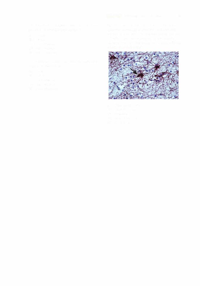
12. This tissue section from a tumor has been immunochemically stained for the intermedi ate filament protein glial fibrillary acidic protein (GFAP). Based on the reddish-brown staining observed (arrow), the tumor has originated from which of the following?
(A)Oligodendrocytes
(B)Chondrocytes
(C)Neurons
(D)Endothelial cells
(E)Fibroblasts
Answers and Explanations
1 . C. A peroxisome originates from preexisting peroxisomes. It imports specific cytosolic proteins and then undergoes fission. The other organelle that divides by fission is the mitochondrion (see receptor-mediated Chapter 3 II A 10).
2.D. Cells import cholesterol by the receptor-mediated uptake of LDLs in coated vesicles. Certain individuals inherit defective genes and cannot make LDL receptors, or they make defective receptors that cannot bind to clathrin-coated pits. The result is an inability to internalize LDLs, which leads to high levels of LDLs in the bloodstream. High LDL levels predispose a person to premature atherosclerosis and increase the risk of heart attacks (see Chapter 3 III A 1 Clinical Considerations).
3.D. Transport of protein fromthe RER to the VTC occurs via (COP-II) coatomer-coated vesicles (see Chapter 3 III B c).
4.B. Membrane recycling after exocytosis of the contents of a secretion granule occurs via clathrin-coated vesicles (see Chapter 3 III A 3).
5.C. Transfer of material among the cisternae ofthe Golgi complex in a retrograde direction takes place via (COP-I} coatomer-coated vesicles (see Chapter 3 III B e).
6.E. The uncoupling of ligands and receptors internalized by receptor-mediated endocytosis occurs in the early endosome (see Chapter 3 II A 9 b).
7.B. Proteins targeted for lysosomes (via late endosomes) leave the TGN in clathrin-coated vesi cles (see Chapter 3 II A 8 a).
8.D. Kinesin is a force-generating protein associated with microtubules. It serves as a molecular motor for the transport of organelles and vesicles outward, away from the centrosome (see Chapter 3 II C 1).
9.C. Globular actin monomers (G actin} polymerize into a double helix of filamentous actin (F actin}, also called a microfilament, in response to the regulatory influence of a number of actin binding proteins (see Chapter 3 II C 2}.
10.D. A microtubule consists of a- and -tubulin dimers polymerized into a spiral around a hollow lumen to form a fairly rigid tubule. When cross-sectioned, the microtubule reveals l3 protofila ment strands, which represent the tubulin dimers present in one complete turn of the spiral (see Chapter 3 II C 1).
11 . E. Glial filaments are a type of intermediate filament composed of GFAP and present in fibrous astrocytes. These filaments are supportive, but they may play additional roles in both normal and pathologic processes in the central nervous system (see Chapter 3 II C 3}.
12.A. The intermediate filament protein GFAP is present in glial cells, including microglia, oligo dendrocytes, fibrous astrocytes, and Schwann cells. Vimentin is found in cells of connective tissue origin, which include fibroblasts, chondrocytes, and endothelial cells. Neurons contain intermediate neurofilaments, which would not stain for either GFAP or vimentin (Chapter 3 II C 3 Clinical Considerations).
60