

I!1Jllt!llllijLymphoid Tissue 205
(see Chapter 14 Section II B 2 c), and force T cells that would be triggered by these proteins to initiate an immune response into apoptosis.
f.The T cells that survive this final test are permitted to leave the thymic medulla to be distributed bythe circulatory system to various regions ofthe body.
g.The entire process of T cell development is directed by a variety of growth factors and signaling molecules released by the various types ofepithelial reticular cells ofthe thymus.
3.T lymphocyte subtypes. There are three types of T cells: na'ive, memory, and effector T cells. Some have a number of subtypes.
a.Naive T cells are immunologically competent cells that have not as yet been activated (i.e., they have not come into contact with their designated antigen outside the thymic cortex), but possess CD45RA surface markers. Once activated, they undergo cell division and form both memory T cells and effector T cells.
b.Memory T cells, derived from naive T cells, possess CD45RO surface molecules. There are two classes of these cells, central memory T cells (CR7 + ) and effector memory T cells (CR7-).
(1 ) Central memory T cells (TCMs) reside in the paracortex of lymph nodes. Ifthey interact with an epitope that they recognize when APCs present it to them, they cause the APC to release IL12, which induces TCMs to undergo cell division, and the newly formed cells differentiate into effector T memory cells.
(2)Effector T memory cells migrate to inflammatory sites, where they undergo cell division and form effector T cells.
c.Effector T cells are derived from effector T memory cells and are capable of initiating an immune response. The subtypes ofeffector T cells areT helper cells, cytotoxic T lymphocytes (CTLs, T killer cells), and regulatory T cells (T reg cells).
(1 ) T helper cells (THO, TH1, TH2, Tl7, and THa ) are CD4+ cells. After activation, these cells synthesize and release numerous growth factors known as cytokines (lymphokines).
THO cells differentiate into the other four types of T helper cells.
(a)TH1 cells regulate responses against viral or bacterial invasions and instruct macrophages in killing bacteria.
(b)TH2 cells regulate humoral responses against parasitic or mucosal attacks by stimulating B cells to produce antibodies.
(c)T17 cells manufacture and release IL17, which recruits neutrophils and enhances their bactericidal activities.
(d)THa cells are activated by IL10 as well as interferon a or (INF-a or INF- ) to act against viruses.
(e)The cytokines IL-4, IL-5, and IL-6 released by TH2 cells induce B cells to proliferate and mature and thus respond to an antigenic stimulus, whereas IL-10 inhibits the formation ofTH1 cells, IL-9 enhances the proliferation ofTH2 cells as well as mast cell responses, and IL13 retards TH1 cell development and induces the development of B cells.
(f)Other cytokines produced by TH1 cells, such as IL-2, IFN-y, and others, modulate the immune response in diverse ways (Table 12.2). Macrophages synthesize and release IL12, which restricts the development of TH2 cells and encourages the formation ofTH1 cells.
Acquired immunodeficiency syndrome (AIDS) is caused by infection with human immunodeficiency virus, which preferentially invades TH cells,
causing a severe depression in their number and thus suppressing both cell-mediated and humoral immune responses.
1.AIDS is characterized by secondary infections by opportunistic microorganisms that cause pneumonia, toxoplasmosis, candidiasis, and other diseases.
2.It is also characterized by the development of certain malignancies, such as Kaposi sarcoma and non-Hodgkin lymphoma.

206 |
BRS Cell Biology and Histology |
|
|||
t a b I |
e |
12.2 |
Biological Activities of Selected Cytokines in the Immune Response* |
||
Cytokine |
|
Secreted by |
|
Target Cell |
Action |
I L-1 a, IL-1 b |
|
Macrophages and |
T cells, macrophages |
Activates both T lymphocytes |
|
|
|
epithelial cells |
|
and macrophages |
|
IL-2 |
|
T" 1 cells |
|
Activated T cells, B cells |
Induces mitosis of activated T and B cells |
IL-4 |
|
T"2 cells |
|
B cells |
Induces mitosis of B cells, their transformation |
|
|
|
|
|
into plasma cells; promotes isotype switching |
|
|
|
|
|
from lgM to lgG and lgE |
IL-5 |
|
T"2 cells |
|
B cells |
Induces mitosis, maturation of B cells; promotes |
|
|
|
|
|
isotype switching from lgM to lgE |
IL-6 |
|
Antigen-presenting cells |
T cells, activated B cells |
Activates T cells, induces maturation of B cells |
|
|
|
and T"2 cells |
|
to lgG-forming plasma cells |
|
IL10 |
|
T"2 cells |
|
T"1 cells |
Inhibits formation of T"1 cells; retards their |
|
|
|
|
|
ability to manufacture cytokines |
IL12 |
|
B cells, macrophages |
N K cells, T cells |
Activates N K cells, facilitates formation |
|
|
|
|
|
|
of T"1 -like cells |
IL1S |
|
Macrophages |
T"1 cells and N K cells |
Acts on T"1 cells to produce IFN-y and |
|
|
|
|
|
|
enhances NK cell activity |
IL-23 |
|
Macrophages |
CDS+ T cells |
decreases the motility of CDS+ T cells |
|
TN F-a |
|
Macrophage |
Macrophages |
Macrophages self-activate to manufacture, |
|
|
|
|
|
|
release IL12 |
|
|
T" 1 cells |
|
Activated macrophages |
Promotes production of oxygen radicals |
|
|
|
|
|
facilitating bacterial killing within endosomes |
|
|
|
|
|
of activated macrophages |
IN F-a |
|
Virally attacked cells |
N K cells, macrophages |
Activates macrophages, N K cells |
|
INF-p |
|
Virally attacked cells |
N K cells, macrophages |
Activates macrophages, N K cells |
|
IFN-y |
|
T" 1 cells |
|
Macrophages, T cells |
Activates cytotoxic T cells to kill altered and/ |
|
|
|
|
|
or foreign cells; promotes phagocytosis by |
|
|
|
|
|
macrophages |
|
|||||
lg, immunoglobulin; IL. interleukin; INF. interferon; NK. natural killer; T", T helper; TNF, tumor necrosis factor. |
|||||
*See Chapter 10 for discussion of cytokines involved in hemopoiesis. |
|
||||
|
(2) |
T cytotoxic cells are CDS+ cells. After priming by an antigenic stimulus via an APC, |
|||
|
|
T cells are induced by IL-2 to proliferate, forming new CTLs, which mediate (via |
|||
|
|
perforins and granzymes) apoptosis of foreign cells as well as virally altered self-cells |
|||
|
|
(Figure 12.1). |
|
|
|
(3)T reg cells (previously called T suppressor cells) are CD4+ cells. T reg cells are of two types, natural and inducible.
(a)Natural T reg cells (nT reg cells) originate in the thymus, possess, in addition to the CD4 marker, CD25 and Foxp3 markers (forkhead family transcription factor box p3), and suppress the immune response in a non-antigen-specific manner. Naive T cells are converted to natural T reg cells by dendritic cells of the thymus under the influence of the cytokine thymic stromal lymphopoietin (TSLP). Interestingly, among the richest sources ofTSLP are the cells ofHassall corpuscles ofthe thymus, which has been implicated in inducing nT reg cell formation.
(b)Inducible T reg cells (also known as TH3 cells) originate outside the thymus from naive T cells. They inhibit the formation ofTH1 cells, thus suppressing the immune response.
(4) Natural T killer cells are similar to NK cells except that they have to go to the thymus to become immunologically competent. They are unusual in that they recognize lipid antigens that are presented to them by APCs in conjunction with a CD1 molecule. Natural T killer cells manufacture and secrete IL-4, IL10, and INF-y, and sport a limited range of TCRs on their plasma membranes.

I!1Jllt!llllijLymphoid Tissue 207
C.B lymphocytes
1. Overview-B lymphocytes
a.B lymphocytes originate and mature into immunocompetent cells within the bone marrow. They are responsible for the humoral immune response. These cells go through a series of developmental steps going from pre-B cells through immature and finally transitional B cells, where they express different light and heavy chains of immunoglobulins on their cell membranes. Transitional B cells travel to the spleen where they either die or undergo final maturation and become mature B cells.
b.Mature B cells express immunoglobulins IgD and the monomeric form of IgM on the
external aspect of their plasma membranes (known as surface immunoglobulins [slgs]); all ofthe immunoglobulin molecules on a particularand willB cellberecognizereferred toandasbind"B cells"to the same antigenic determinant (epitope). There are several types ofB cells, but B-2 B cells and B-1 B cells are the most predominant forms.
(1 ) B-2 B cells are the "conventional" B cells in this
textbook; they are the most numerous and are present throughout the body; they must interact with T cells to propagate (in the bone marrow) and to become activated; they recognize an immense number of epitopes; and they are able to switch isotypes (i.e., switch in the synthesis of antibody types) and establish memory B cells. They are considered to be firmly related to the adaptive immune system.
(2)B-1 B cells are much fewer in number than B cells and are usually limited to the gastrointestinal and respiratory systems; they very seldom require T-cell interaction; they do not have to regenerate in the bone marrow but proliferate peripherally; they target carbohydrates rather than proteins; they very seldom switch isotypes; and they do not establish memory B-1 B cells. They cross over between the innate and adaptive immune systems.
c.CD40 molecules are present on the plasmalemmae of B cells. They interact with CD40 receptors on TH2 cells, causing release of cytokines that
(1 ) facilitate proliferation and transformation of B cells into B memory and plasma cells and
(2)inhibit TH1 cell proliferation.
d.The specific cytokines that are released by T helper cells depend on the invading pathogen.
(1 ) IL-4 and IL-5 are released by T helper cells in response to parasitic worms causing B cells to switch to IgE formation.
(2)IL-6 and IFN-y are released by T helper cells in response to bacteria and viruses in the connective tissue, causing B cells to switch to IgG formation.
(3)Transforming growth factor (TGF- ) is released by T helper cells in response to the presence of bacteria and viruses on mucosal surfaces causing B cells to switch to IgA formation.
e.B lymphocytes can present epitopes complexed with class II human leukocyte antigen (HLA) to TH1 cells; therefore, they are considered to be APCs.
f.When activated, B lymphocytes release IL-12 to induce TH1 cell formation and NK cell activation.
g.During a humoral immune response to an antigenic challenge, B lymphocytes proliferate and differentiate to form plasma cells and B memory cells (Figure 12. 1A).
2.Plasma cells lack surface antibody and actively synthesize and secrete antibody specific against the challenging antigen.
3.B memory cells are long-lived committed immunocompetent cells that are formed during proliferation in response to an antigenic challenge. They do not react against the antigen but remain in the circulation or in specific regions ofthe lymphoid system. Since they increase the size of the original clone, they provide a faster and greater secondary response (anamnestic response) against a future challenge by the same antigen.
4.There are also two groups of B cells in the spleen: the more populous follicular B cells and fewer marginal zone B cells.
a.Follicular B cells are more populous and are located in the primary and secondary follicles ofthe spleen (see VI C 3 ofthis chapter); they sport IgM, IgD, and CD21 surface markers; and

I!1Jllt!llllijLymphoid Tissue 209
1 . The MHC (major histocompatibility complex) is a large genetic complex with many loci1), that encode two main classes of integral membrane molecules: class I molecules (MHC which are expressed by nearly all nucleated cells, and class II molecules (MHC II), which are expressed by the various cells that function as APCs.
2.In humans, the MHC is referred to as the HLA complex (human lymphocyte antigen). Therefore, MHC I is class I HLA, and MHC II is class II HLA. As described below, MHC I molecules bind epitopes derived from endogenous proteins, whereas MHC II molecules bind epitopes derived from exogenous proteins. Both types of MHC molecules present their epitopes to T cells.
B.lmmunogens are molecules that are capable of inducing an immune response.All immunogens are antigens, that is, molecules whose epitopes can react with an antibody or a TCR (T cell receptor).
Most, but not all, antigens are immunogens.
1.Exogenous immunogens are derived from proteins that were endocytosed or phagocytosed by APCs and degraded intracellularly, yielding antigenic peptides containing an epitope that enter the trans-Golgi network (TGN).
2.In the TGN, the complex is sorted into specialized antigenic peptides (containing an epitope) that associate with class II HLA molecules (MHC II molecules).
a.These epitopes are relatively long, composed of 13 to 25 amino acids.
b.Class II HLA molecules are synthesized on the rough endoplasmic reticulum (RER) and are loaded within the RER cisternae with a protein known as CLIP (class II-associated invariant protein).
c.The class II HLA-CLIP complex enters the Golgi apparatus, where it is delivered to MIIC
vesicles (MHC class II compartment vesicles that already contain epitopes derived from exogenous immunogens), where the CLIP is exchanged for the epitope.
d.The epitope-class II HLA complexes are transported to and displayed on the cell surface, where they are presented to T cells.
3.Endogenous immunogens are derived from proteins that were produced within host cells (these may be viral proteins synthesized in virus-infected cells or tumor proteins synthesized in cancerous cells).
a.Class I HLA molecules (MHC I molecules), synthesized on the RER surface, enter the RER cisternae.
b.Endogenous immunogens are degraded by organelles of the host cells, known as proteasomes, into short polypeptide fragments. These fragments are antigenic peptides (8- 12 amino acids in length) known as epitopes.
c.The epitopes are transported by TAP1 and TAP2 (transporter proteins l and 2) into the RER cisternae.
d.Within the RER cisternae, the epitopes derived from endogenous immunogens are loaded on the class I HLA.
e.The peptide-class I HLA complexes are transported to the Golgi complex for sorting and eventual delivery within clathrin-coated vesicles to the cell surface, where they are presented to T cells.
C.MHC (HLA) restriction-T lymphocytes. Each subtype of T lymphocytes (except T memory cells) recognizes only epitopes that are associated with either MHC I or MHC II (class I or class II HLA) molecules as follows:
1. THl and TH2 cells (CD4+ cells) recognize MHC II (class II HLA) molecules.
2.Cytotoxic T cells (CDS+ cells) recognize MHC I (class I HLA) molecules.
3.T memory cells (CD45RO cells) recognize both MHC I and MHC II (class I and class II HLA) molecules.
I!1Jllt!llllijLymphoid Tissue |
21 1 |
2.The different isotypes exhibit functional differences.
a.lgA is secretory immunoglobulin that is released, in the form of dimers, into tears, bile, saliva, milk, and as part of the nasal discharge to protect the body (and the nursing infant) from pathogenic microorganisms and invading antigens. It is protected from digestive enzymes by its secretory component synthesized by epithelial cells and hepatocytes.
b.IgO and lgE are reaginic antibodies; IgE binds to IgE receptors of mast cells and basophils and prompts the release of pharmacologic agents from these cells to initiate an immediate hypersensitivity response. IgD binds to B-cell plasma membranes and permits these cells to recognize antigens and thus initiate a response against antigenic challenges by prompting B cells to differentiate into antibody-secreting plasma cells.
c.lgG, the most abundant serum immunoglobulin, crosses the placental barrier to shield the fetus-a process known as passive immunity. It attaches to antigenic sites on invading microorganisms, thus opsonizing these pathogens, making them available for phagocytosis by macrophages and neutrophils. These antibodies activate NK cells, thus initiating ADCC.
d.lgM forms pentamers and is the firstisotype to be formedinanimmune response. Itactivates the complement system. Its monomeric form binds to the B-cell plasma membrane.
Diffuse lymphoid tissue is considered to be a secondary lymphoid tissue. It is especially prominent in the mucosae of the gastrointestinal and respiratory systems. It is organized as nonencapsulated clusters of lymphoid cells or as lymphoid (lymphatic) nodules. Diffuse lymphoid tissue is collectively called mucosa-associated lymphoid tissue (MALT).
A.MALTconsists oftwo major types: bronchus-associated lymphoid tissue (BALl) and gut-associated lymphoid tissue (GALT). Both types possess lymphoid nodules that are isolated from one another, except in the case of Peyer patches.
B. Peyer patches are aggregates of lymphoid nodules found in the ileum. They are components of the GALT.
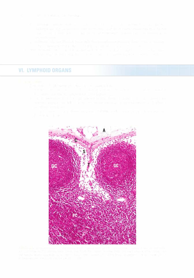
C.Lymphoid (lymphatic) nodules are transitory dense spherical accumulations of lymphocytes (mostly B cells). The dark, peripheral region of nodules (corona) is composed mainly of small, newly formed lymphocytes. Lymphoid nodules of the GALT are isolated from the lumina of their respective tracts by microfold (M) cells, which transfer antigens from the lumen and present them (without processing them into epitopes) to lymphocytes and macrophages lying in deep invaginations oftheir basal cell surfaces. From here, an appropriate immune response is mounted by lymphoid tissue in the underlying lamina propria.
1 . Secondary nodules, formed in response to an antigenic challenge, have a lightly staining central area called the germinal center, which is composed of B lymphocytes (lymphoblasts
[centroblasts] as well as centrocytes). A darker region, known as the mantle (corona), is composed of resting B cells that are being displaced from the germinal center by the newly formed B cells. In addition to centroblasts and centrocytes, the germinal center houses B memory cells, plasma cells, migrating dendritic cells, follicular dendritic cells, macrophages, and reticular cells.
a.Centroblasts do not display surface immunoglobulins (sigs), whereas centrocytes have expressed sigs.
b.Centrocytes that express sigs against self are forced into apoptosis.
c.Surviving centrocytes become B memory cells or plasma cells.
d.Migrating dendritic cells, derived from the bone marrow, are located in various regions of the body, and when they encounter an antigen, they travel to a nearby lymphoid nodule or lymph node to precipitate an immune reaction.

I!1Jllt!llllijLymphoid Tissue 213
d.Function. Lymph nodes filter lymph, maintain and produce T and B cells, and possess memory cells (especially T memory cells). Antigens delivered to lymph nodes by APCs are recognized by T cells, and an immune response is initiated. Additionally, some of the stromal cells and some of the dendritic cells of lymph nodes express the transcription factor AIRE (autoimmune regulator), which selectively induces anti-self-T cells that escaped destruction in the thymus to go into apoptosis.
2.Structure-Lymph nodes. Lymph nodes are divided into three regions: the outermost cortex, the middle paracortex, and the innermost medulla (Figure 12.2). Stromal cells of these regions release a variety of chemotactic chemokines lymphocytes (CCLs) that attract lymphocytes. Depending on the CCL released, B cells or T cells are attracted to a particular region ofthe lymph node. In order to attract both B cells and T cells, CCLs are conveyed to the luminal endothelial cell membranes ofpostcapillary venules, the region where lymphocytes leave the circulatory system to enter the substance ofthe lymph node. Once in the lymph node stroma, both B cells and T cells are segregated within the cortex ofthe lymph node by those CCLs to which each responds.
a.The cortex of lymph nodes
(1 ) lies deep to the capsule, from which it is separated by a subcapsular sinus.
(2)is incompletely subdivided into compartments by connective tissue septa derived from the capsule.
(3)contains lymphoid nodules and sinusoids.
(a)Lymphoid nodules are composed mainly ofB cells but also ofsome T cells, follicular dendritic cells, macrophages, and reticular cells. They may possess a germinal center and then they are known as secondary lymphatic nodules.
(b)Sinusoids are endothelium-lined lymphatic spaces that extend along the capsule and trabeculae and are known as subcapsular and cortical sinusoids, respectively.
b.The paracortex of the lymph node is located between the cortex and the medulla.
(1 ) It is composed of a non-nodular arrangement of mostly T lymphocytes (the thymus dependent area of the lymph node) and dendritic cells.
(2)The paracortex is the region where circulating lymphocytes gain access to lymph nodes via postcapillary (high endothelial) venules (HEVs).
c.The medulla of a lymph node lies deep to the paracortex and cortex, except at the region of the hilum. It is composed of medullary sinusoids and medullary cords.
(1 ) Medullary sinusoids are endothelium-lined spaces supported by reticular fibers and reticular cells. They frequently contain macrophages and receive lymph from the cortical sinuses.
(2)Medullary cords are composed of lymphocytes and plasma cells.
B. The thymus is a primary lymphoid organ.
1.Overview-Thymus
a.The thymus is a bilobed structure located in the neck. It is derived from both endoderm (epithelial reticular cells), derived from the third pharyngeal pouch of the embryo, and mesoderm (lymphocytes [thymocytes]). The development of the epithelial reticular cells of the thymus (especially in the medulla) is dependent on the formation and release of lymphotoxins, members of the tumor necrosis factor family of cytokines. In the absence of these proteins the thymus does not develop or function properly. The thymus begins to involute near the time of puberty.
b.A connective tissue capsule surrounds the thymus. The septa of this capsule divide the parenchyma into incomplete lobules, each of which contains a cortical and medullary region (Figure 12.3). The thymus does not possess lymphoid nodules.
2.Structure-Thymus
a.The thymic cortex is supplied by arterioles in the septa; these arterioles provide capillary loops that enter the substance of the cortex. The cortex is the region in which T-cell maturation occurs.
(1 ) Epithelial reticular cells (Figure 12.3)
(a)There are six types of epithelial reticular cells (see Table 12.4). They are pale cells (derived, during embryogenesis, from the third and perhaps fourth pharyngeal pouches) andhave a large ovoid lightlystaining nucleus that often displays a nucleolus.


I!1Jllt!llllijLymphoid Tissue 21 5
(b)They possess long processes that surround the thymic cortex, isolating it from both the connective tissue septa and the medulla. These processes, which are filled with bundles oftonofilaments, form desmosomal contacts with each other.
(c)They manufacture thymosin, serum thymic factor, thymopoietin, and thymic stromal lymphopoietin, hormones that function in the transformation of immature T lymphocytes into immunocompetent T cells.
(2)Thymocytes
(a)Thymocyte plasmalemma possesses Notch-1 receptors that permit these cells to respond to cytokines released by epithelial reticular cells to become T cells. Once committed to the T-cell lineage, theyare known as immature T lymphocytes and are noted to be present within the thymic cortex in different stages of differentiation.
(b)Thymocytes are surrounded by processes of epithelial reticular cells, which help segregate thymocytes from antigens during their maturation.
(c)They migrate toward the medulla as they mature. Most T cells die in the cortex, and the dead cells are phagocytosed by macrophages.
(d)Surviving T cells are naive. Theyleave the thymus and are distributed to secondary lymphoid organs by the vascular system.
(3)Blood-thymus barrier
(a)This barrier exists in the cortex only, making it an immunologically protected region.
(b)It ensures that antigens escaping from the bloodstream do not reach developing T cells in the thymic cortex.
(c)It consists of the following layers: endothelium of the thymic capillaries and the associated basal lamina, perivascular connective tissue and cells (e.g., pericytes and macrophages), and type I epithelial reticular cells and their basal laminae.
b.Thymic medulla
(1 ) The thymic medulla is continuous between adjacent lobules and contains large numbers of epithelial reticular cells and mature T cells, which are loosely packed, causing the medulla to stain lighter than the cortex (Figure 12.3).
(2)It also contains whorl-like accretions of type VI epithelial reticular cells called Hassall corpuscles (thymic corpuscles). These structures display various stages of keratinization
and increase in number with age. It has been shown that these epithelial reticular cells manufacture TSLP (thymic stromal lymphopoietin), a cytokine that facilitates dendritic cell maturationwhich, in turn, elicit the transformation of naive T cells into natural T reg cells.
(3)Mature T cells exit the thymus via venules and efferent lymphatic vessels from the thymic medulla. The T cells then migrate to secondary lymphoid structures.
(4)Hormones acting on the thymus
(a)Thymosin, thymopoietin, thymulin (thymic factor), somatotropin, thymic stromal lymphopoietin, and thymic humoral factor promote the formation of immunocompetent T cells.
(b)Thyroxin encourages thymulin production by epithelial reticular cells.
(c)Adrenocorticosteroids depress T-cell formation in the thymus.
DiGeorge syndrome, also called congenital thymic aplasia, is character ized by the congenital absence of the thymus and parathyroid glands,
resulting from abnormal development of the third and fourth pharyngeal pouches.
1. This syndrome is associated with abnormal cell-mediated immunity but relatively normal humoral immunity.
2.It usually results in death from tetany or uncontrollable infection.
C. Spleen
1.Overview-Spleen
a.A simple squamous epithelium (peritoneum) covers the dense irregular collagenous connective tissue capsule of the spleen, which sends trabeculae into the substance of the spleen to form a supportive framework (Figure 12.4).

21 6 |
BRS Cell Biology and Histology |
FIGURE 12.4. This low-power photomicrograph displays the smooth capsule IC) of a human spleen. Observe that the spleen is not divided into a cortex and a medulla; instead, it has red pulp IRP) and white pulp. Note the presence of a lym phoid nodule with a germinal center IGC) composed of B lymphocytes. The lymphoid nodule is invested by the T-cell-rich region of the spleen, known as the periarterial lymphatic sheath IPALS). The arrow points to the marginal zone located between the white pulp and the RP I X 1 32).
b.The spleen is similar to lymph nodes in that it possesses a hilum, but differs from both the thymus and the lymph nodes in that it lacks a cortex and medulla. It further differs from lymph nodes because it has no afferent lymphatic vessels.
c.The spleen is divided into red pulp and white pulp; the latter contains lymphoid elements.
These two regions are separated from each other by the marginal zone (Figure 12.4).
d. Function-Spleen. The spleen filters blood, phagocytoses damaged and aged erythrocytes, and is a site of proliferation of B and T lymphocytes and the production of antibodies by plasma cells. In some animals, such as the dog but not in humans, it stores red blood cells.
2.Vascularization of the spleen is derived from the splenic artery, which enters the hilum and gives rise to trabecular arteries.
a.Trabecular arteries leave the trabeculae, become invested by a periarterial lymphatic sheath (PALS, described later), and are known as central arteries.
b.Central arteries branch but maintain their lymphatic sheath until they leave the white pulp to form several straight penicillar arteries.
c.Penicillar arteriesenterthe redpulp.Theyhave three regions: pulp arterioles, macrophage sheathed arterioles, and terminal arterial capillaries. These last named vessels either drain directly into the splenic sinusoids (closed circulation) or terminate as open-ended vessels within the splenic cords of the red pulp (open circulation).
d.Splenic sinusoids are drained by pulp veins, which are tributaries of the trabecular veins; these in turn drain into the splenic vein, which exits the spleen at the hilum.
3.Structure-Spleen
a.White pulp of the spleen includes all of the organ's lymphoid tissue (diffuse and nodular), such as lymphoid nodules (mostly B cells) and PALS (mostly T cells) around the central arteries. It also contains macrophages and other APCs.

I!1Jllt!llllijLymphoid Tissue 21 7
b.The marginal zone (MZ) of the spleen
(1 ) is a sinusoidal region between the red and white pulps at the periphery of the PALS
(Figure 12.4).
(2)receives blood from capillary loops derived from the central artery and is thus the first site where blood contacts the splenic parenchyma.
(3)is richly suppliedbyavidlyphagocytic macrophages and otherAPCs. There are two types of macrophages in the marginal zone: the MZ macrophages and the MZ metallophilic macrophages.
(a)MZ macrophages express molecules on their surface that recognize pathogens such as bacteria, viruses, and yeasts.
(b)MZ metallophilic macrophages express molecules that bind to cell-bound
oligosaccharide ligands; these macrophages also interact with marginal zone B cells (see Section II C 4 b in this chapter), promotingthem to migrate to the white pulp ofthe spleen and present blood-borne antigens to T cells.
(4)The MZ is the region where circulating T and B lymphocytes enter the spleen before becoming segregated to their specific locations within the organ and where interdigitating dendritic cells are able to display their MHC-epitope complex for recognition by T cells.
(5)Stromal cells ofthe MZ release a variety ofCCLs (chemotactic chemokines lymphocytes) that attract lymphocytes. Depending on the CCL released, B cells or T cells are attracted to a particular region of the spleen. In order to attract both B cells and T cells to the MZ, CCLs are conveyed to the luminal endothelial cell membranes of MZ sinusoids. Once in the spleen parenchyma, both B cells and T cells are segregated within the white pulp of the spleen by those CCLs to which each responds.
c.Red pulp of the spleen (Figure 12.4) is composed of an interconnected network of sinusoids supported by a loose type of reticular tissue (splenic cords).
(1 ) Sinusoids
(a)are lined by long fusiform endothelial cells separated by relatively large blood containing intercellular spaces.
(b)have a discontinuous basal lamina underlying the endothelium and circumferen tially arranged ribs of reticular fibrils.
(2)Splenic cords (cords of Billroth) contain plasma cells, stellate reticular cells, blood cells, and macrophages enmeshed within the spaces ofthe reticular fiber network. Processes of the macrophages enter the lumina of the sinusoids through the spaces between the endothelial cells.
D.Tonsils are aggregates of lymphoid tissue, which sometimes lack a capsule. All tonsils are in the upper section of the digestive tract, lying beneath but in contact with the epithelium. B and T cells in the three sets oftonsils are localized in differentregions ofany particular tonsil. The mechanism of distribution is similar to that which occurs in lymph nodes and the spleen, and depends on the various chemotactic chemokines for lymphocytes (CCLs) secreted by stromal cells. Tonsils assist in combating antigens entering via the nasal and oral epithelia.
1 . Palatine tonsils
a.possess crypts, deep invaginations of the stratified squamous epithelium covering of the tonsils, frequently containing debris;
b.possess primary and secondary (with germinal centers) lymphoid nodules;
c.are separated from subjacent structures by a connective tissue capsule.
2.The pharyngeal tonsil is a single tonsil in the posterior wall of the nasopharynx.
a.It is covered by a pseudostratified ciliated columnar epithelium.
b.Instead of crypts, it has longitudinal pleats (infoldings).
c.Possesses primary and secondary (with germinal centers) lymphoid nodules.
3.Lingual tonsil
a.is on the dorsum ofthe posterior third ofthe tongue and is covered by a stratified squamous nonkeratinized epithelium.
b.possesses deep crypts, which frequently contain debris. Ducts of the posterior mucous glands of the tongue often open into the base of these crypts.
c.possesses primary and secondary (with germinal centers) lymphoid nodules.