


Extracellular Matrix
A.Structure. The extracellular matrix (ECM) is an organized meshwork of macromolecules surrounding and underlying cells. Although it varies in composition, in general it consists of an amorphous ground substance (containing primarily glycosaminoglycans [GAGs], proteoglycans, and glycoproteins [multiadhesive glycoproteins]) and fibers (Figure 4.1).
B. Functions. The ECM, along with water and other small molecules (e.g., nutrients, ions), constitutes the extracellular environment. By affecting the metabolic activities of cells in contact with it, the ECM may alter the cells and influence their shape, migration, cell-cell interactions, cell division, and their differentiation. Additionally, the ECM provides physical support against compressive as well as tensile forces.
A.Glycosaminoglycans (GAGs), are long, unbranched polysaccharides composed of repeating identical disaccharide units.
1.An amino sugar, either N-acetylglucosamine or N-acetylgalactosamine, is always one of the repeating disaccharides.
2.Because GAGs are commonly sulfated and usually possess a uronic acid sugar, which has a carboxyl group in the repeating disaccharide unit, they have a strong negative charge.
3.GAGs are generally linked to a core protein.
4. The attraction of osmotically active cations (e.g., Na+) to GAGs results in a heavily hydrated matrix that strongly resists compression.
5.Their extended random coils occupy large volumes of space because they do not fold compactly.
6.GAGs may be classified into four main groups on the basis of their chemical structure (Table 4.1).
a.Hyaluronic acid (hyaluronan) is a very large unsulfated molecule up to 20 1-!m inlength and as much as 10,000 kDa in weight; but it is not attached to a core protein.
b.The other three GAG groups are chondroitin sulfate and dermatan sulfate, heparin and heparan sulfate, and keratan sulfate.
61


l!il!lJ'dQijExtracellular Matrix |
63 |
B.Proteoglycans consist of a core protein from which many GAGs extend. These large molecules are shaped like a bottlebrush (Figure 4. 1A).
1.Proteoglycans may attach to hyaluronic acid via their core proteins to form large complex aggregates.
2.Their core proteins, their molecular size, and the number and types of GAGs they contain show marked heterogeneity.
3.Function. Proteoglycans act as binding sites for growth factors (e.g., fibroblast growth factor) and other signaling molecules. They confer unique attributes on the ECM in certain locations (e.g., selective permeability in the filtration barrier of the glomerulus).
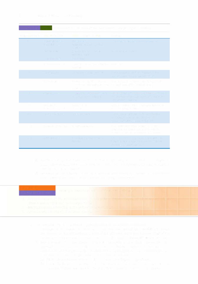
C.Glycoproteins (multiadhesive glycoproteins) are multifunctional molecules whose domains bind to components of the ECM and to receptors on the cell surface, thereby promoting adhesion between the cell and the matrix (Table 4.2).
1.Fibronectin
a.Types and location
(1 ) Matrix fibronectin forms fibrils in the ECM.
(2)Cell-surface fibronectin is a protein that transiently attaches to the surface of cells.
(3)Plasma fibronectin is a circulating plasma protein that functions in blood clotting, wound healing, and phagocytosis.
b.Function. Fibronectin is a multifunctional molecule.
(1 ) Fibronectin has domains for binding collagen, heparin, various cell-surface receptors, and cell adhesion molecules (CAMs).
(2)It mediates cell adhesion to the ECM by binding to fibronectin receptors on the cell surface.
CLINICAL |
Wound healing in adults involves the formation of fibronectin tracks |
|
CONSIDERATIONS |
||
along which cells migrate to their destinations. |
||
|
1 . In connective tissue, wound healing is often characterized by migration of fibroblasts across blood clots, where they adhere to fibronectin.
2. In epithelia, wound healing involves reepithelialization, which depends on the basal lamina serving as a scaffold for cell migration to cover the denuded a rea; epithelial cell proliferation and replacement then occur.
t a b I e |
4.2 |
Major Glycoproteins of the Extracellular Matrix |
|
Glycoprotein |
Location |
|
Function |
Fibronectin |
Most connective tissues |
Binds collagen, heparan sulfate, various cell-surface receptors, |
|
|
|
|
and CAMs, thus mediating cell adhesion to the ECM. |
Laminin |
Basal laminae of epithelial cells and |
|
external laminae of muscle cells and |
|
Schwann cells |
Entactin |
Basal laminae of epithelial cells and |
|
external laminae of muscle cells and |
|
Schwann cells |
Anchors cells to their basal laminae (and external lamina), thus assisting in adhering epithelial cells to the underlying connective tissue.
Links laminin to type IV collagen of the basal laminae (and external laminae).
Tenascin |
Embryonic connective tissue |
Facilitates cell-matrix adhesion, thus assisting in cell migration. |
Chondronectin |
Cartilage |
Assists cartilage cells in adhering to their matrix. |
Osteonectin |
Bone |
Assists bone cells in adhering to their matrix. |
|
|
Influences calcification of the bone matrix. |
|
|
|
CAMs. c e l l adhesion molecules;

64BRS Cell Biology and Histology
2.Laminin is located in basal laminae, where it is synthesized by adjacent epithelial cells, and in external laminae surrounding muscle cells and Schwann cells.
a.The arms of this large cross-shaped glycoprotein have binding sites for cell-surface receptors (integrins), heparan sulfate, type IV collagen, and entactin.
b.Function. Laminin mediates interaction between epithelial cells and the ECM by anchoring the cell surface to the basal lamina.
3.Entactin is a component of all basal (and external) laminae. a. This sulfated adhesive glycoprotein binds laminin.
b.Function. Entactin links laminin with type IV collagen in the lamina densa.
4.Tenascin is an adhesive glycoprotein most abundant in embryonic tissues. a. Tenascin is secreted by glial cells in the developing nervous system.
b.Function. Tenascin promotes cell-matrix adhesion and thus plays a role in cell migration.
5.Chondronectin, a glycoprotein in cartilage, attaches chondrocytes to type II collagen.
a.This multifunctional molecule has binding sites for collagen proteoglycans and cell-surface receptors.
b.Function. By influencing the composition of its ECM, chondronectin plays a role in the development and maintenance of cartilage.
6.Osteonectin
a.This ECM calcium-binding glycoprotein found in bone is synthesized by osteoblasts.
b.It has binding sites fortype I collagen and for integrins of osteoblasts and osteocytes.
c.Function. Osteonectin plays a role in bone formation and remodeling and in maintaining bone mass by influencing calcification.
D. Fibronectin receptors, which belong to the integrin family of receptors, are transmembrane proteins consisting oftwo polypeptide chains.
1.Because they enable cells to adhere to the ECM, they are known as CAMs.
2.They bind to fibronectin via a specific tripeptide sequence (Arg-Gly-Asp; RGD sequence); other extracellular adhesive proteins also contain this sequence.
3.Function. They link fibronectin outside the cell to cytoskeletal components (e.g., to actin) inside the cell (Figure 4.2) and may activate cell-signaling pathways that determine the cell's behavior; conversely, the cell can enhance or inhibit its ability to bind to the ECM.
A.Collagen is the most abundant structural protein of the ECM. It exists in at least 25 molecular types, which vary in the amino acid sequence of their three a-chains (Table 4.3). There are four major categories of collagens into which the 25 or so different molecular types of collagens may be classed. They are fibril-forming, fibril-associated, network-forming, and transmembrane collagens. Before the discussion of the members of these major categories, fibril-forming collagen synthesis and network-forming collagens have to be described.
1.Collagen synthesis and assembly into fibrils occur via a series of intracellular and extracellular events (Figure 4.3).
a. Intracellular events in collagen synthesis occur in the following sequence:
(1 ) Preprocollagen synthesis occurs at the rough endoplasmic reticulum (RER) and is directed by messenger ribonucleic acid (mRNA) that encodes the different types of a-chains to be synthesized.
(2)Hydroxylation of specific proline and lysine residues of the forming polypeptide chain occurs within the RER. The reaction is catalyzed by specific hydroxylases that require vitamin C as a cofactor.
(3)Attachment of sugars (glycosylation) to specific hydroxylysine residues also occurs within the RER.
(4)Procollagen triple-helix formation takes place in the RER and is precisely regulated by propeptides (extra nonhelical amino acid sequences) at both ends of each a-chain. The three a-chains align and coil into a triple helix.

l
Extracellularr |
Fibronectin |
lntegrin subunits |
|
FIGUREspace4.2. |
l ntegrin receptors such as the |
||
|
|
fibronectin receptor link molecules outside the cell with components inside |
|
the cell. This is common at focal contacts (adhesion plaques), where the integrins serve as transmembrane linkers that mediate reciprocal interactions between the cytoskeleton and the extracellular matrix. IAdapted from Gartner LP, Hiatt JL. Color
Textbook of Histology. 2nd ed. New York, NY: Saunders; 2001 :46 ) |
|
|||
t a b I e |
4.3 |
Characteristics of Some of the Best Known Collagen Types |
||
Molecular Type |
Cells Synthesizing |
Major Locations in Body |
Function |
|
|
Fibroblasts |
Dermis of skin, tendons, |
Resists tension |
|
|
|
|
ligaments, fibrocartilage, |
|
|
|
|
capsules of some organs |
|
|
Dsteoblasts |
Bone matrix |
The arrangement of collagen fibers in compact |
|
|
|
|
|
bone reduces the presence of cleavage planes |
|
Ddontoblasts |
Dentin matrix |
Structural support and provides a degree of |
|
|
|
|
|
elasticity to dentin |
I I |
Chondroblasts |
Hyaline and elastic cartilages |
Resists intermittent pressure |
|
I l l |
Fibroblasts |
Dermis of skin and capsules of |
Forms structural framework |
|
|
|
|
some organs |
|
|
Reticular cells |
Lymph nodes, spleen |
|
|
|
Smooth muscle cells |
Smooth muscle |
Forms external lamina |
|
|
Schwann cells |
Nerve fibers |
|
|
|
Hepatocyte |
Liver |
Forms reticular fibers |
|
(continued)
65
66 |
BRS Cell Biology and Histology |
|
|||
|
t a b I e |
4.3 |
Characteristics of Some of the Best Known Collagen Types (continued) |
||
Molecular Type |
Cells Synthesizing |
Major Locations in Body |
Function |
||
|
IV |
Endothelial cells |
Blood vessels |
Forms lamina densa of the basal lamina |
|
|
|
Epithelial cells |
Epidermis and lining of body |
|
|
|
|
|
|
cavities |
|
|
|
Muscle cells |
Skeletal muscles, smooth |
Forms external lamina |
|
|
v |
|
|
muscles, heart |
|
|
Schwann cells |
Nerve fibers |
|
||
|
Mesenchymal cells |
Placenta and dermal-epidermal |
Unknown |
||
|
|
|
|
junction |
|
|
VII |
Keratinocytes |
Dermal-epidermal junction |
Forms anchoring fibrils that secure lamina |
|
|
|
|
|
|
densa to underlying connective tissue |
IX |
Chondrocytes |
Hyaline and elastic cartilages |
|
|
(associated with collagen types |
|
|
II and XI) |
Binds to type I I collagen and affixing itto the proteoglycans of the cartilage matrix
XI |
Chondrocytes |
Hyaline and elastic cartilages |
Acts to stabilize the type I I and type IX collagen |
|
|
as well as type I collagen |
substructure ofthe cartilage matrix; forms the |
|
|
|
core of type I collagen |
XII |
Fibroblasts |
Dermis of skin |
Binds to surface of type I collagen and assist it |
|
Mesenchymal cells |
Placenta |
in resisting tensile forces |
XIII |
Various cell types |
In various tissues |
Assists in the formation of focal adhesions |
|
|
|
by binding to fibronectin, integrins, and |
|
|
|
components of the lamina reticularis |
XVI I |
Epidermis of the skin |
Hemidesmosomes |
It has domains that are embedded in the |
|
|
|
epidermal cell membrane binding both to |
|
|
|
keratins as well as to integrins and laminin |
XVI I I |
Epithelial cells |
Basal lamina of the retina of |
|
|
the eye |
When degraded enzymatically, it inhibits the formation of new blood vessels and induces apoptosis of endothelial cells
(5)Addition of carbohydrates occurs in the Golgi complex, to which procollagen is transportedvia transfervesicles. With the addition ofcarbohydrates, the oligosaccharide side chains are completed.
(6)Secretion of procollagen occurs by exocytosis after secretory vesicles from the trans Golgi network are guided to the cell surface along microtubules.
CLINICAL |
Scurvy is associated with a deficiency of vitamin C. |
|
CONSIDERATIONS |
||
|
1 . Scurvy is caused by the synthesis of poorly hydroxylated tropocollagen, which is unable to form either a stable triple helix or collagen fibrils.
2.Symptoms include bleeding gums and eventual tooth loss.
3.Administration of vitamin C reverses the disease.
b. Extracellular events in collagen synthesis occur in the following sequence:
(1 ) Cleavage of procollagen is catalyzed by the enzymes procollagen peptidases, which are located on the extracellular aspect of the cell membrane and remove most of the propeptide sequences at the ends of each a-chain, yielding tropocollagen molecules.
(2)Self-assembly of tropocollagen occurs as insoluble tropocollagen molecules that aggregate near the cell surface. The cell establishes longitudinal furrows in its membrane, and the procollagen molecules are discharged into these furrows and are converted into tropocollagen molecules (as just described).
(a)Fibrils characteristic of types I, II, III, V, and VII collagen are produced.
(b)These fibrils have a transverse banding periodicity of 67 nm in types I, II, and III collagen (Figures 4.4 and 4.5); the periodicity varies in other types of collagen.


68 |
BRS Cell Biology and Histology |
Each collagen fiber bundle is composed of smaller fibrils, which in turn consist of aggregates of tropocollagen molecules. Tropocollagen molecules self-assemble in the extracellular environment in such a fashion that there is a gap between the tail of the one and the head of the succeeding molecule of a single row. As fibrils are formed, tails of tropocollagen molecules overlap the heads of tropocollagen molecules in adjacent rows. Additionally, the gaps and overlaps are arranged so that they are in register with those of neighboring (but not adjacent) rows of tropocollagen molecules. When stained with a heavy metal, such as osmium, the stain preferentially precipitates in the gap regions, resulting in the repeating light and dark banding of collagen.
FIGURE 4.4. The levels of organization in collagen fibers. As seen by light microscopy, collagen fibers consist of collagen fibrils, which typically reveal a 67-nm cross-banding when observed by electron microscopy. The periodicity along the
collagen fibril is due to the precise arrangement of tropocollagen molecules, which overlap each other,& producing gap regions where electron-dense stains penetrate and produce a transverse banding across the fibril. (Reprinted with permission from Gartner LP. Hiatt JL. Color Atlas and Text ofHistology. 6th ed. Baltimore. MD: Wolters Kluwer Health/Lippincott Williams Wilkins; 201 3:66.)

l!il!lJ'dQijExtracellular Matrix |
69 |
FIGURE 4.5. Electron micrograph showing a number of collagen fibrils with their characteristic 67-nm cross-banding. The large black structures represent calcium phosphate deposits.
2.Synthesis of network-forming collagens, specifically type IV collagen, is unique in that it assembles into a meshwork rather than fibrils.
a.TypeIV collagen constitutes most ofthe lamina densa ofbasal laminae and external laminae.
b.It differs from other collagen types as follows:
(1 ) The propeptide sequences are not removed from the ends of its procollagen molecules.
(2)Its triple-stranded helical structure is interrupted in many regions.
(3)It forms head-to-head dimersthat interact to form lateral associations, creating a sheet like meshwork.
Alport syndrome is the result of genetic defects in the genes that are responsible for the formation of collagen types Ill, IV, and V, which result in
the abnormal type IV collagen assembly. Since the lamina densa of the basal lamina is formed mostly by type IV collagen, individuals afflicted by AIport syndrome present with glomerulonephritis that results in end-stage kidney disease. These individuals also have hearing disorders and an anomaly of the lens of their eyes known as lenticonus, a bulge that occurs in the lens during development; the consequence of the bulge may be a herniated lens. Since the gene that codes for type IV collagen is
located on the X chromosome, AIport syndrome occurs much more frequently in males than in females.
3.Classes of Collagens
a.Fibril-forming collagens (types I, II, III, V, and XI) are the most common of the collagens; their subunits are the tropocollagen molecules that assemble to form long, flexible fiber bundles whose tensile strength is greater than that of stainless steel of the same diameter. This class of collagen, because of their staggered arrays of tropocollagen molecules,
70 |
BRS Cell Biology and Histology |
display a 67-nm cross-banding. Each tropocollagen molecule, as mentioned above, is composed of three a-chains wrapped around each other, where each a-chain is about 1,000 amino acids long. Because every third amino acid in each a-chain is a glycine, the smallest of the amino acids, the three chains can form a very tight helix by bending around these amino acids. Moreover, hydroxyproline and hydroxylysine molecules also abound in all three a-chains; the hydroxyprolines of each a-chain form tight bonds with each other, assisting in the maintenance of the tight helix. The hydroxylysines bind to each other across neighboring tropocollagen molecules, thus assisting in the formation of collagen fiber bundles.
b.Fibril-associated collagens (types IX and XII) are bound to the surfaces of fibril forming collagens and thereby they stabilize the collagen framework of the tissues in which they reside by adhering not only to the collagen fibers but also to the molecules of the ground substance. Type IX collagens are associated with type II collagens of cartilage, and type XII collagens are localized on the surface of type I collagens of the dermis and placenta.
c.Network-forming collagens (types IV and VII), unlike the other types of collagen, have procollagen as their subunits. As described above, procollagen possesses the propeptide sequences at both ends of the molecule, which, in fibril-forming collagens, are removed by procollagen peptidase in the extracellular environment. Procollagen molecules are unable to assemble in the style of tropocollagen molecules; hence, they do not form fibers. Instead, the procollagen molecules form head-to-head dimers that interact forming lateral associations, creating a sheet-like meshwork. Type IV collagen forms the lamina densa of the basal lamina, and type VII collagens form the anchoring fibrils that secure the lamina densa to the lamina reticularis of the underlying connective tissue.
d.Transmembrane collagens (types XIII, XVII, and XVIII) are associated with focal adhesions, hemidesmosomes, and the basal laminae, respectively.
Knobloch syndrome (type I) is an inherited disease resulting from the malformation of type XVI I I collagen. The symptoms include encephalo
cele, the formation of large brain vesicles that jut out through a defect ofthe bony skull due to incom plete fusion of the neural tube during embryonic development, as well as sporadic detachment of the retina, and nearsightedness that may become evident by the time the child is a year old.
B.Elastic fibers
1 . Components
a.Elastin, an amorphous structural protein, imparts remarkable elasticity to the ECM; 90% of elastic fibers or elastic sheets are composed of elastin.
(1 ) Elastin is unusual in that its lysine molecules form unique linkages with one another.
(2)Lysine residues of four different chains form covalent bonds called desmosine cross- links to create an extensive elastic network.
(3)Like a rubber band, after being stretched, the elastin returns to its original shape once the tensile force is released.
b.Fibrillin-1, a glycoprotein, organizes elastin into fibers and is the main component of the peripheral microfibrils of elastic fibers.
(1 ) The amino terminus of one fibrillin-1 interacts with the carboxyl terminus of another fibrilin- 1 molecule to form pencil-like head-to-tail assembly of fibrilin- 1 molecules to form microfibrils.
(2)Fibrillin- 1 possesses binding sites for tropoelastin, which form cross-links with each other.
(3)Heparin competes with tropoelastin for binding sites on fibrillin-1, which probably has a regulatory effect on elastic fiber formation.

l!il!lJ'dQijExtracellular Matrix |
71 |
c. Fibulin-5, a protein, forms bonds with integrin molecules ofcells such asvascular smooth muscle cells and endothelial cells ofblood vessels, and also facilitates the formation of elastic fibers. (1 ) Fibulin-5 also binds to microfibrils and tropoelastin.
(2)It has been shown that during wound healing fibulin-5 is present in an increased concentration than in undamaged blood vessels.
d.Type VIII collagen is often associated with elastic fibers, most probably to limit the extent of elastic fiber stretching, protecting the elastic fibers from being damaged by overstretching.
2.Synthesis of elastic fibers is carried out by fibroblasts in elastic ligaments, smooth muscle cells in large arteries, and chondrocytes and chondroblasts in elastic cartilage.
a.Synthesis begins with the elaboration offibrillin microfibril templates that are arranged in a parallel array near regions of the cell surface.
b.These cells manufacture and exocytose a soluble form of elastin, known as tropoelastin, and fibulin-5 which bind not only to each other but also to fibrillin- 1 .
c.Tropoelastin molecules cross-link with each other to form insoluble, mature elastin.
d.Additional factors, such as heparan sulfate, microfibril-associated glycoproteins, and fibrillin-2, have been implicated in the assembly of elastic fibers.
CLINICAL |
Marfan syndrome results from mutations in the genes encoding fibrillin, |
|
CONSIDERATIONS |
||
a critical component of elastic fibers. |
||
|
1.Patients with this condition have unusually long, slender limbs and long fingers.
2.The lens of the eye often dislocates; cardiovascular problems are common; and the aorta may rupture, causing death.
3.Treatment includes drugs that decrease blood pressure, and in severe cases surgery replacing the aorta.

Review Test
Directions: Each of the numbered items or incomplete statements in this section is followed by answers or by completions of the statement. Select the ONE lettered answer or completion that is BEST in each case.
1 . Which one of the following statements about the fibronectin receptor is true?
(A)It is located exclusively in the basal lamina.
(B)It is a cross-shaped glycoprotein.
(C)It mediates the linkage of molecules out side the cell with cytoskeletal elements inside the cell.
(D)It belongs to the entactin family of receptors.
(E)Its absence is associated with scurvy.
2.Which one of the following events in colla gen synthesis occurs outside of the cell?
(A)Synthesis of preprocollagen
(B)Hydroxylation oflysine residues
(C)Triple-helix formation
(D)Carbohydrate addition to procollagen
(E)Cleavage of procollagen by procollagen peptidases
3.A medical student goes to the emergency department and is diagnosed with a ruptured bowel, the result of a genetic condition called Ehlers-Danlos type IV syndrome. Which one of the following statements about this patient's condition is true?
(A)He has a defect in the synthesis of mRNA encoding type I collagen.
(B)He has a defect in the genes encoding type IV collagen.
(C)He has defective type II collagen.
(D)He has an increased riskofbreaking his bones.
(E)He has a defect in the translation ofmRNA for type III collagen.
4.Which one of the following statements about hyaluronic acid is true?
(A)It is a component ofelastic fibers.
(B)It is a glycosaminoglycan.
(C)It is a proteoglycan with a shape resem bling a bottlebrush.
(D)It is sulfated.
(E)It is a small molecule.
5.Which one of the following statements about osteonectin is true?
(A)It is present in the lacunae of bone.
(B)It is a proteoglycan.
(C)It binds to type II collagen.
(D)It influences calcification of bone.
(E)It is synthesized by osteoclasts.
6.Which of the following statements about scurvy is true?
(A)One of its symptoms is bowlegs.
(B)It is caused by excessive glycosylation of tropocollagen.
(C)It is caused by a deficiency ofvitamin A.
(D)It is associated with structurally defective elastic fibers.
(E)It is alleviated by eating citrus fruits.
7.Which one of the following is a glycoprotein across which fibroblasts migrate during wound healing?
(A)Fibrillin
(B)Fibronectin
(C)Elastin
(D)Entactin
(E)Laminin
72

8.Which of the following is an adhesive glyco protein that links type IV collagen with laminin in the lamina densa?
(A)Fibrillin
(B)Fibronectin
(C)Elastin
(D)Entactin
(E)Tenascin
9.Which one of the following is a main com ponent of peripheral microfibrils in an elastic fiber?
(A)Fibrillin
(B)Fibronectin
(C)Elastin
(D)Entactin
(E)Laminin
l!i1fftlllilExtracellular Matrix |
73 |
10. Which one ofthe following is present in the basement membrane and is manufactured by connective tissue cells?
(A)Fibrillin
(B)Fibronectin
(C)Elastin
(D)Entactin
(E)Laminin

Answers and Explanations
1 . C. The fibronectin receptor is a transmembrane protein that enables cells to adhere to the ECM. Laminin is a cross-shaped glycoprotein in the basal lamina, where entactin is also present (see Chapter 4 II D).
2.E. In the extracellular space, peptidases cleave off end sequences ofprocollagen to yield tropo collagen, which self-assembles to form collagen fibrils (see Chapter 4 III A 1).
3.E. Ehlers-Danlos type IV syndrome is associated with a defect in the synthesis and translation of mRNA for type III reticular collagen (see Chapter 4 III A 1 Clinical Considerations).
4.B. Hyaluronic acid is a glycosaminoglycan, not a proteoglycan. The core protein of proteogly cans can attach to hyaluronic acid forming large aggregates (see Chapter 4 II A).
5.D. Osteonectin synthesized by osteoblasts influences the calcification of bone and binds to type I collagen in the bone matrix. Type II collagen is found in cartilage (see Chapter 4 II C).
6.E. Scurvy is caused by a deficiency of vitamin C, a necessary cofactor in the hydroxylation of preprocollagen. Citrus fruits are rich in vitamin C (see Chapter 4 III A 1 Clinical Considerations).
7.B. Fibronectin forms tracks along which cells migrate. During wound healing in connec tive tissue, fibroblasts adhere to fibronectin in blood clots, facilitating the healing process (see Chapter 4 II C 1 Clinical Considerations).
8.D. Entactin is a sulfated adhesive glycoprotein in basal and external laminae that binds both type IV collagen and laminin (see Chapter 4 II C).
9.A. Fibrillin is the major component of the peripheral microfibrils of elastic fibers (see Chapter 4 III B).
10.B. Fibronectin is synthesized by cells of the connective tissue, usually fibroblasts, and is located in the lamina reticularis near the lamina densa (see Chapter 4 II C 1).
74