

Molecular and Cellular Signaling - Martin Beckerman
.pdfReferences and Further Reading |
19 |
Saraste M [1999]. Oxidative phosphorylation at the fin de siecle. Science, 283: 1488–1493.
Vale RD, and Milligan RA [2000]. The way things move: Looking under the hood of molecular motor proteins. Science, 288: 88–95.
Ubiquitin-Preotosome Complex
Ciechanover A [1998]. The ubiquitin-proteosome pathway: On protein death and cell life. EMBO J., 17: 7151–7160.
Organelle Function and Trafficking
Mellman I, and Warren G [2000]. The road taken: Past and future foundations of membrane traffic. Cell, 100: 99–112.
Presley JF, et al. [1997]. ER-to-Golgi transport visualized in living cells. Nature, 389: 81–85.
Quality Control
Ellgaard L, Molinari M, and Helenius A [1999]. Setting the standards: Quality control in the secretory pathway. Science, 286: 1882–1888.
Lindahl T, and Wood RD [1999]. Quality control by DNA repair. Science, 286: 1897–1905.
Wickner S, Maurizi MR, and Gottesman S [1999]. Posttranslational quality control: Folding, refolding and degrading proteins. Science, 286: 1888–1893.

2
The Control Layer
Eukaryotic cell organization impacts signaling by the control layer in several ways. One of the most important of these is to provide flexibility by increasing the number of control points. The expression of a gene can be regulated through interactions with the DNA and the transcription machinery, with the histones, and with the splicing machinery. This property of the eukaryotic control layer is explored further in the first part of this chapter. The nucleosome, the basic repeating unit of chromatin, will be discussed first and then nuclear architecture will be examined. Chromosomes are not randomly distributed through the nucleus. Rather, there is a considerable amount of nuclear order in support of transcription and splicing. The arrangement of chromosomes into distinct structures and compartments will be looked at.
Signaling proteins are highly modular, and most signaling proteins appear to have been built by the forming of different arrangements of a set of building block domains. Modularity is a good example of efficient coding— if proteins are constructed from almost independent pieces, their parts can be reused to make other proteins. This method of design facilitates genetic rearrangements in which new proteins can be created without the body having to first design new building blocks. There is a hierarchy of protein structures from primary to quaternary. This organization will be the presented in the middle part of the chapter.
Proteins belonging to the control layer are themselves controlled through the addition and removal of small groups of atoms. These changes are reversible. They are referred to as post-translational modifications since they occur subsequent to translation. They serve several signaling purposes. The modifications influence the location of the signaling proteins within the cell; they regulate their signaling activities by turning catalytic activities on and off, and exposing and hiding interfaces, and they alter the messages they convey. The main types of posttranslational modifications will be presented in the last part of the chapter.
21

22 2. The Control Layer
2.1Eukaryotic Chromosomes Are Built from Nucleosomes
The double-stranded DNA molecules of the eukaryotic cell nucleus form associations with proteins called histones. The resulting DNA-histone material is known as chromatin. It was initially thought that DNA molecules were uniformly wrapped in histones. The main function of the largely undifferentiated histones was in support of supercoiling, which allowed the chromatin to pack tightly into the nucleus. The current picture differs radically from the earlier one. In the new picture, histones are not distributed uniformly. Instead, there is a fundamental repeating unit of DNA plus histone called the nucleosome, and these nucleosomes are strung together much like individual beads on the string. The histones have a quite specific stoichiometry and act in a dynamic manner to regulate gene transcription. They are not merely a passive set of girders for the DNA.
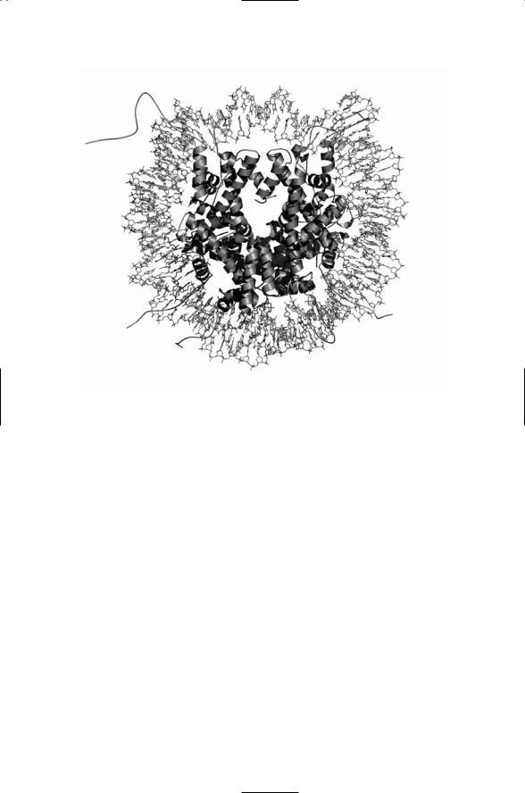
Nucleosomes are composed of 146 base pairs of DNA wrapped about a histone octamer. The duplex DNA is wrapped about a histone core composed of H2A, H2B, H3, and H4 histone pairs. The core nucleosome unit (shown in Figure 2.4) is connected by a linker segment (called H1) of chromatin to the next nucleosome core unit. Each nucleosome has the DNA wrapped about 1.75 times about the histone core forming a bead-like structure roughly 11 nm in diameter. The nucleosomes are themselves organized into 30-nm diameter solenoidal structures, and these next larger structural units are further wound into loops, and then into minibands, and finally into tightly coiled chromosomes. By this means a meter or so of DNA is organized into a compact unit that fits into a region a few microns in each linear dimension.
Growing crystals suitable for use in X-ray crystallography is often the most difficult step in applying the technique to biomolecules, especially large ones. In the case of the nucleosome, the challenge was in getting the phases aligned. The exact positioning of the nucleosome along a length of DNA is known as the phase. In order to create a crystal suitable for highresolution X-ray crystallography the DNA and histone core of each nucleosome must have the same phase. This problem was solved through the development of a bacterially derived palindrome (“reading” the same forwards and backwards) DNA sequence that is able to attach to the histone core in a repeatable (same phase) way. Multiple copies of these sequences were inserted into bare histone cores taken from chicken red blood cell nuclei. The resulting nucleosome and histone core structures are displayed in Figures 2.1 and 2.2.
The transcription machinery and regulatory elements must have access to DNA in order for transcription to occur. In its fully wound form, most of the DNA binding sites are blocked, and are inaccessible. The DNA material that is to be transcribed or replicated must be unwound and separated
2.2 The Highly Organized Interphase Nucleus |
23 |
FIGURE 2.1. Structure of the nucleosome revealed by X-ray crystallography at 2.5 Å resolution: Shown in the figure is the double-stranded DNA wrapped around a histone core. The DNA molecule is depicted in a stick model highlighting the base pairings between complementary strands, and the histones are shown as ribbons and strings. [From Harp et al. [2000]. Acta Cryst. D, 56: 1513–1534. Reprinted with permission from the authors.]
sufficiently to expose the binding sites. Steric blockages are relieved, or alternatively enhanced, by proteins that interact with and modify DNA structure, and also by proteins that alter histone structure. In eukaryotes, the ability to influence gene expression by modifying histone structure adds a further layer of control to that available through the protein-DNA binding. Multiple control points are situated on histone tails that extend out from the histone core beyond the DNA. As can be seen in Figure 2.2, these sites are well exposed, thereby providing regulatory elements with ease of access.
2.2 The Highly Organized Interphase Nucleus
The interphase nucleus is a highly organized structure. The cycle of cell growth and division passes through four stages. The first of these stages (G1) is a cellular growth stage that prepares the way either for entry into a cell

24 2. The Control Layer
FIGURE 2.2. Structure of the histone octamer revealed by X-ray crystallography at 2.5 Å resolution: Depicted are the H2A, H2B, H3, and H4 core histones without the surrounding DNA. Tails from the H2A, H2B, H3, and H4 histones are represented by strings. These structures extend out from the core and interact with DNA and with neighboring nucleosomes. [From Harp et al. [2000]. Acta Cryst. D 56: 1513–1534. Reprinted with permission from the authors.]
division series of stages (S, G2, and M), to growth arrest (G0), or to apoptosis (cell death). Cells that do not undergo growth arrest or apoptosis enter a synthesis (S) stage where the DNA is replicated in preparation for mitosis. This stage is followed by a further mitosis preparatory stage (G2) where RNAs and proteins required for mitosis are synthesized. Finally, the cell enters into mitosis (M) where the cell divides to produce two offspring. The three stages preceding mitosis are collectively referred to as interphase.
Chromosomes are not randomly distributed in the nucleus during interphase. Instead, they occupy distinct chromatin territories and are positioned in a way that reflects their replication status. The chromatin territories are partitioned into ~1Mbp-chromatin domains. Interchromatin spaces separate the chromosome territories from one another. Gene-rich early replicating chromatin is sequestered from gene-poor chromatin, and from mid-to-late replicating chromatin. Early replicating chromosomes are located in the nuclear interior, while inactive and late-replicating chromo-
2.2 The Highly Organized Interphase Nucleus |
25 |
somes are situated at the periphery of the nucleus near or in contact with the nuclear envelope.
The nuclear architecture facilitates transcription and splicing. Highly condensed chromatin serves to repress transcription while an open formation allows access of transcription and splicing factors to the sites of transcription. Chromosomes containing active regions of transcription are more open in their organization than those not actively undergoing transcription. The active regions typically lie on the outer portion of the chromosome territories. They extend into the interchromatin spaces to permit a maximal exposure of the surface of active genes to transcription and splicing factors; the two activities, transcription and splicing, are synchronized with one another.
The nucleus has several other kinds of structures in addition to chromatin territories. The additional structures—nucleoli, coiled (Cajal) bodies, and interchromatin granule clusters—are nonmembrane-associated organelles. They are used for sequestering and storage, and for preparation and assembly of materials used for gene transcription, pre-mRNA splicing, and protein synthesis. They are highly dynamic structures that form and reform about local concentrations of materials that process RNA molecules. In more detail:
•The nucleolus is the site of ribosome assembly, and forms in response to transcription of ribosomal DNA (rDNA), the genes that encode ribosomal RNA. Preribosomal RNA (pre-rRNA) units associate with the small nucleolar RNAs (snoRNAs), and are processed to form mature rRNAs. Pretransfer RNAs (pre-tRNAs) are imported into the nucleolus and processed there. The mature rRNAs associate with the tRNAs and a large ensemble of ribosomal proteins to form ribosomes. Once assembled the ribosomes are exported to the cytoplasm.
•Cajal bodies (CBs) are the sites of assembly of transcription complexes, and contain high concentrations of RNA Pol I, Pol II, and Pol III. Recall that there are three kinds of eukaryotic polymerases. RNAP Pol II transcribes pre-mRNAs and most splicing RNAs. The other RNA polymerases transcribe rRNA subunits, pre-tRNAs, and the U6 splicing RNAs. A cell may contain up to ten Cajal bodies. Once assembled within a CB, pol I complexes translocate to the nucleoli, where they transcribe rRNA genes. Pol III assemblages diffuse to the U6 small nuclear RNA (snRNA) involved in splicing, 5S rRNA and tRNA transcription sites, and Pol II complexes diffuse out of the CBs and over to mRNA and snRNA transcription sites.
•Interchromatin granule clusters (IGCs), or speckles, are distributed throughout the inter-chromatin spaces. These organelles are enriched in U1, U2, U4/U6, and U5 small nucleolar ribonuclear particles (snRNPs) and other components of the splicing machinery. (These will be discussed in detail in Chapter 15.) The localization of splicing factors within the
26 2. The Control Layer
IGCs is not static, but instead changes in time in a way that reflects the transcriptional activity underway. Splicing factors diffuse in and out of the granules to and from sites of transcription activity.
2.3Covalent Bonds Define the Primary Structure of a Protein
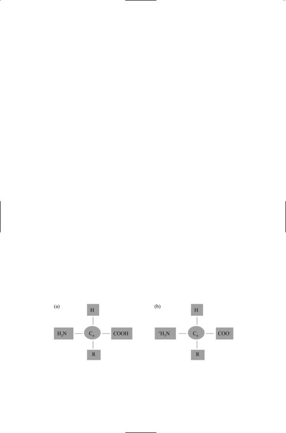
Proteins have a primary structure, a secondary structure, and a tertiary structure. Proteins constructed from more than one polypeptide chain have a quaternary structure, as well. A protein is a linear chain of amino acids, connected to one another by means of peptide bonds. Twenty different amino acids contribute to the formation of proteins. All have the same basic architecture shown in Figure 2.3. There is a central carbon atom, called an alpha carbon. Tied to it are an amino group, a carboxyl group, and a hydrogen atom. The fourth bond is with atoms belonging to the side chain (R). Two forms, uncharged and charged, are depicted in Figure 2.3.
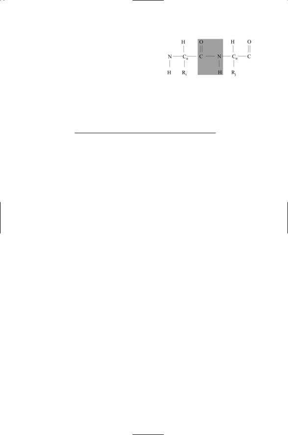
The starting point in determining a protein’s structure is the linear sequence of amino acids. These sequences are unique for each type of protein. In a protein, each amino acid residue in a sequence is linked to the next amino acid residue by means of a covalent peptide bond, and for that reason proteins are commonly referred to as polypeptides. The primary structure of a protein macromolecule is its covalent structure, including all covalent disulfide bonds that form during folding. The core of the amino acid is the repeating NCaC unit. These units, covalently linked to one another by peptide bonds, form the main chain, or backbone, of the protein in which a carboxyl group of one amino acid and an amino group of the next amino acid in the sequence are linked together by a peptide bond. The result of joining amino acids by means of peptide bonds is depicted in
Figure 2.4. By comparing Figures 2.3 and 2.4 one can see that two hydrogen atoms and an oxygen atom are removed during peptide bond formation. These are restored during hydrolysis of the peptide bond.
FIGURE 2.3. Amino acid structure: In amino acids, the bound organic groups, denoted by the “R” symbols, are termed side chains. (a) Form of an uncharged amino acid. (b) Form of a dipolar, or zwitterion, amino acid.
2.4 Hydrogen Bonds Shape the Secondary Structure |
27 |
FiGURE 2.4. Two amino acids joined together by means of a peptide bond.
TABLE 2.1. Properties of common secondary structure: The second column lists the psi (y) values and the third column the phi (j) values for the ideal structural elements. The last column list the number of residues per helical or strand turn.
Secondary structure |
psi |
phi |
n |
Alpha helix |
-57.8 |
-47.0 |
3.6 |
3.10 helix |
-74.0 |
-4.0 |
3.0 |
Pi helix |
-57.1 |
-69.7 |
4.4 |
Beta strand |
-139.0 |
+135.0 |
2.0 |
|
|
|
|
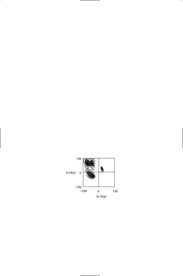
The peptide CN link is partially double bond in character. Consequently, the atoms cannot rotate freely about the CN bond axis. Instead, the four atoms highlighted in Figure 2.4 form a rigid planar peptide unit. The single bonds on each side of the peptide bond, that is, the CaC and the NCa bonds, are quite flexible with respect to rotations (torsions) about the bond axes. The angle of rotation about the CaC axis is usually denoted as psi (Y) and that about the NCa axis as phi (j).The identification of (Y, j) for each amino acid residue completes the specification of the main chain conformation. Not all values of Y and j are allowed, due to steric constraints associated with the van der Waals radii. Instead, a Ramachandran plot of the (Y, j) values for a given protein will have areas of high density of points indicative of that protein’s b sheet and a helix secondary structural content. Large regions of the Ramachandran plot will be empty corresponding to (Y, j) values that are sterically forbidden. Characteristic psi and phi values for several kinds of helical structures, and for the beta sheet, are presented in Table 2.1. The tabulated values differ considerably from one another, and data points falling about these values will be well separated in a Ramachandran plot.
2.4 Hydrogen Bonds Shape the Secondary Structure
Portions of the polypeptide chain that occur near one another tend to form geometrically regular, repeating structures during the process of folding into a three-dimensional functional form. The most commonly encountered regular structures are alpha helices (a helices), beta sheets (b sheets) and
28 2. The Control Layer
turns. Sequences that do not form one of these kinds of regular structures are grouped together in a general category called random coils. The arrangement of these features within the protein constitutes the protein’s secondary structure.
Alpha helices and beta sheets are stabilized by hydrogen bonds between amide (NH) and carbonyl (CO) groups. In alpha helices, the hydrogen bonds form between a carbonyl group on the ith residue and the amide group on the (i + 4)th residue lying below it. In beta sheets, the hydrogen bonds form between the carbonyl group lying on one strand and the amide group situated immediately adjacent to it on the other strand. The beta strands can be oriented in either a parallel or an antiparallel manner. Some proteins are mostly alpha helix; other mostly beta sheet, and some are a mixture of the two. These are referred to as a/b if the two types of structural element are mixed and a + b if they remain distinct.
Alpha helices are about 10 residues in length and these structures account for about a third of the amino acid residues in a typical protein. Beta sheets are typically 6 amino acid residues in length and they account for a quarter of the residues. Turns allow the chain to reverse direction. They along with loops are usually located on the protein surface and thus contain polar and charged residues. Protein structures tend to be compact with little space left open in the interior. Hydrogen bonds formed in the protein interior neutralize buried polar groups. Structural compactness and the use of hydrogen bonds to neutralize interior polarity drive the formation of the alpha helices and beta sheets.
FIGURE 2.5. Ramachandran plot: Shown is a stereotypic contour plot of the distribution of main chain torsion angles j and Y determined from high-resolution X- ray crystallographic data. Darker shading denotes more highly favored torsion angle combinations. Blank (white) regions represent conformations that are sterically forbidden. The most favorable conformations for alpha helices and beta sheets are concentrated into the three main regions. Beta sheet torsion angles appear as a double-peaked distribution in the upper left quadrant of the plot. The distribution of torsion angles for right handed alpha helices peaks in the lower left quadrant, and that for left handed alpha helices peaks in the upper right quadrant. Each of the 20 amino acids populates similar, but slightly different, portions of the Ramachandran plot.
2.6 Protein Secondary Structure Elements and Chain Topology |
29 |
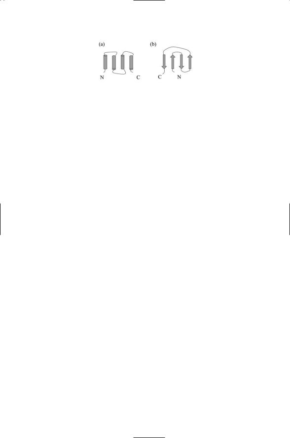
FIGURE 2.6. Topology of two common structural motifs: (a) The four-helix bundle, and (b) the Greek key. In the plots, cylinders represent alpha helices, and arrows oriented in the amino-to carboxyl-terminal direction denote beta strands. The helices and strands are connected by short turns and longer loops. The four-helix bundle can be regarded as a pair of helix-turn-helix structures. The Greek key is assembled from two pairs of antiparallel beta strands.
2.5Structural Motifs and Domain Folds: Semi-Independent Protein Modules
In the alpha helix, the backbone forms the inner portion of structure while the side chains rotate outward. Several different arrangements of helices can be formed. One arrangement is as a stand-alone helix.Another arrangement is as coiled coils in which two or more helices are twisted about one another to make a particular stable structure.This type of structural motif is common in muscle and hair. Yet another kind of structure is formed when two helices are connected by a flexible loop. This structure, a helix-loop-helix, is commonly encountered in DNA-binding proteins. More generally, when secondary structure elements associate with other secondary structures they form supersecondary structures. In this process, strands, loops, and turns connect the alpha helices and beta sheets, and form different kinds of stereotypic compact structures. When these structures involve sequential arrangements of two or three elements they are referred to as structural motifs.
Domains are structural units formed by sequential combinations of more than three secondary structures, and by stable associations of two or more structural motifs. Typical examples of a domain is the four-helix bundle formed by two pairs of alpha helices connected by a loop; another typical domain is the five-element, alternating arrangement of beta sheets and alpha helices known as a Rossman fold, and still another is a two pairs of beta sheets known as a Greek key. Domains are semi-independent folding units, and for that reason they are often referred to as domain folds.
2.6Arrangement of Protein Secondary Structure Elements and Chain Topology
A number of empirical rules can be formulated that summarize the packing of amino acid sequences into three-dimensional folding domains. These rules are quite general and summarize the observation that secondary struc-