

The Elisa guidebook
.pdfFig. 2.
Indirect ELISA. Antibodies from a particular species react with antigen attached to the solid phase. Any bound antibodies are detected by the addition of an antispecies antiserum labeled with enzyme. This is widely used in diagnosis.
The indirect system offers the advantage that any number of antisera can be examined for binding to a given antigen using a single antispecies conjugate. Such systems have been heavily exploited in diagnostic applications, particularly when examining (screening) large numbers of samples. One problem that
Page 17
such systems have is the varying degree of nonspecific binding in individual sera. This tends to widen the dispersion (variability) in assay results and, therefore, increases the need to process many sera to assess confidence.
2.3¡ª
Sandwich ELISA
Sandwich ELISA can be divided into two systems, which have been named the direct sandwich ELISA and the indirect sandwich ELISA.
2.3.1¡ª
Direct Sandwich ELISA
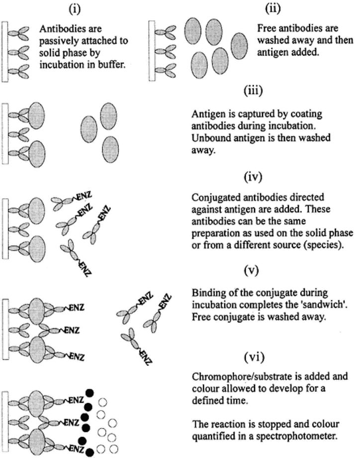
The direct sandwich ELISA illustrated as follows and in Fig. 3.

The direct sandwich ELISA, involves the passive attachment of antibodies to the solid phase (stages i and ii). These antibodies (capture antibodies) then
Page 18
Fig. 3.
Direct sandwich ELISA. This system exploits antibodies attached to a solid phase to capture antigen. The antigen is then detected using serum specific for the antigen. The detecting antibody is labeled with enzyme. The capture antibody and the detecting antibody can be the same serum or from different animals of the same species or from different species. The antigen must have at least two different antigenic sites.
bind antigen(s) that are added in stage iii. The antigen(s) are diluted in a blocking buffer to avoid nonspecific attachment to the solid phase. Here, the components of the blocking buffer should not contain any antigens that might bind to
Page 19
the capture antibodies. After incubation and washing, an antibody¨Cantigen complex is attached to the solid phase (stage iv).
The captured antigen (sometimes referred to as trapped) is then detected by the addition and incubation of enzyme-labeled specific antibodies in blocking buffer (stage v). Thus, this is a direct conjugate binding with the antigenic targets on the captured antigen. This second antibody can be the same as that used for capture, or be different in terms of specific animal source or species in which it was produced. After incubation and washing (stage vi), the bound enzyme is developed by the addition of substrate/chromogen (stage vii), then stopped (stage viii), and finally read using a spectrophotometer (stage ix).
Since a single enzyme-conjugated antibody is used, the system is limited to the specificities and properties inherent in that particular antibody set. This limits the versatility of the test¡ªe.g., each antibody preparation used must be labeled (for different antigens)¡ªin the same way as the direct ELISA was limited to single antibody preparations.
The system also is limited in that antigens must have at least two antigenic sites (epitopes), since both the capture and the detecting antibodies need to bind. This can limit the assay to relatively large antigenic complexes.
The capture antibody (on the solid phase), and the detecting antibody, can be against different epitopes on an antigen complex. This can be helpful in orienting the antigenic molecules so that there is an increased chance that the detecting antibodies will bind. It can also be an advantage when investigating small differences between antigenic preparations by the use of different detecting antibodies and a common capture antibody, and more versatile and hence appropriate systems are dealt with in Subheading 2.3.2. The use of exactly the same antibodies for capture and detection (e.g., mAbs) can lead to problems whereby there is a severe limitation of available binding sites for the detector. The size and the spatial relationship (topography) of the epitopes on the antigenic target is also critical and can greatly affect the assay.
2.3.2¡ª
Indirect Sandwich ELISA
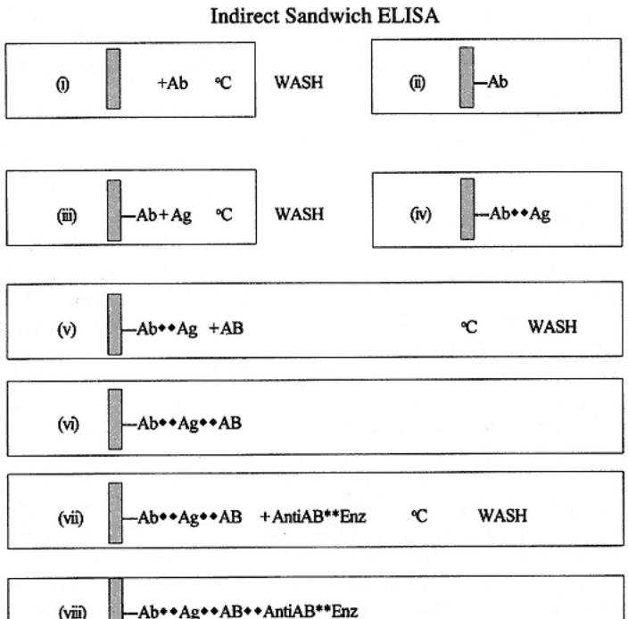
Indirect sandwich ELISA is illustrated as follows and in Fig. 4. In indirect sandwich ELISA assay stages i¨Civ are quite similar to those of the direct sandwich ELISA. Thus, antibodies are passively attached to the solid phase and antigen(s) are captured. However, stage v involves the addition of detecting antibodies. In this case, the antibodies are not labeled with enzyme. After incubation and washing (stage vi), the detecting antibodies are themselves detected by addition and incubation with an antispecies enzyme conjugate (stage vii). The bound conjugate is then processed as described in the other systems (stages xiii¨Cix).
The advantage to this assay is that any number of different sources of antibodies (samples) can be added to the captured antigen, provided that the spe-
Page 20
cies in which it was produced is not the same as the capture antibody. More specifically, the enzyme conjugated antispecies antibody does not react with the antibodies used to capture the antigen. It is possible to use the same species of antibody if immunochemical techniques are used to select and produce particular forms of antibodies and with attention to the specificity of the enzyme conjugate used. Thus, as an example, the capture antibody could be processed to a bivalent molecule without the Fc portion (also called F(ab')2 fraction). The detecting antibodies could be untreated. The enzyme conjugate could then be
Page 21

Fig. 4.
Indirect sandwich ELISA. The antigen is captured by a solid-phase antibody. Antigen is then detected using antibodies from another species. This in turn is bound by an antispecies conjugate. Thus, the species of serum for the coating and detecting antibodies must be different; the antispecies cojugate cannot react with the coating antibodies.
an antispecies anti-Fc portion of the Ig molecule. Thus, the conjugate would react only with antibodies containing Fc (and therefore not the capture molecules). The need to devise such assays depends on the reagents available.
Page 22
It may be that a mAb is available that confers a desired specificity as compared to polyclonal sera or that one wishes to screen a large number of mAbs against an antigen that must be captured (it may be at a low concentration or in a mixture of other antigens). In this the case use of F(ab')2 polyclonal sera is unsuccessful; therefore, the preparation of fragments for the capture antibody is worthwhile, and in fact, relatively easy-to-use kits are available for this purpose. The use of a commercially available antimouse Fc completes the requirements.
2.4¡ª
Competition/Inhibition Assays
The terms competition and inhibition, describe assays in which measurement involves the quantification of a substance by its ability to interfere with an established pretitrated system. The systems involve all the other ELISA configurations already described. The assays also can be used for the measurement of either antibody or antigen. The terminology used in the literature can lead to confusion the term blocking-ELISA is also frequently used to describe such assays. This section describes the possible applications of such methodologies, indicating the advantages and disadvantages. C-ELISA (competition ELISA) and I-ELISA (inhibition ELISA) are used to describe generally the assays involving the elements described in Subheading 2.1.¨C2.3. and the particular application of competitive or inhibition assay dealt with specifically for each different system examined. Reference should be made to the preceding descriptions of the basic systems for direct, indirect, and sandwich ELISAs, which are the basis of the C-I assays.
2.4.1¡ª
Direct C-ELISA: Test for Antigen
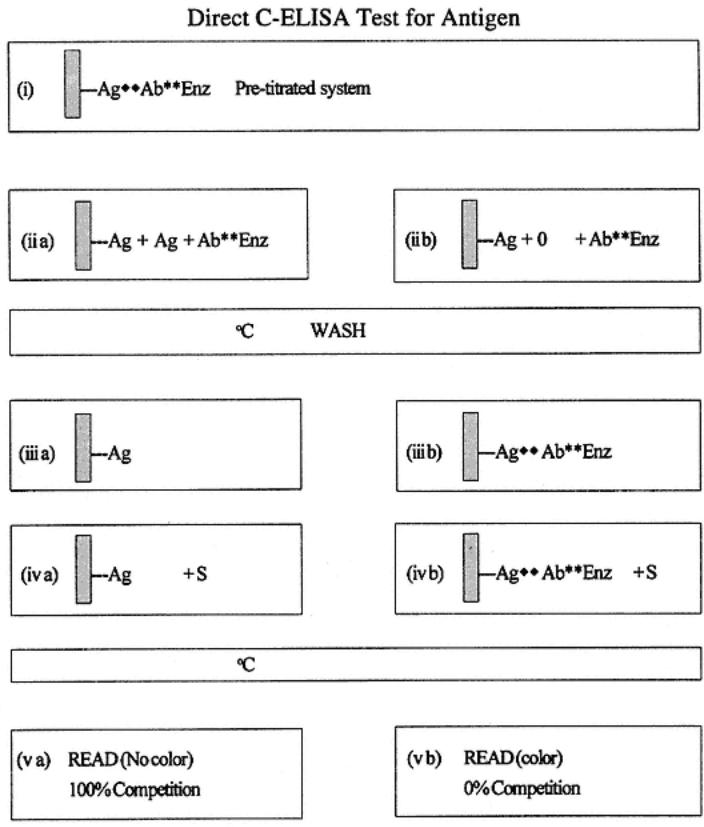
Direct C-ELISA testing for antigen is described and shown in the following diagram and in Fig. 5. A pretitrated, direct system is challenged by the addition of antigen. The effect of the addition is measured by a decrease in expected color of the pretitrated system (used as a control). Thus, the competition stages proper start at stage iii, in which a sample is added to a solid phase that has the system antigen already passively attached. This sample is diluted in blocking buffer to prevent antigen binding to the solid phase nonspecifically. At this stage, nothing should happen in terms of binding. The pretitrated dilution of labeled antibody (specific for the solid-phase antigen) is then added. The competitive phase now begins where, if the test antigen introduced is the same or similar to the solid-phase antigen, it will bind with the introduced labeled antibodies (stage ii a). The degree of competition in time depends on the relative concentration of molecules of the test and solid-phase antigen (and to the degree of antigenic similarity). After incubation and washing, the amount of labeled antibodies in the test is quantified after addition of substrate, and so forth. When
Page 23

there is no antigen in the test sample, or when the antigenic similarities are limited, there is no binding with the labeled antibodies (stage ii b); thus, there is nothing to prevent (compete with) the binding of the labeled antibodies (stage iii). The net result is that for samples containing antigen, there is competition affecting the pretitrated expected color, whereas in negative samples there is no effect on the pretitrated color.
2.4.2¡ª
Direct C-ELISA: Test for Antibody
Direct C-ELISA testing for antibody is illustrated in the diagram at the top of page 25 and in Fig. 6. The system here is the same as that for the test of antigen; however, the measurement or comparison of antibodies is being made.
Page 24